
Abstract
Introduction
S
Low-level laser therapy (LLLT) has been used by physical therapists in the treatment of cutaneous ulcers in order to accelerate the healing process, 9 being described as an adjuvant for regulation of cell proliferation 10 and inflammatory process. 11 It is important to emphasize that bacterial colonization of ulcers can interfere with the healing process. In an early experiment, Lanzafame et al. 12 inoculated methicillin-resistant S. aureus in a murine pressure ulcer model (Balb/C mice) and observed no positive results following blue-laser irradiation at 455±5 nm (350 mW, 1 cm diameter, for 15 min). There are scant studies demonstrating the effect of blue laser on bacterial control, and among the existing studies, few used this light spectrum in different bacterial species. 13 –19
One of the few studies found in the literature using violet laser in bacterial cultures was the in vitro work performed by Guffey et al., 15 who combined wavelengths of 405 and 880 nm (1, 3, 5, 10, and 20 J/cm2) to find a dose-dependent effect for inhibiting P. aeruginosa and S. aureus in all dosages tested. In parallel, the same authors 14 conducted a similar study with violet and blue lasers at 405 and 470 nm (1, 3, 5, 10, and 15 J/cm2) in P. aeruginosa and S. aureus, demonstrating inhibitory effects regarding both wavelengths. Enwemeka et al. 17 assessed the effect of a 405 nm laser (1–60 J/cm2) on in vitro bacterial growth of two strains of S. aureus resistant to methicillin, obtaining an almost complete inhibition in both. In a further study, these authors performed a similar experiment with 470 nm laser (1–60 J/cm2) at the same fluences, achieving similar results. 16
Determining the most efficient fluence for inhibiting bacterial cultures is the main goal of all researchers and practitioners in this area. However, no study was found in the literature that has evaluated, in only one experiment and at different incubation periods, the effects of blue laser on the main bacterial species that usually colonize ulcers. Therefore, the objective of the present study was to analyze the influence of blue laser on the in vitro bacterial growth of S. aureus, E. coli, and P. aeruginosa at different fluences and incubation times.
Materials and Methods
Samples
Strains of S. aureus ATCC 25923 (gram-positive), P. Aeruginosa ATCC 27853 (gram-negative), and E. coli ATCC 25922 (gram-negative) were obtained from the American Type Culture Collection (ATCC) and provided by the Department of Medical Clinics – Infectious Diseases Division of the Ribeirão Preto School of Medicine at the University of São Paulo. Bacterial cultures were kept on Petri plates (90×15 mm) containing Mueller–Hinton culture medium (BD®, New Jersey, PA), seeded, and incubated for 24 h at a temperature of 37°C. After this period, all the strains were stored in TSB culture medium (Trypticase Soy Broth, BD®, New Jersey, PA) at 4°C with 20% glycerol.
Experimental procedure
After bacterial growth, the cells were suspended in saline solution with turbidity of 0.5 according to the McFarland scale [1.5×10⁸ colony forming units (CFU)/mL]. Next, five serial dilutions were made until a concentration of 1.5×10³ CFU/mL was achieved. An aliquot of 300 μL of this suspension was transferred to the wells of the microtitulation plate (diameter of the wells: 6.4 mm; depth: 17 mm; volume: 360 μL) and then were irradiated. Next, the samples were shaken for homogenization and a fraction of 100 μL of each suspension was spread over the surface of the Petri plates (90×15 mm) containing Mueller–Hinton culture medium and incubated at 37o C. The counts of CFU were performed after 24 and 48 h by means of the software ImageJ® (
Each laser fluence was applied to three different samples, with each experiment being repeated three times on consecutive days (n=9). The experiments were divided into blocks of only one bacterial species, totaling 162 samples (6 intensities×3 repetitions×3 blocks×3 bacterial species). There was contamination in 23 samples; therefore, it was necessary to repeat four blocks of the experiment.
For the laser irradiation, the Laserpulse equipment (Ibramed®, Amparo, São Paulo, Brazil) was developed by the manufacturer specifically for conducting this study (Table 1), and was applied with continuous emission, average power of 70 mW, and at a wavelength of 450 nm. The fluence presented by LLLT devices takes into account the diode irradiation area as a parameter for calculation, which may vary depending upon the manufacturer or equipment. In order to standardize this important parameter, for our experiment, we considered the area of irradiation of 1 cm2 for calculating the fluences of 0 (control), 3, 6, 12, 18, and 24 J/cm2, resulting in irradiation times of 0, 43, 86, 172, 257, and 343 sec, respectively. The control wells were not irradiated, remaining in the same experimental conditions of room light.
The energy density (fluence) irradiation was calculated according to the formula below:
Because the samples were irradiated in a colorless liquid medium (saline solution), the experiments were performed before the beginning of the study to evaluate the laser transmissivity in the microtitration plate, saline solution, and bacterial cultures (at the same concentration and volume used throughout the experiment). The samples were positioned between the diode and the power meter. Therefore, it was observed that the initial average power of the laser was 71.9±0.1 mW, which was attenuated after crossing over the bottom of the plate (63.7±0.46 mW), saline solution (69.4±0.4 mW), and bacterial lines (S. aureus: 67.2±1.03 mW; P. aeruginosa: 67.1±0.3 mW; and E. coli: 68.4±0.4 mW), separately (p<0.05). These findings demonstrate a small attenuation of irradiation after being applied to the bacterial cultures.
All irradiation occurred directly, punctually, and perpendicularly to the plate, with the LLLT device mounted over the area to be irradiated by means of a support at a distance of 2 mm. The LLLT device was previously calibrated before irradiation of each series of plates by using a power meter (average power) with PM3 sensor head of 0.5 mW to 2W and broadband sensor (RoHS) (Coherent®, Staunton, VA) as suggested by Guirro et al. 20
All data were tested for normality by using the Shapiro–Wilk test. Data on CFU counts converted into log values (logarithm) and showing nonparametric distribution were submitted to Kruskal–Wallis and Dunn's post-hoc tests. Analysis of power and transmissibility of the diodes showed parametric distribution and were submitted to repeated measure ANOVA and Tukey's post-hoc tests. For analysis of the correlation between CFU and laser fluence, the Spearman's correlation test was used. All the tests were performed by using the statistical software SPSS 17.0 (IBM®, Chicago, IL) at significance level of 5% (p<0.05)
Results
The blue laser light inhibited the growth of all bacterial species at fluences >6 J/cm2 compared with controls (p<0.05), but no differences were found when they were compared among themselves (Table 2). It was also observed that there was a correlation ranging from moderate (S. aureus and P. aeruginosa) to strong (E. coli) between the fluences used and bacterial inhibition (Table 3). With regard to the experimental times following irradiation, we can state that there was no difference in bacterial growth between 24 and 48 h (Table 2 and Fig. 1).
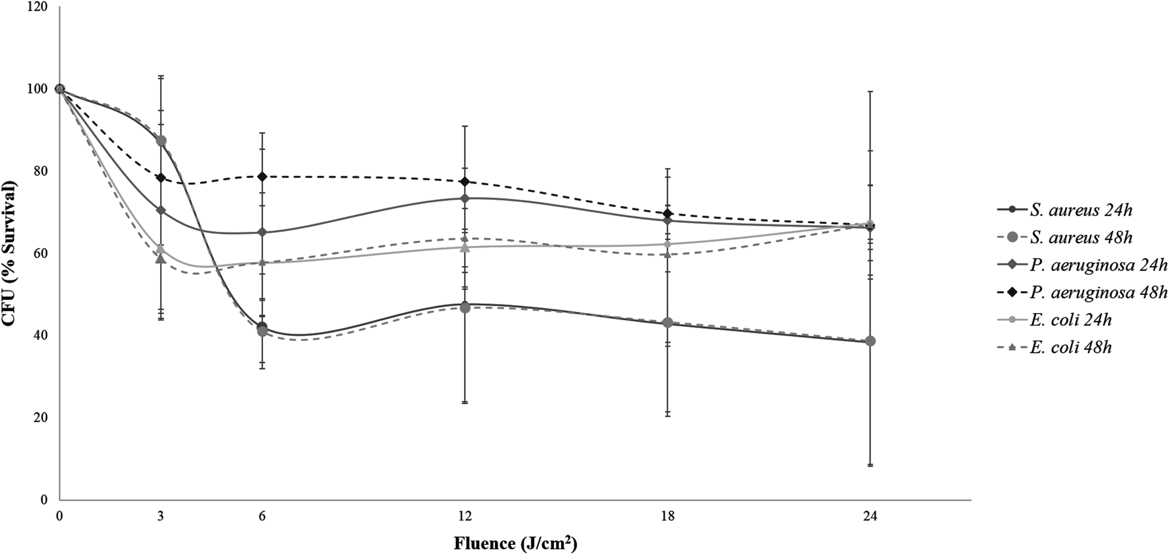
Effect of blue laser 450 nm, at fluences of 0, 3, 6, 12, 18, and 24 J/cm2, on Staphylococcus aureus, Pseudomonas aeruginosa, and Escherichia coli, after 24 and 48 h of irradiation.
p<0.05
vs. control for the same time.
vs. 3 J/cm2 for the same time.
CFU, colony-forming units.
Discussion
In the present study, we used blue laser (450 nm, 70 mW) for inhibiting the growth of S. aureus at fluences >6 J/cm2, with an inhibition rate of 57.9%. However, Guffey and Wilborn 14 found no significant result by using blue laser at higher power and lower fluences (470 nm; 150 mW, at 1, 3, and 5 J/cm2), even reporting a stimulation of 15.5% at a fluence of 3 J/cm2. Paradoxically, inhibitions of 61.7% and 56.1% were found at fluences of 3 and 5 J/cm2, respectively, for a wavelength of 405 nm (160 mW). Nevertheless, this wavelength is close to the ultraviolet light spectrum, whose antibacterial effects are well known. 21
Corroborating the present study, Enwemeka et al. 17 used 405 nm violet laser at high power (500 mW) and achieved significant results by inhibiting bacterial growth in two strains of S. aureus (MRSA), namely, US-300 and IS-853. In a further study, Enwemeka et al. 22 performed a similar experiment with 470 nm blue laser (150 mW) at the same fluences (0, 1, 3, 5, 7, 9, 11, 13, 15, 17, 19, 25, 30, 35, 40, 45, 50, 55, and 60 J/cm2), reporting a bacterial inhibition similar to that of their previous study, that is, 91.7% (US-300) and 94.8% (IS-853) at an energy density of 55 J/cm2.
In a recent study, Guffey et al. 19 analyzed the effect of the violet laser therapy (405 nm, 160 mW) with daily irradiations of 9 J/cm2 on cultures of S. aureus for 7 days consecutively. Cultures grown within 24 h following irradiation were inoculated into the plate and then irradiated again, with this procedure being repeated for 7 days. The results indicated greater inhibition (59.49%) after the 4th day of irradiation, with the inhibitory effect decreasing from the 5th to the 7th day of treatment. The authors stated that bacteria could become resistant to irradiation as a result of the treatment, proliferating more over time. We believe that such a hypothesis should be considered with reservation, because these effects must not be associated with the repetition of treatment, but with the possible increase in bacterial concentration during the therapeutic process, as the author did not mention any maintenance of the initial dilution for all irradiations. Corroborating this statement, Bumah et al. 18 assessed the effect of violet laser therapies of 405 nm (18 mW) and blue laser 470 nm (25 mW) at fluences of 0, 1, 3, 45, 50, 55, 60, and 220 J/cm2 on three different concentrations of S. aureus (3×10⁶; 5×10⁶ and 7×10⁶ CFU/mL), reporting lower inhibition at higher bacterial concentrations and demonstrating that bacterial density is relevant for a successful treatment.
In the present study, we have assessed the bacterial behavior within 24 and 48 h following laser irradiation, and no relevant alterations in their growth were observed after 24 h. The 48 h period was considered because Marius et al. 23 had reported that the species analyzed in their study showed growth capacity of up to 48 h, a fact not confirmed in our work, however. Therefore, our study demonstrated that the pattern of bacterial growth within the 24 h period was enough for analysis; therefore, it was not necessary to follow it over a longer period of time.
In the present study, our best results were obtained at a fluence of 6 J/cm2, whose values were maintained until the highest dosage tested (24 J/cm2). Similarly, Guffey and Wilborn 14 observed inhibition of P. aeruginosa at fluences of 5, 10, and 15 J/cm2 by using violet lasers of 405 nm (160 mW) and 470 nm (150 mW). In a parallel study, the same authors 15 evaluated the effect of the association between violet lasers (405 nm, 200 mW) and infrared light (880 nm, 250 mW) on the growth of P. aeruginosa, obtaining an almost complete inhibition at a fluence of 20 J/cm2, differently from the present study. These findings reinforce the hypothesis that irradiation parameters can influence the results directly, mainly regarding the power.
Within this context, we can cite the study by Nussbaum et al., 24 who assessed the effect of laser therapy (810 nm, 15 mW) depending upon different pulse regimes (continuous; pulsed at 26, 292, 1000, and 3800 Hz). The authors reported growth of E. coli by using laser operated in continuous and pulsed mode at 1000 Hz and fluences of 1, 2, 5, and 10 J/cm2. In a parallel study, the same authors found similar results by using laser in continuous mode at same wavelength and fluence to irradiate cultures of E. coli. 25 On the other hand, our study had different results, as the growth of E. coli was inhibited by using blue laser (450 nm, 70 mW) at all fluences. Up to now, however, no study addressing the effect of blue laser on this bacterial species was found, which makes it impossible to lead a discussion based on light spectrum.
In the present study, we can observe that the cultures of E. coli and P. aeruginosa showed a similar behaviour at all dosages tested, with the growth of S. aureus being more inhibited. This fact can be explained by the morphological difference between gram-positive (S. aureus) and gram-negative (E. coli and P. aeruginosa) bacteria cells. Gram-positive bacteria have thicker cell walls, whereas gram-negative ones have thinner cell walls. 26 These structural divergences may be factors determining the penetration of laser irradiation, including biological effects. In this way, we suggest that such aspects of each bacterial species should be investigated morphologically and physiologically. Therefore, it is expected that a consensus can be reached about the results in order to establish the standardization of the best parameters to be used for each species.
Conclusions
Irradiation with blue laser light at 450 nm was capable of inhibiting the growth of S. aureus, E. coli, and P. aeruginosa strains, with the results maintained for up to 48 h following irradiation. Also, no dose-dependent relationship was found. In this way, a low fluence of 6 J/cm2 was found to be effective in inhibiting these bacteria isolated in vitro.
Footnotes
Acknowledgments
We thank the Coordination for Improvement of Higher Education Personnel (CAPES) and the São Paulo Research Foundation (FAPESP) for financial support (2011/22170-0) and the grant of the scientific initiation scholarship (2013/02974-2).
Author Disclosure Statement
No competing financial interests exist.