
Abstract
Introduction
E
Different substances may be used as canal irrigants, in an attempt to minimize the negative effects of this smear layer. 6 Sodium hypochlorite (NaOCl) solutions are commonly used because of their antimicrobial activity and ability to dissolve organic tissues. 7 Chelating agents, such as ethylenediaminetetraacetic acid (EDTA), have often been used in association with NaOCl to supplement its cleaning action and help in the removal of inorganic components of the smear layer. 8
To improve the action of irrigating agents, studies have evaluated the possibility of enhancing the contact of these substances with the root canal walls. 9,10 Agitation of these solutions, using different techniques and materials, can promote changes in their chemical and physical properties, thus improving penetration and their effects on root dentin. 11,12
Different studies have evaluated the effects of laser irradiation on removal of the root dentin smear layer, 10,13 fracture resistance, 14 and disinfection. 15 Favorable results were observed with Nd:YAG at 1320 and 1064 nm, 2 diode at 980 nm, 14 and Er:YAG 16 lasers. However, there is still no consensus on the protocol and type of laser most suitable for promoting changes in the root dentin, without adversely compromising its microstructure. Considering its use as an alternative protocol in endodontic therapy, it would be interesting to evaluate how irrigation with different laser devices affects root dentin.
To evaluate different protocols of biomechanical preparation of root canals, it is essential that minor changes in the mineral content of the structure be identifiable. Among the methods developed for the measurement of dentin mineral changes, the most satisfactory and reproducible results can be observed with measurement of cross-sectional microhardness. 5,17,18 Confocal laser scanning microscopy (CLSM) has recently emerged as an interesting method that allows morphological analysis and quantification of surface roughness of different materials and substrates. 19,20
Therefore, the aim of this in vitro study was to evaluate the effect of Nd:YAG (1064 nm) and diode (980 nm) lasers, in association with irrigating protocols, on the roughness and morphology of the root canal lumen dentin, as well as root dentin cross-sectional microhardness.
Materials and Methods
Study design
The variables being examined were root canal lumen dentin superficial roughness and cross-sectional microhardness. There were five experimental groups (n=10 each): (1) deionized water, (2) 17% EDTA, (3) 17% EDTA with 60 sec manual agitation, (4) 17% EDTA with 50 sec 980 nm diode laser (2 W) agitation, and (5) 17% EDTA with 50 sec 1064 nm Nd:YAG (1.5 W) laser agitation. The roots were not instrumented beforehand, to allow evaluation of the effects of the irrigation protocols on superficial dentin microhardness and roughness only.
Laser systems
A 980 nm wavelength gallium-aluminum-arsenide diode laser (SIROlaser 2.2, SIRONA Dental, Bensheim, Germany) system equipped with a 20 W power source was used. The laser delivery system used in the study was a 300 μm sized fiberoptic cable, at 2 W in pulse mode (10 ms–100 Hz). The actual power of the parallel fiberoptic tip was 272.60 J/cm2.
A 1064 nm wavelength Nd:YAG laser (SmartFile, DEKA, Italy) system was also used. The laser delivery system consisted of a fiberoptic cable with a 300 μm quartz fiber, at 1.5 W and in pulse mode (100 μs–15 Hz). The actual power of the parallel fiberoptic tip was 124.3 J/cm2. 21
Specimen preparation
Freshly extracted, single-rooted, bovine mandibular anterior teeth were stored in 0.1% thymol for disinfection and then washed in running water for 24 h. The teeth were examined under a stereomicroscope to select those with similar size, similar root morphology, and absence of cracks and structural defects. Periodontal tissues and calculus were removed mechanically from the root surfaces with a periodontal scaler. Twenty-five specimens were selected and sectioned transversally near the cementoenamel junction with a water-cooled diamond disc (KG Soresen, Barueri, SP, Brazil) at slow speed, so as to remove the crown and standardize the root lengths at 18 mm. To perform the test procedures, the specimens were horizontally embedded in autopolymerizing acrylic resin (Vipi Cril Plus—Vipi, Pirassununga, Brazil). Each root was sectioned longitudinally using a water-cooled diamond saw in a cutting machine (ISOMET 1000—Buehler, Lake Forest, IL), and a total of 50 specimens were prepared. Similarly to Arslan et al., 17 the acrylic blocks were drilled with adjusting screws to help stabilize the two halves of each root during execution of the irrigation protocols. The size of the block was ∼20×30×30 mm (Fig. 1). The surfaces of the specimens were ground using 800, 1000, and 1200 grit abrasive papers (Norton Abrasivos Ltda, São Paulo, SP, Brazil) with a grinding machine (MetaServ 250—Buehler, Dusseldorf, Germany) under running water, and were polished with diamond paste (Arotec, Cotia, Brazil). They were then cleaned with deionized water and an ultrasonic cleaner (Alpha 3L Plus, Ecel Indústria e Comércio LTDA, Ribeirão Preto, Brazil).
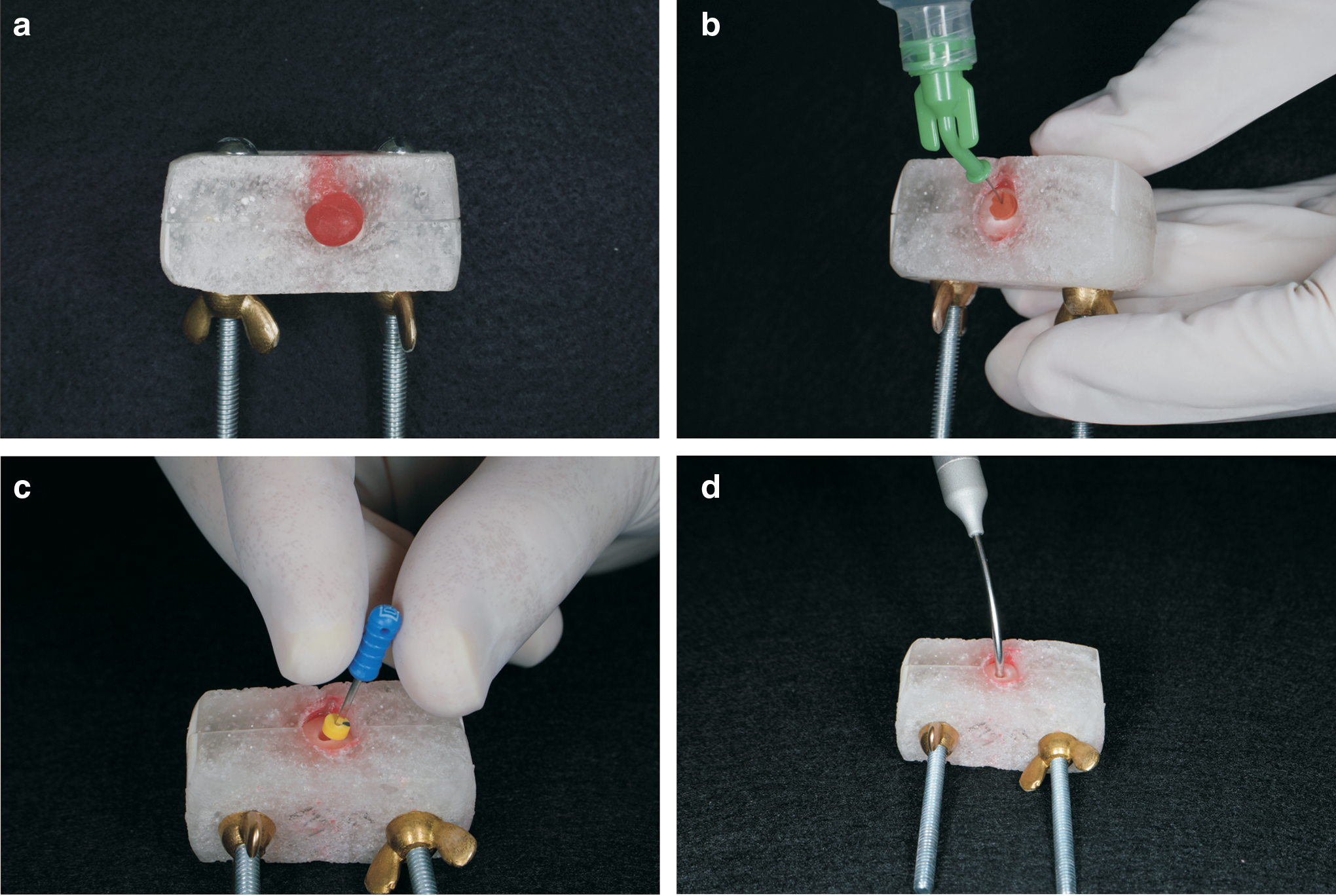
The acrylic block used to stabilize the two halves of each root during the application of irrigation protocols.
Microhardness/roughness tests were performed before and after irrigation protocols.
The percentage of hardness lost (%KHN) was calculated using the following formula, according to Carvalho et al.: 22
kNm%=100 (KHN[I]–KHN[F])/KHN(I), where KHN(I) is the average of the initial hardness measurements, and KHN(F) is the average of the final hardness values.
The percentage of roughness increase (%Sa) was calculated using the following formula:
Sa%=100 (Sa[I]–Sa[F])/Sa(I), where Sa(I) is the average of the initial roughness measurements, and Sa(F) is the average of the final roughness values.
Microhardness measurements
Dentin microhardness was measured with a Knoop indenter at 40× magnification (HMV-2000; Shimadzu Corporation, Kyoto, Japan), under a 10 g load and 15 sec dwell time. The specimens were individually fixed in the device, such that the test surfaces were perpendicular to the microindenter tip. In each root third, three indentations were made along lines parallel to the edge of the root canal lumen. The first indentation was made 30 μm from the root canal edge, and two other indentations were made at a distance of 30 μm from each other. The representative hardness value for each third was obtained by calculating the average of the results for the three indentations. 8
Superficial roughness analysis
The analysis of surface roughness was performed with an OLS4000 LEXT (Olympus Corporation, Tokyo, Honshu, Japan) CLSM. The specimens were fixed on glass slides, maintaining the surface of the root canal perpendicular to the microscope objective. For this procedure, a parallelometer and an adhesive material (Pritt, Henkel, Mexico) were used. The three root canal thirds were scanned with a 20× objective, which magnified the original size of the sample 200 times. The microscope had a 405 nm semiconductor laser that allowed for three-dimensional reading of the surface roughness (μm2). For each scanned root third, a central area of 0.04 mm2 was selected, and a measurement of surface roughness was obtained (Sa—conforms to ISO 25178). Representative images of each group were selected for qualitative topography analysis.
Irrigation protocols
After the acrylic block of each root was adapted, the root apex was closed with wax to prevent the irrigant from overflowing (Fig. 1). The root was then dried with paper points (CellPack—Dentisply, Petrópolis, Brazil) and treated as follows: 1. Deionized water (negative control group): The root canal was irrigated for 120 sec with 20 mL of deionized water as a final flush, and then dried with paper points (CellPack, Dentsply, Petrópolis, RJ, Brazil). The total volume of irrigant was 20 mL. 2. 17% EDTA only (positive control group): The root canal was irrigated for 120 sec with 10 mL 17% EDTA (Biodinâmica, Ibiporã, PR, Brazil) as a final flush. Then the specimens were irrigated with 5 mL 2.5% NaOCl (Farmácia da Terra, Ribeirão Preto, SP, Brazil) for 120 sec. This was followed by a final rinse with 5 mL distilled water. The total volume of EDTA solution was 10 mL, the total exposure time to EDTA solution was 120 sec, and the total volume of all irrigants was 20 mL. 3. 17% EDTA with manual agitation: The root canal was filled with 5 mL 17% EDTA, a size 30 file (Dentsply, Rio de Janeiro, Brazil) was placed, and the solution was agitated for 60 sec. The specimens were then irrigated with 5 mL 17% EDTA for 60 sec, followed by 5 mL 2.5% NaOCl for 120 sec. A final rinse was performed with 5 mL of deionized water. The total volume of EDTA solution was 10 mL, the total exposure time to EDTA solution was 120 sec, and the total volume of all irrigants was 20 mL. 4. 17% EDTA with 980 nm diode laser: The root canal was filled with 1 mL 17% EDTA, and the laser fiber agitated the solution with 2 W in continuous waves for 10 sec. This procedure was repeated five times, so that the total agitation was 50 sec. The laser fiber had helical motion from the apical to cervical third. The specimens were then irrigated with 5 mL 17% EDTA for 70 sec, followed by 5 mL 2.5% NaOCl for 120 sec. A final rinse was performed with 5 mL of deionized water. The total volume of EDTA solution was 10 mL, the total exposure time to EDTA solution was 120 sec, and the total volume of all irrigants was 20 mL. 5. 17% EDTA with 1064 nm Nd:YAG laser: The root canal was filled with 1 mL 17% EDTA, and the laser fiber then agitated the solution with 1.5 W at 10 Hz for 10 sec. This procedure was repeated five times, so that the total agitation was 50 sec. The laser fiber had helical motion from apical to cervical third. The specimens were then irrigated with 5 mL 17% EDTA for 70 sec, followed by 5 mL 2.5% NaOCl for 120 sec. A final rinse was performed with 5 mL of deionized water. The total volume of EDTA solution was 10 mL, the total exposure time to EDTA solution was 120 sec, and the total volume of all irrigants was 20 mL.
Statistical analysis
The Kolmogorov–Smirnov statistical test for normality revealed normal distribution only for microhardness data (p<0.05). Two-way ANOVA and Tukey's tests were performed for statistical comparison of percentage change in dentin hardness (α=0.05). Change in roughness was statistically analyzed using Kruskal–Wallis and Student–Newman–Keuls post-hoc tests (α=0.05). The paired t test was used to compare changes in dentin hardness and roughness within the same groups. All statistical analyses were performed using the SigmaStat software, version 3.5 (Systat Software Inc., Chicago, IL).
Results
Microhardness measurements
Deionized water presented no significant changes in dentin microhardness (p=0.093). All EDTA groups showed significant reduction in dentin microhardness (p<0.05), and Groups 2 (EDTA only) and 3 (EDTA with manual agitation) presented similar microhardness values (p>0.05). The Nd:YAG and 980-nm diode laser groups caused greater reduction of dentin microhardness and were significantly different from groups 2 and 3 (p<0.05). The microhardness reduction did not vary between the root thirds (p>0.05). Table 1 presents the Knoop microhardness mean and standard deviations for all studied groups and root thirds.
Same lowercase letters indicate that there was no significant difference between initial and final kNm values of each agent (paired t test, p>0.05).
Same uppercase letters indicate that there was no significant difference among the protocols and root thirds (two way ANOVA and Tukey's tests, p>0.05).
EDTA, ethylenediaminetetraacetic acid.
Superficial roughness analysis
Deionized water presented no significant changes in dentin roughness (p=0.113). All EDTA groups significantly increased dentin roughness (p<0.05), and Groups 2 (EDTA only) and 3 (EDTA with manual agitation) presented similar dentin roughness values (p>0.05). Higher values were observed with the Nd:YAG and diode lasers groups, and these were significantly different from groups 2 and 3 (p<0.05). The change in roughness change did not differ between root thirds (p>0.05). Table 2 presents the dentin roughness mean and standard deviations for all studied groups.
Same lowercase letters indicate that there was no significant difference between initial and final Sa values of each agent (paired t test, p>0.05).
Same uppercase letters indicate that there was no significant difference among the protocols and root thirds (Kruskal–Wallis and Student–Newman–Keuls tests, p>0.05).
EDTA, ethylenediaminetetraacetic acid.
Figure 2 shows representative CLSM images of the root canal surface topography for each irrigation protocol. The EDTA groups, regardless of agitation procedure used, showed irregular dentin surfaces compared with the control group. However, the laser-activated irrigation (LAI) groups presented higher numbers of widely opened dentinal tubules, indicating a higher degree of dentin erosion.
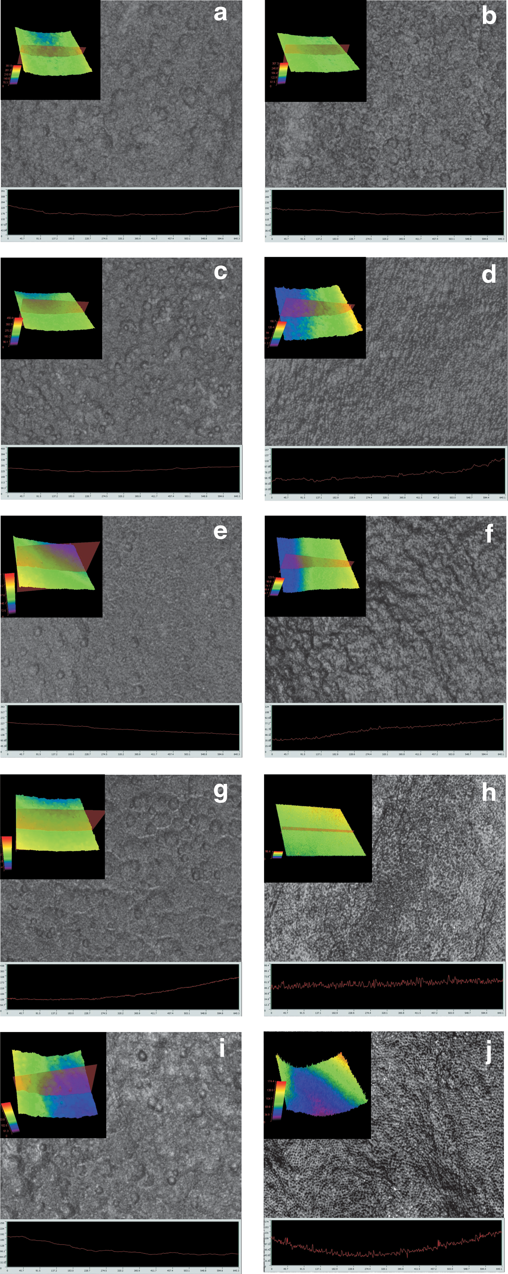
Photomicrograph representative of the confocal laser scanning microscopy (CLSM) analysis (20×). Before treatment samples: deionized water
Discussion
Irrigating solutions are used in endodontics to complement the mechanical action of endodontic instruments, when cleaning and disinfecting root canals. 23 They act by causing physical and chemical changes in the ultrastructure of the root dentin, thus decreasing microhardness and increasing dentin permeability. 8,24 Laser irradiation has been proposed in endodontics with the aim of reducing the number of microorganisms within the root canal system, 15 removing the smear layer, and improving the adhesion of sealers to the root canal walls. 13,14 The concept of LAI is based on cavitation, caused by absorption of water with midinfrared wavelength lasers, which in turn generates vapor-containing bubbles, and exerts shear force on the dentinal walls. 2,25 Therefore, LAI has been widely used to improve the chemical and mechanical effects of endodontic irrigation solutions on root dentin ultrastructure properties.
Previous studies have already evaluated the effects of LAI on root dentin microhardness, 17 smear layer removal, 10 dentinal debris removal, 26 and disinfection. 27 However, they did not evaluate the changes in root dentin roughness caused by LAI. An increase in roughness contributes to removal of the dentin with endodontics files, and promotes micromechanical interlocking of root sealers to dentin. Therefore, understanding the characteristics of the irrigated dentin surface is essential in obtaining adhesion of endodontic sealers that have different physicochemical characteristics. 28 In this study, the effects of EDTA laser agitation with different wavelengths on root dentin microhardness and roughness were evaluated and then compared with EDTA alone and EDTA with manual agitation.
It was previously reported that Nd:YAG laser at 100 Hz/15 W caused an average temperature elevation of <9°C on the root surfaces and, according to the authors, this temperature change would cause minimal damage in bone and periodontal tissues (Strakas et al 2013). 29 As the present study used 15 Hz/1.5 W for Nd:YAG lasers, the temperature increase on the root surface woud have been minimal. Similar results could be expected for diode lasers, as both lasers used similar wavelengths and tissue interaction.
As dentin microhardness may vary considerably between different teeth, the present study measured hardness values for each specimen before as well as after application of irrigation protocol. 17 Microhardness parameters were measured in a manner similar to that of Cruz-Filho et al., 8 where a Knoop hardness indenter was used with a load of 10 g, for 15 sec. This allowed good visualization of the pre- and post-treatment indentations on the most superficial layer of dentin of the root canal lumen. Arslan et al. 17 reported that 808 nm diode laser agitation of EDTA for only 40 sec caused more reduction in root dentin microhardness than did EDTA associated with ultrasonic agitation. Therefore, in corroboration with Arslan et al., 17 the present study used 50 sec of laser agitation with a 980 nm diode laser. This was seen to induce greater reduction in microhardness than EDTA alone or associated with manual agitation. It must also be mentioned that the two wavelengths used in the present study caused similar reductions in microhardness. Probable explanations include light/irrigant interaction and similar degrees of water absorption caused by Nd:YAG (1064 nm) and diode (980 nm) lasers.
In the present study, it was also observed that although the root thirds were structurally different, the dentin microhardness and roughness values, after use of irrigating solutions, were statistically similar between the thirds. These results are in accordance with those of Tartari et al., 30 who reported that despite differences in the structure of the roots thirds, the resulting alterations were found to be similar.
The effects of chemical solutions on root dentin roughness have been previously evaluated. According to Ballal et al., 31 maleic acid reduced the microhardness of root dentin similarly to EDTA, but increased the surface roughness significantly more than EDTA. Tartari et al. 30 observed that EDTA, etidronic (HEBP), and citric acid, associated with different irrigation regimens, significantly increased the root dentin roughness. Similar to these results, we observed that EDTA caused a greater increase in dentin roughness than did deionized water. However, the effects of LAI were not evaluated in previous studies. In this study, it was demonstrated that LAI significantly increases the dentin roughness to a greater extent than EDTA only or EDTA with manual agitation. Furthermore, similar to the microhardness results, the present study showed no differences in dentin roughness with the two tested lasers. These results could also be because of the similar wavelengths that promoted the same laser/solution interactions.
Although neither wavelength exhibited good interactions with water, the increased roughness and decreased microhardness with laser agitation could be explained by the laser/root dentin interaction. According to Esteves-Oliveira et al., 21 these wavelengths increase root dentin permeability. Therefore, the demineralizing effect of the EDTA used could also be increased, with deeper mineral removal.
Overall, in the present study, better results were seen when EDTA was laser agitated, as these groups presented lower dentin microhardness and higher surface roughness. According to previous studies, 2,5,10,13 these results could be related to the laser cavitation process, which improves the demineralizing effects of EDTA on the dentin walls. Therefore, this cavitation effect may also play a role in the efficient cleaning of canal walls achieved by LAI protocols. However, the CLSM roughness results also indicated a higher degree of dentin erosion in LAI groups. There has been some concern about the erosive effects of irrigation solutions, as the alterations in the dentin surface might affect its interactions with root canal filling materials and could also decrease the resistance to penetration of bacteria and apical leakage. 5 It is interesting to mention that when root canals are irrigated with NaOCl followed by EDTA, the collagen degradation with a consequent decrease in flexural strength is caused by the action of hypochlorite on the protein content of root dentin and has no association with demineralization promoted by the final rinse with EDTA. 8,32 Therefore, further studies are still required to assess the effects of critical values of roughness and lasers/irrigant interactions on the dentin bond strength of endodontic sealers and fracture resistance of the root canal, using different solution regimes.
Conclusions
Within the limitations of this study, it is possible to conclude the following:
Nd:YAG and 980 nm diode laser EDTA irradiation caused greater reduction in microhardness and increased the roughness of all root thirds, compared with EDTA only or EDTA with manual agitation.
Manual EDTA agitation had no additional effects on root dentin microhardness or surface roughness, compared with EDTA alone, regardless of the root third.
Footnotes
Acknowledgments
The authors are grateful for Fundação de Apoio à Pesquisa do Estado de São Paulo (FAPESP) #2013/23535-7, and thank Regina Guenka Palma Dibb for her contribution with CLSM analysis.
Author Disclosure Statement
No competing financial interests exist.