
Abstract
Introduction
C
Research on experimental animals has shown bone remodeling in the condyle in response to mandibular advancement. 6 Some researchers studied new methods for mandibular condyle growth stimulation and acceleration with or without functional appliances in experimental animals. Condylar cartilage, which is a secondary cartilage, responds to environmental factors, such as ultrasound application, functional therapies, and laser application. 7,8 In recent years, the application of low-level laser therapy (LLLT) has become a popular way to stimulate tooth movement, condylar growth, and mandibular advancement. 9 Many researchers studied the laser biostimulation effects in the tissues of experimental animals before clinical use, and claimed that laser irradiation does not have any side effects. 8 The cellular bases of the biostimulatrory effect of LLLT on chondrocytes and bone formation have been studied by many researchers. Khadra et al. revealed that LLLT might improve bone formation in calvarial bone defects in experimental rats. 10 Seifi et al. performed a research study, and concluded that LLLT could stimulate condylar growth and cause mandibular advancement in rats. 8 Abtahi et al. published a research article showing that irradiation of LLLT during mandibular advancement in rabbits increased bone formation in the condylar area. 2 However, the specific mechanism and laser energy density values for condylar biostimulation and bone cell activities are still unknown.
Mandibular growth studies were usually analyzed with cephalometric radiography. Lateral radiography is used widely in detecting mandibular morphology changes in orthodontics. This method is reliable, and is less costly than other methods. 11
The present study was designed to evaluate the effects of different energy dosages of LLLT with an intraoral appliance on mandibular advancement in rats.
Materials and Methods
The sample consisted of 48 8-week-old male Wistar Albino rats weighing between 260 and 280 g, divided randomly into four groups. Group I (n=12) was the control group; group II (n=12) was the appliance group; group III (n=12) was the 8 J laser irradiation with appliance group; and group IV (n=12) was the 10 J laser irradiation with appliance group (Table 1). The rats were caged in a 23°C temperature, 12 h night/12 h day environment and regulated humidity conditions. They were fed ad libitum with a standard soft diet to prevent any damage to the orthodontic appliances. Experimental rats were stimulated with a low-level laser in the temporomandibular joint region bilaterally 15 times over 30 days. Morphological changes in the mandible were evaluated with lateral radiographs on days 0 and 30. An intramuscular injection of 20% Ketamine hydrochloride (Ketalar-Eczacıbaşı/Turkey) and 80% Xylazine (Rompun-Bayer/Germany) was used before the lateral radiography and functional appliance applications of the anesthesia.
Cephalometric analysis
Lateral radiographs were taken before the experiment on day 0 (T0) and after the experiment on day 30 (T1) with the same digital machine (DX3000, Dexcowin, Seul/Korea). The right sides of the rats' heads were placed in contact with Digital Phosphor plate films (Digora Imaging Plates Size 2, Soredex, Finland). The distance between the radiographic focus of the device and the film was 10 cm, and radiographic images were obtained (60 kVp, 1 mA and 1.30 sec). The criteria for the selection of radiographs were high quality and sharpness. In addition, the condylar area, the posterior border of the ramus, and the lower border of the mandible had to be clearly readable on the cephalometric radiographs to define skeletal landmarks. Each digital radiograph was traced with a digital tracing program (Universal Desktop Ruler, AVPSoft) by the same investigator. The angular and linear measurements are shown in Table 2 and Fig. 1. 12
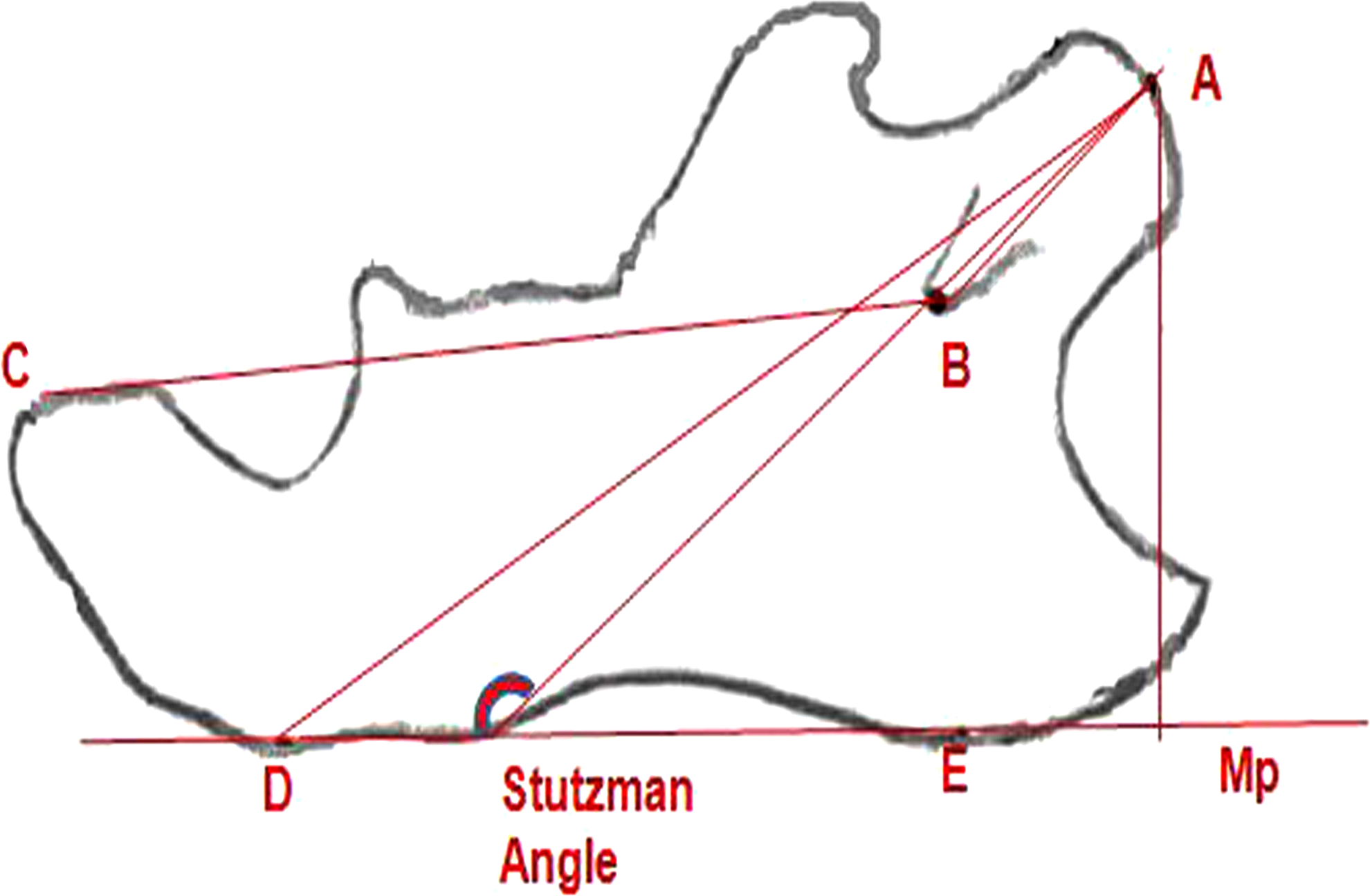
Skeletal landmarks, linear and angular measurements. 12
Functional bite-jumping appliances
The functional bite-jumping appliance performed in this study was similar to that developed for growing rats in earlier studies, 6,12 Silicon impressions were obtained from mandibular incisors, and plaster models were made. Functional appliances were constructed for the mandibular incisors, and identical inclination planes were fitted to the lower incisors of rats in the experimental groups and bonded to lower incisors with the self-etch bonding system (Transbond Plus, 3M, Monrovia, USA). These functional appliances produced 2.5–3 mm continuous forward mandibular advancement in the experimental groups (Fig. 2).

Bite-jumping appliance.
Low-level laser irradiation
In this study, a 810 nm GaAs diode laser (Cheese, Wuhan, China) with a probe diameter of 0.625 cm2 was used. Experimental rats in groups III and IV were stimulated with low-level laser in the temporomandibular joint region, applied in direct contact to the skin bilaterally 15 times over 30 days. The rats in group III received 8 J/cm2 (0.25 W, 20 sec) energy density on each side per session. The rats in group IV received 10 J/cm2 (0.25 W, 25 sec) energy density on each side per session.
Statistical analysis
The statistical analysis was processed with SPSS for Windows (Statistical Package for Social Science, SPSS Inc. Chicago, IL, Windows version 20.0). All the linear and angular measurements were repeated for 10 randomly selected samples 15 days later by the same investigator. The Pearson correlation test was used to define the difference between two registrations (Table 3). The nonparametrical Friedman test, Kruskal–Wallis, and Mann–Whitney U test were applied to examine the intragroup and intergroup significant differences in angular and linear measurements (Tables 4 and 5).
See Table 2 for definition of variables.
p<0.05.
See Table 2 for definition of variables.
p<0.05.
Results
Body weight
The body weight of the samples did not change during the 30-day interval in any group. In this process, the increase of the weight in the control group was more than that of other experimental groups (Table 6).
Linear and angular measurements
Differences among the groups
There were significant changes in mandibular morphology in the control group. On day 30, A–B (the length of condylar process), A–D (the distance from point A to menton), and B–C (the length of the mandibular base) had increased significantly (p<0.05). In group II A–B, A–C (mandibular length), and A–D, parameters increased significantly (p<0.05) in the study period. All linear and angular measurements had increased significantly in group III by day 30 (p<0.05). In group IV, all linear and angular measurements except the B–C variable increased significantly during the study period (p<0.05) (Table 4).
Differences between groups
All variables according to the Kruskal–Wallis statistical test results showed significant differences between groups (p<0.05). The Mann– Whitney U test results showed that the growth of A–B between T0 and T1 in groups II, III, and IV were significantly greater than in the control group (p<0.05). The linear growth of A–C at T0–T1 in groups III and IV was significantly greater than in the control group (p<0.05), and there were statistically significant differences found between groups III and IV (p<0.05). The dimension change in A–D at 30 days in groups II, III, and IV was significantly greater than in the control group (p<0.05). The growth of B–C in the study period in groups II and IV was significantly lower than in the control group (p<0.05), and group III showed greater mean values than group IV (p<0.05). At the same time, sagittal growth of mandible A-Mp (the perpendicular distance from A [the the most posterosuperior point of the condyle] to Mp [the mandibular plane]) showed vertical length changes in the mandible. The linear growth of A-Mp at T0–T1 in group III was significantly greater than in group II and the control group (p<0.05) (Table 5).
The Stutzman angle's results in the T0–T1 period in groups III and IV was significantly greater than in control group and group II (p<0.05) (Table 5).
Discussion
The results of this research showed that condylar growth was significantly increased with LLLT and a mandibular advancement device during the 30 day experimental period.
The rat model was chosen for this study because the morphological, histological, and cellular structures of the rat condyle are similar to those of humans. 6 In this study, we distinctly evaluated the biostimulatory effects of a low-level 810 nm GaAs laser on condylar growth activity during mandibular advancement in rats. A fixed bite-jumping device was also used to generate mandibular advancement in growing rats for the 4-week experimental period.
Among the groups
The control group results during T0–T1 showed significant mandibular growth without any external stimulus. This result showed the role of the condyle in the process of mandibular growth. Rabie et al. reported that mesenchymal cells in the posterior part of the condyle are significantly higher than the anterior and middle parts, and that this causes new bone formation in the posterior region of the condyle in growing rats. Because of this, without any external stimuli growing, rats showed a natural growth period. 13
Mandibular advancement devices in growing patients can treat mandibular retrognathia by the acceleration of mandibular growth. 14 Our study supports these findings, as group II showed statistically significant growth in three mandibular growth parameters during T0–T1. Recent studies reported that mandibular growth was enhanced by new bone formation in the condyle with functional application in growing rats. 11 Our results also showed that continuous mandibular advancement with a bite-jumping appliance stimulates mandibular growth in growing rats.
Pinheiro et al. reported that laser-irradiated regions exhibited new bone formation, collagen deposition, and an osteoblastic increase. Laser irradiation affected the vascular reaction as a result of this new bone formation, and condylar growth was achieved. 15 We found similar results in other laser biostimulation studies. 8 According to our study, laser- irradiated groups with an intraoral device (groups III and IV) showed better results than the control and appliance group during T0–T1. In particular, the 8 J/cm2 laser irradiation group showed the best increase in mandibular advancement scores during the study period.
Between the groups
Our results showed that the growth of the condylar process (A–B) in groups II, III, and IV between T0 and T1 were significantly greater than in the control group. In their research, Xiong et al. revealed that the condyles of the experimental group animals were elongated. 12 Abtahi et al. supported these findings, and declared that there was an enhanced bone formation in the condylar region. 2 According to these results, the growth of the condyle in growing rats could be increased by external stimuli, such as LLLT and bite-jumping appliances.
The LLLT groups showed a greater increase in mandibular length (A–C) growth than the device and control groups. Group III also demonstrated greater mandibular length increase than group IV. Oyonarte et al. found that low-intensity ultrasound application could modify mandibular growth in growing rats. 16 Seifi et al. showed greater mandibular length growth from 910 nm diode laser irradiation in rats' condyles. 8 Our findings showed that LLLT with a bite-jumping device was effective for mandibular growth biostimulation. The 8 J/cm2 energy density dose was more efficient than 10 J/cm2.
The distance from point A to menton (A–D) is known as an effective mandibular length. Mandibular growth in this dimension in groups II, III, and IV between T0 and T1 was significantly greater than in the control group; group III showed a greater increase in mandibular growth than did other groups. According to Taira et al., mandibular advancement with a fixed functional appliance effectively accelerated growth in this parameter in growing rats in a 30-day period. 11 LLLT with functional appliances affects the organic matrix formation, chondroblastic activity, and condylar cartilage layers, and this stimulation causes condylar growth and mandibular advancement. Although the difference between the three experimental groups was not statistically significant, the best effective mandibular length growth was observed with the 8 J/cm2 laser-irradiated group.
The control group showed the greatest growth in the length of the mandibular base (B–C). This may be because the mandibular base was far away from the laser application region and because the C point was behind the lingual part of the anterior incisor to which the appliance was fitted. Because of this, the C point may have been affected by chewing forces in the anterior region.
Changes in the perpendicular distance from A to Mp showed the vertical growth in groups. Group III showed the greatest vertical growth, similar to the results of other variables, and 8 J/cm2 laser irradiation with bite-jumping device application was seen as more efficient than in the other experimental groups. Similar to our results, El-Bialy et al. found that low-intensity pulsed ultrasound for 4 weeks stimulated mandibular condyle growth and increased mandibular height. 7
The growth of the mandibular condyle was also evaluated by the change of the Stutzman angle in growing rats; the opening of this angle showed the increase in mandibular length. 17 In this study, laser-irradiated groups showed greater growing values than the control and appliance groups. The 8 J/cm2 laser irradiation with bite-jumping appliance application group was seen to be more efficient than the other experimental groups. New bone formation may be shifted substantially lower and to the posterior part of mandibular condyle. As a result of this condition, the A point was shifted similarly, which may have caused an increase in the Stutzman angle.
This is the first cephalometrical evaluation report on two different energy-dose LLLT devices and functional bite-jumping devices used together in rats' mandibular condyle. In our study, we irradiated the mandibular condyle as an active growth region. We believe that LLLT irradiations with bite-jumping appliances can help to solve the duration and stabilization problems in functional treatments of class II malocclusions. This experimental animal study might be used as guidance for future studies on skeletal class II patients.
This study could be performed using histological evaluations in addition to cephalometric evaluations in the experimental process, to analyze the cellular and condylar cartilage changes. Another limitation of this study was the absence of LLLT-only groups without any functional appliances, for evaluating the naked laser effect on condylar growth.
Conclusions
This experimental research showed that LLLT irradiation with a bite-jumping device application had a significant impact on the increase of mandibular growth in growing rats. LLLT applications in condylar regions might be useful for shortening functional appliance therapy in skeletal class II patients.
Footnotes
Acknowledgments
This study was supported by the Scientific and Technological Research Council of Turkey (1135447).
Author Disclosure Statement
No competing financial interests exist.