
Abstract
Introduction
E
However, Er:YAG laser irradiations require relatively high-energy densities to vaporize enamel and dentine. 5 This study examined the influence of energy density of Er:YAG laser. Therefore, it is worth exploring the optimal setting of the laser prior to testing the microleakage after the restoration with the self-etch adhesive system.
The aims of the present study were to evaluate the surface characteristics treated at different energy density parameters of Er:YAG laser on the cementum and dentine surface by scanning electron microscopy (SEM) and to compared microleakage of the cementum and dentine margin with the self-etch adhesive system for class V restoration among the laser surface preparation groups, with the diamond bur preparation as the control.
Materials and Methods
Sample collection and preparation
The experiment sample consisted of 80 extracted human premolars diagnosed as having advanced periodontal disease. All samples were washed and stored in 0.2% thymol. The teeth were scaled by using an ultrasonic scaler (Cavitron 2000, USA) for removing residual tissues. All teeth were examined macroscopically for defects in the cementum, 4 A class V cavity was prepared with a high-speed turbine (Aston Mini2, J. Morita, Tokyo, Japan) and a No. 401 diamond bur (Shofu Inc., Japan) on the midbuccal surface with the occlusal margins located 1 mm apical to the cementoenamel junction (CEJ) in the cementum and the gingival margins located 3 mm apical to the CEJ in each sample. The dimensions of cavities were standardized by using a clear acrylic template 3 mm wide and 2 mm high. The cavity depth was calibrated to be 2 mm using a periodontal probe (Hu-Friedy CP-11.5B Dental Screening Probe) for measurement. 6 The experimental study was composed of two parts, namely, part I: morphological studies, and part II: microleakage evaluation.
Part I: Morphological studies
The 80 teeth were randomly allocated into four groups (20 teeth per group).
Group 1: the mechanical cavities were prepared using a high-speed turbine with a no.401 diamond bur (the control group)
Group 2: Er:YAG laser at 50 mJ/pulse, 15 Hz, energy density 3.77 J/cm2
Group 3: Er:YAG laser at 75 mJ/pulse, 15 Hz, energy density 5.65 J/cm2
Group 4: Er:YAG laser at 100 mJ/pulse, 15 Hz, energy density 7.53 J/cm2
For groups 2, 3, and 4, the laser cavities were prepared using an Er: YAG laser system (Fotona, LightWalker AT, Fidelis, Slovenia) with a wavelength of 2.94 μm, and a laser handpiece H14-N with a cylindrical sapphire fiber tip with a 1.3 mm diameter (code 71766). The laser irradiation was performed in a noncontact mode to the margin of the cementum removal and treated dentine layer at the cavity floor with a focused beam of 50 mJ (energy density 3.77 J/cm2), 75 mJ (energy density 5.65 J/cm2), and 100 mJ (energy density 7.53 J/cm2) with a repetition rate of 15 Hz, pulse duration of 200 μs, under a continuous water mist (6 mL/min) in each group. The beam spot size was maintained at 1.3 mm from a distance of 2–3 mm, and the laser beam was kept perpendicular to the root surface (buccal). 7,8 Then the samples were observed by SEM.
For the SEM evaluation, these samples were bisected and immersed in 70% ethanol concentration for 10 min. The specimens were dried in a desiccator at a critical point. The dried specimens were mounted on a metal stand and gold coated (0.2 μm) (Quorum Emitech K500X Sputter coater, England) by cathode atomization under vacuum, and then examined with SEM (S3000, Hitashi, Japan). Photographs were taken at 100×, 500×, and 3000× magnification. 9,10
Part II: Microleakage evaluation
For the microleakage test, 60 samples were randomly allocated into four groups (15 teeth per group) after laser irradiation of the root surface. All samples were restored with the Adper™ Prompt™ L-Pop™ self-etch adhesive (3M/ESPE) system. The Adper bonding was applied to the cementum and dentine surface with a light brushing pressure for 15 sec. A gentle stream of air was directed at the root surface to thoroughly spread the adhesive into a thin film. The surface appeared glossy and then was cured with halogen light for 10 sec (according to the manufacturer's instructions). After application of the self-etch adhesive system, all the cavities were filled with a nano-hybrid composite resin Filtek™ Z350 XT A3 (3M/ESPE) in one layer with cervical matrices No 856 s, Anterior/premolarX (Kerr Hawe SA, Switzerland) and light cured for 40 sec. The restorations were finished with fine-grit finishing diamond burs (Diatech Dental AG), then polished with a series of sandpaper disks (Soflex, 3M ESPE, USA). The same operator performed preparation, restoration, and finishing–polishing procedures. All samples were stored in distilled water at 37°C for 24 h. After undergoing thermocycling (Thermocycling, HWB 332 R, Thailand) 10,000 cycles at temperatures between 5°C and 55°C and with a dwell time of 30 sec each cycle, the teeth were sealed with sticky wax at the root apices and two coats of nail varnish were applied to the tooth except for the 1 mm rim of the restoration margins. The teeth were then immersed in 0.2% methylene blue solution for 24 h. 11 After removal from the dye, the teeth were rinsed with tap water for 5 min and dried at room temperature. Next, the specimens were sectioned longitudinally on the buccolingual trough the center of the restoration vertically with a water-cooled, slow-speed diamond saw (Isomed, Buehler Ltd, Lake Bluff, IL). The sectioned teeth were observed under a polarizing microscope (×40 magnification) (Nikon, Japan). Two blinded examiners scored the restorations independently and calculated the degree of dye penetration with the NIS-Elements program (Nikon, Japan). All data were analyzed with SPSS 13.0 for Windows statistical package. Statistical analyses were performed with one way ANOVA (p<0.05) and Mann–Whitney U tests with Bonferroni correction (p<0.05).
Results
There were two main results comprising the descriptive evaluation from SEM (Fig. 1) and the measurement of microleakage in micrometers. At ultrahigh magnification, the diamond bur surface exhibited a smooth surface and a debris-like smear layer (Fig.1A–C). In the laser group, the surface of cementum showed micro-irregular surfaces and also roughness with numerous long-thin projections resulting from cementum ablation (Fig. 1F, I, and L). The results of surface micro-irregularities produced after laser treatment with three different energy densities (energy density 3.77 J/cm2 in Fig. 1E and F, energy density 5.65 J/cm2 in Fig. 1H and I, and energy density 7.53 J/cm2 in Fig. 1K and L) showed similar fine micro-irregularities and an absence of smear layer (500× and 3000×).
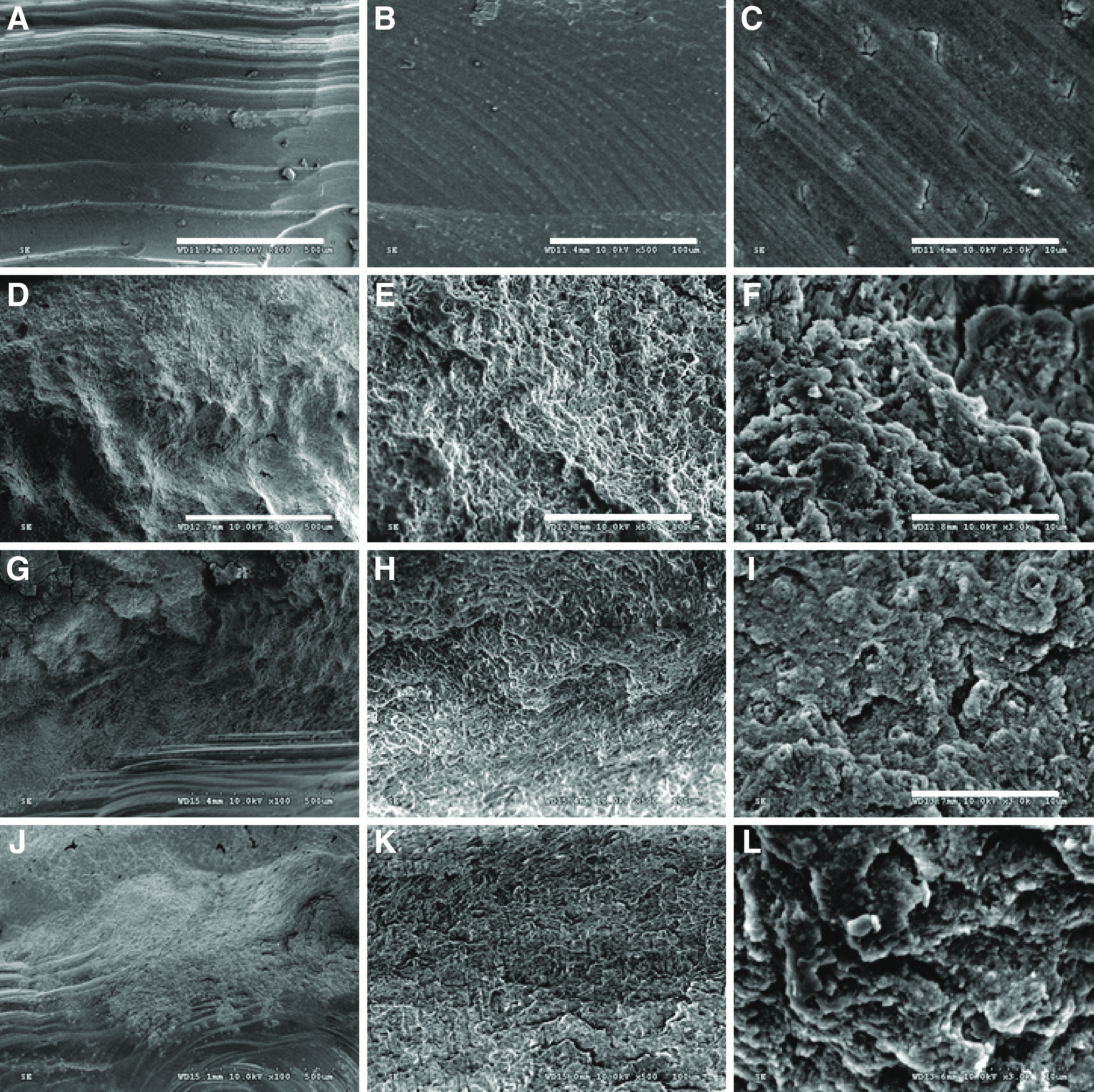
Scanning electron microscopic (SEM) image of representative Erbium: Yttrium Aluminum Garnet (Er:YAG) laser-prepared cavity.
The pattern of cementum root surfaces were dependent on Er:YAG laser parameters. All of the Er:YAG energy densities showed a noninvasive selective grinding cementum root surface. Morphological findings by SEM indicated that Er:YAG laser irradiation with water irrigation could create an irregular surface without smear layer and carbonization. On the other hand, cavity surfaces treated with diamond bur had a flat appearance and smooth cutting cavity with a debris-like smear layer. SEM analysis revealed sharp and long thin projected micro-irregularities of the Er:YAG-lased surface.
This study demonstrated that the diamond bur surface exhibited a smooth surface and debris-like smear layer (Fig. 1A–C). Therefore, the surface that resulted from use of Er:YAG lasers was rougher than the diamond bur treated surface.
The microleakage studies were assessed using the dye penetration method (Fig. 2).
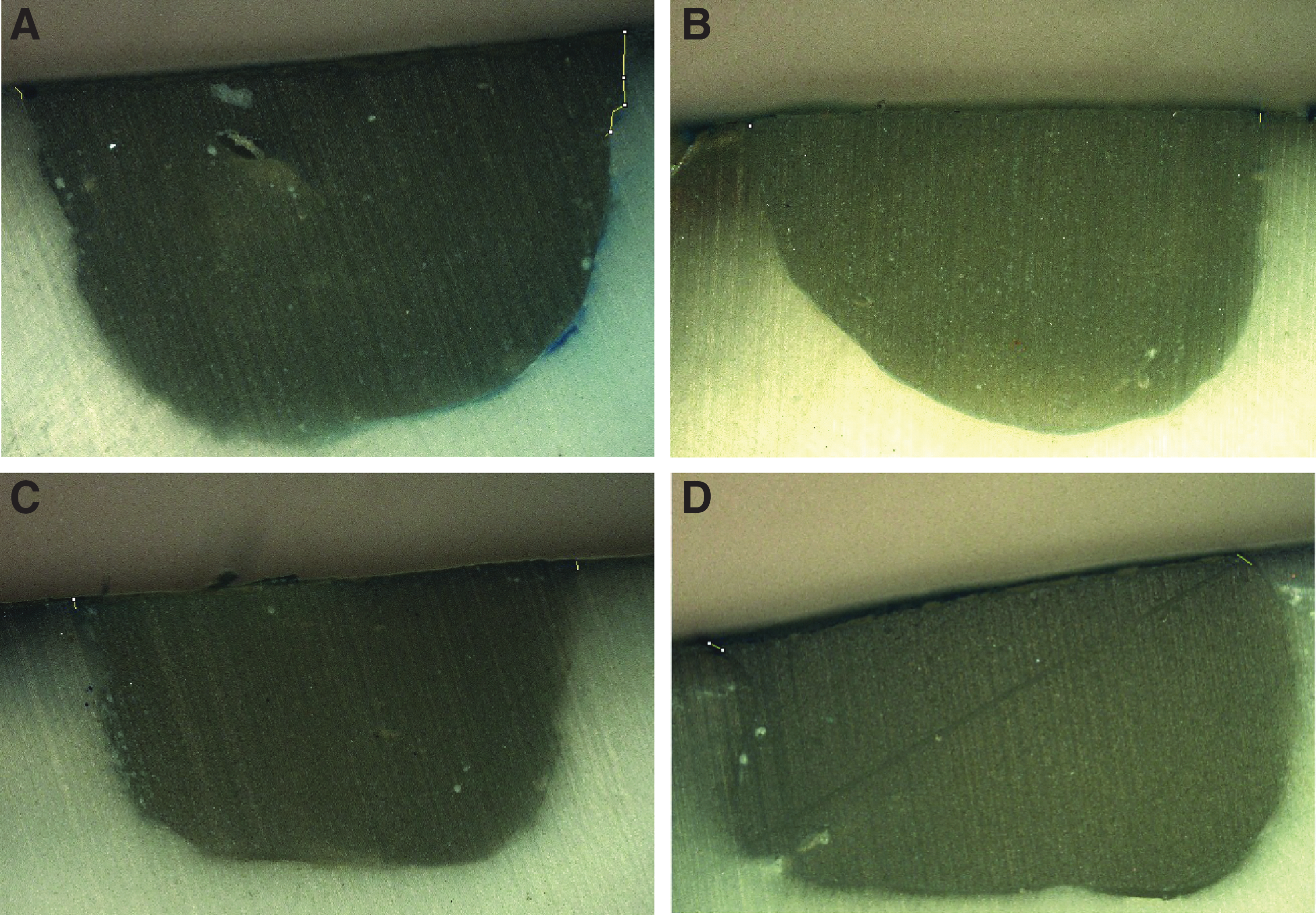
Polarization evaluated marginal microleakage of different energy of Erbium: Yttrium Aluminum Garnet (Er:YAG) laser.
Table 1 shows that the diamond bur cavity preparation gives the highest value (159.16 μm on the occlusal surface and 163.06 μm on the gingival surface) for microleakage, whereas the energy density of 3.77 J/cm2 in the Er:YAG laser groups gives the lowest value (85.15 μm on the occlusal surface and 93.28 μm on the gingival surface) when the cavities were treated with a self-etch bonding system. The results of the gingival wall and occlusal wall of cementum, significant differences of microleakage between the cavities prepared by the Er:YAG laser and those prepared by the diamond bur, were evident. Furthermore, the diamond bur showed more marginal microleakage scores than the three groups treated with Er:YAG laser (p<0.05).
Statistically significant difference (vertical comparison between diamond burs group and the three laser groups on occlusal surface, p<0.05).
Statistically significant difference (vertical comparison between diamond burs group and the three laser groups on gingival surface, p<0.05).
When comparing the occlusal marginal microleakage (Table 2) and gingival marginal microleakage (Table 3) by group, a statistically significant difference in both the occlusal and gingival microleakage was found among the three groups (diamond cavity preparation, and energy densities 3.77 and 5.65 J/cm2 for Er:YAG laser) for the self-etch adhesive system, whereas the energy density of 5.65 J/cm2 for Er:YAG laser compared with the energy density of 7.53 J/cm2 for Er:YAG laser did not indicate a statistically significant difference in either occlusal or gingival microleakage.
Statistically significant difference.
Statistically significant difference.
Table 4 shows a significant difference between the occlusal and gingival microleakage for all the groups (p<0.05). When the three laser groups were compared, the group with an energy density at 3.77 J/cm2 (50 mJ) showed the lowest microleakage of occlusal and gingival margins.
Statistically significant difference.
Discussion
In our study, preparation with various energy densities of the Er:YAG laser could alter morphology of dental hard tissues, and subsequently, the application of the acid material to laser-treated teeth could affect the bonding mechanism of adhesives and the microleakage phenomenon. Therefore, the total etch adhesive could penetrate to the tissue surface and decrease microleakage. 12 Laser-irradiated margins compared with those prepared with a conventional diamond bur have irregular surfaces, and this could create microspaces and, eventually, more leakage. 13 Denaturation of collagen fibers has also been observed, as the action of the Er:YAG laser system relies directly on water molecules. 8,14 Current all-in-one adhesives contain copolymers that prevent phase separation and act as wetting agents and promoters of the diffusion of resin into exposed collagen. 15 As opposed to cavity preparation by bur, which results in a layer of debris at the surfaces, cavity preparation by Er:YAG laser results in surfaces free of smear layers and smear plugs. 16
Moreover, this has a favorable effect depending upon the laser parameters. The ablation threshold of human enamel has been reported to be in the range of 9–14 J/cm2 for the Er:YAG and Erbium, Chromium-doped:Yttrium, Scandium, Gallium, and Garnet (Er,Cr:YSGG) laser wavelengths. 17 In the present study, we used the lower energy density than the previous study (Apel et al.). 17 Similar to our results, Bertrand et al. 18 reported that class V resin restorations placed in cavities prepared with a bur or an Er:YAG laser showed lower microleakage on the occlusal than on the cervical walls.
In most of the reported studies, they used the Er:YAG laser at the pulse energy of >300 mJ. These energy levels induced subsurface damage in the enamel, so that many researchers reported poor marginal adaptation and high degrees of microleakage. 19 When lower energy levels are used for cavity preparation, microleakage of laser- and bur-treated cavities are not significantly different. 15,20 –24
Less microleakage was noticed with low energy pulses. There was a statistically significant difference between the two energy settings of laser preparation and conventional methods at both dentin and enamel. 25 According to our study, Er:YAG laser energy per pulse played a role in cavity preparation, which is significant for microleakage at class V restorations. Low-laser energy per pulse, at a fixed frequency time, contributes to a better bonding than a higher power laser setting, showing better marginal integrity of resin composite restorations with less microleakage.
The degree of microleakage in class V cavities was affected by the type of adhesive restorative materials, type of self-etching adhesive, cavity margin location, and tooth preparation method either by Er:YAG laser or dental bur. Muhammed and Dayem observed that after acid etching 37% phosphoric acid gel and laser-conditioned dentin, the hybridization effectiveness was compromised because of the selective ablation of organic tissue, leading to less collagen left exposed and consequently left to be hybridized. 26
Pashley and Tay 27 reported that self-etch dental adhesives differed in their aggressiveness. Therefore, they are classified into three categories, according to acidity: mild, moderate, and aggressive. Self-etch adhesives whose pH is <1.5 are called aggressive self-etch adhesives. On the other hand, self-etch adhesives that have a pH>1.5 are categorized as mild or moderate. The self-etch adhesives used in this study, of Adper™ Prompt™ L-Pop™, had a pH=0.8. The reason for this finding could be the single step application of this bonding system, which will reduce its technique sensitivity. The primer of this bonding agent has a pH<1.5 and is considered an aggressive acidic primer. This primer could create surface demineralization to a depth of 1 μm, which creates a surface for micromechanical retention. 28 In addition, the more acidic pH of Adper™ Prompt™ L-Pop™ may cause more micromechanical retention and a thicker hybrid layer in dentin. Therefore, using laser surface preparation can enhance a more appropriate morphologic effect for micromechanical retention. The results were a decreased amount of gingival and occlusal microleakage of composite resin restoration.
Conclusions
To summarize, this study demonstrated that cementum and root dentin presented micro-irregularities after Er:YAG laser irradiation. Therefore, the microleakage of Er:YAG laser irradiation was significantly decreased compared with diamond bur cavities.
Footnotes
Acknowledgments
We thank Sajee Sattayut, all staff of the Lasers in Dentistry Research Group and Faculty of Dentistry, Khon Kaen University.
Author Disclosure Statement
No competing financial interests exist.