




Abstract
Introduction
R
The pathogenesis of rheumatic fever (RF) in susceptible individuals is related to autoimmune humoral and cellular responses directed toward human tissues, triggered by the response to β-hemolytic group A streptococci. Cardiac involvement induces pericardial, myocardial, and endocardial inflammation, leading to permanent damage of the heart valves. 3 The most common late injury to the heart valve with rheumatic involvement is mitral stenosis, resulting in commissural fusion and anatomical changes of the subvalvular apparatus, including chordae tendineae thickening, and fibrosis of the submitral apparatus, including the papillary muscles. 4,5
Commissural fusion sectioning with a scalpel blade, known as commissurotomy, is the traditional technique that is still widely used to treat mitral stenosis. Some situations also require the surgical division of fused papillary muscles. Several reports demonstrate the occurrence of late restenosis postcommissurotomy, with reoperation required in ∼11–27% of patients over an 8-year period. 6
The high-power laser (HPL) or surgical laser used for different surgical procedures has many advantages over instrument-based surgery, including the prevention and reduction of bleeding, infection, pain, and swelling, in addition to allowing high-precision cutting. 7 HPL was first used in the cardiovascular field in Brazil by Macruz et al., who studied the laser rays' effects on atheromatous plaques in the human aorta. 8 Twenty years later, Dallan and Oliveira performed transmyocardial laser revascularization surgery (TMLR) as a therapeutic option for patients with end-stage ischemic coronary artery disease secondary to advanced atherosclerotic disease. 9 A few years later, the same authors combined TMLR with cell therapy, which appeared to be a safe technique that may have synergistically acted to reduce myocardial ischemia, with a clinically relevant improvement in the functional capacity of patients with refractory angina. 10 There is no report in the literature on the use of this kind of laser in valvular surgery, specifically for mitral stenosis.
The aim of this study was to compare the histological findings of porcine cusp mitral valves treated in vitro with a scalpel blade to those treated with HPL cutting.
Materials and Methods
Cusp obtainment and preparation
This study design was submitted to and approved by the Ethics and Research Committee of the Heart Institute (InCor) from the University of São Paulo School of Medicine.
This is an experimental and descriptive study in which five explanted mitral valves from young (6–12 months of age), healthy swine of both genders were used; they were obtained from a slaughterhouse, under the supervision of the Inspection Department of the Federal Ministry of Agriculture and the Health Surveillance Department of the State Health Secretariat. The valves were rinsed in saline solution before removal and immediately preserved in a sterile glycerin liquid container at room temperature, according to the recommendations of previous studies. 11 –13 Subsequently, the material was transported to the laboratory of Lasers and Applications Center (CLA) of the Institute of Energy and Nuclear Research (IPEN-USP). In the laboratory, the mitral valves were rehydrated in three different receptacles containing 0.9% saline solution in room air for 30 min each. Once rehydrated, 10 cusps were divided randomly into the following two groups:
Cusp group (G1)—cut with scalpel blade (n = 5);
Cusp group (G2)—cut with laser (n = 5).
Surgical cut
The mitral valve, with its cusps, was positioned on Styrofoam and fixed with metal pins on the flat surface of a workbench. Mitral valve hydration with 0.9% saline solution was performed as needed throughout the experiment.
Additional treatment doses were tested before the present study. We have tested emitting wavelengths (λ) of 1064 and 10,600 nm. We observed less thermal damage associated with the desired effect (cutting and chordae shortening on the same optical fiber) using the following parameters:
G2 cusps were treated using a CW diode laser (MediLaser model -DMC, São Carlos, Brazil) with an emitting wavelength (λ) of 980 nm, a power (P) of 9.0 W, a time (T) of 12 sec, an irradiance of 5625 W/cm2, a spot area of 0.0016 cm2, a spot diameter of 0.0451 cm, and an energy (E) of 108 J in a continuous operation mode with a fiber size of 400 μm. The laser was perpendicularly applied to the cusps in contact mode, only once.
The power was measured using a power meter equipment (Fieldmaster GS; Coherent Instruments) for each use. A single operator, with the aid of three assistants, performed the laser application procedure. All of the researchers used safety goggles that blocked the specific diode laser wavelength as well as other personal protective equipment.
Histological and morphometric methods
The valve samples (submitted to blade or to laser cutting) were sectioned for histological analysis. After fixation in 10% buffered formalin and routine histological processing, 5-μm sections were obtained and stained with hematoxylin–eosin (HE), Masson's trichrome, and Sirius red. The histological slides were observed under an optical microscope for qualitative description of the alterations in staining. The morphometric measurements were carried out with an interactive computer-assisted image analyzer (AxioVision, V.4.7.1.0; Carl Zeiss). The distance between the cutting border to the furthermost point of altered collagen staining (Masson's trichrome and Sirius red methods) was determined in each section.
Statistical analysis
A nonparametric test to compare and evaluate proportions was performed to verify qualitative variables. A chi-square test was carried out and results were considered statistically significant if p-value <0.05.
Results
In the cusps cut with a scalpel blade (G1), no histological change was observed in HE, Masson's trichrome, or Sirius red staining. This means that the normal valve's extracellular matrix with loose collagen in the central cusp and denser collagen on the periphery and its usual homogenous blue color and birefringence were maintained (Fig. 1a–c).
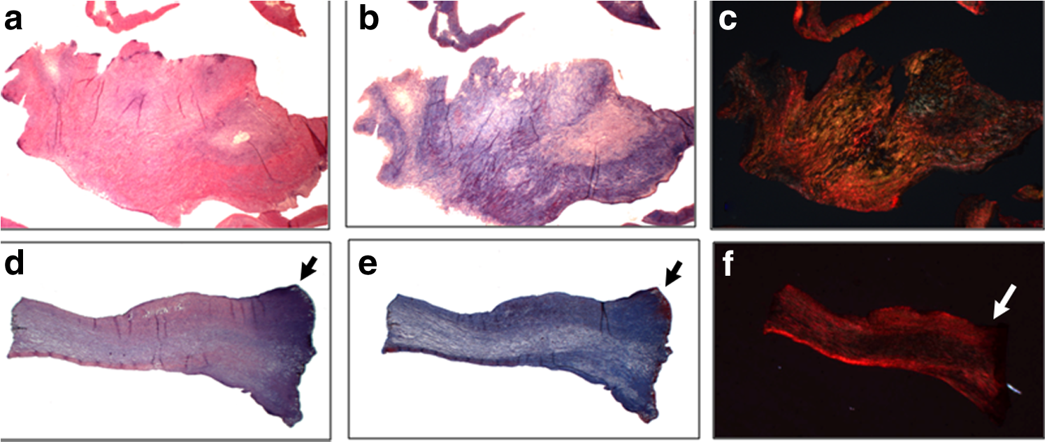
Histological sections of normal porcine cusps cut with a scalpel blade
In cusps cut with the laser (G2), a hyaline basophilic aspect was focally observed in sections stained by HE (Fig. 1d). Using Masson's trichrome staining, we observed a dark red color on the edges; areas of lower birefringence on the tissue edges were seen in sections stained with Sirius red (Fig. 1e, f).
Measures of thermal lesion extension postlaser cutting
The distance between the cutting edge and the end of the thermal lesion showed a greater extent on the slides stained with Sirius red than on those stained with Masson's trichrome (Fig. 2a, b).
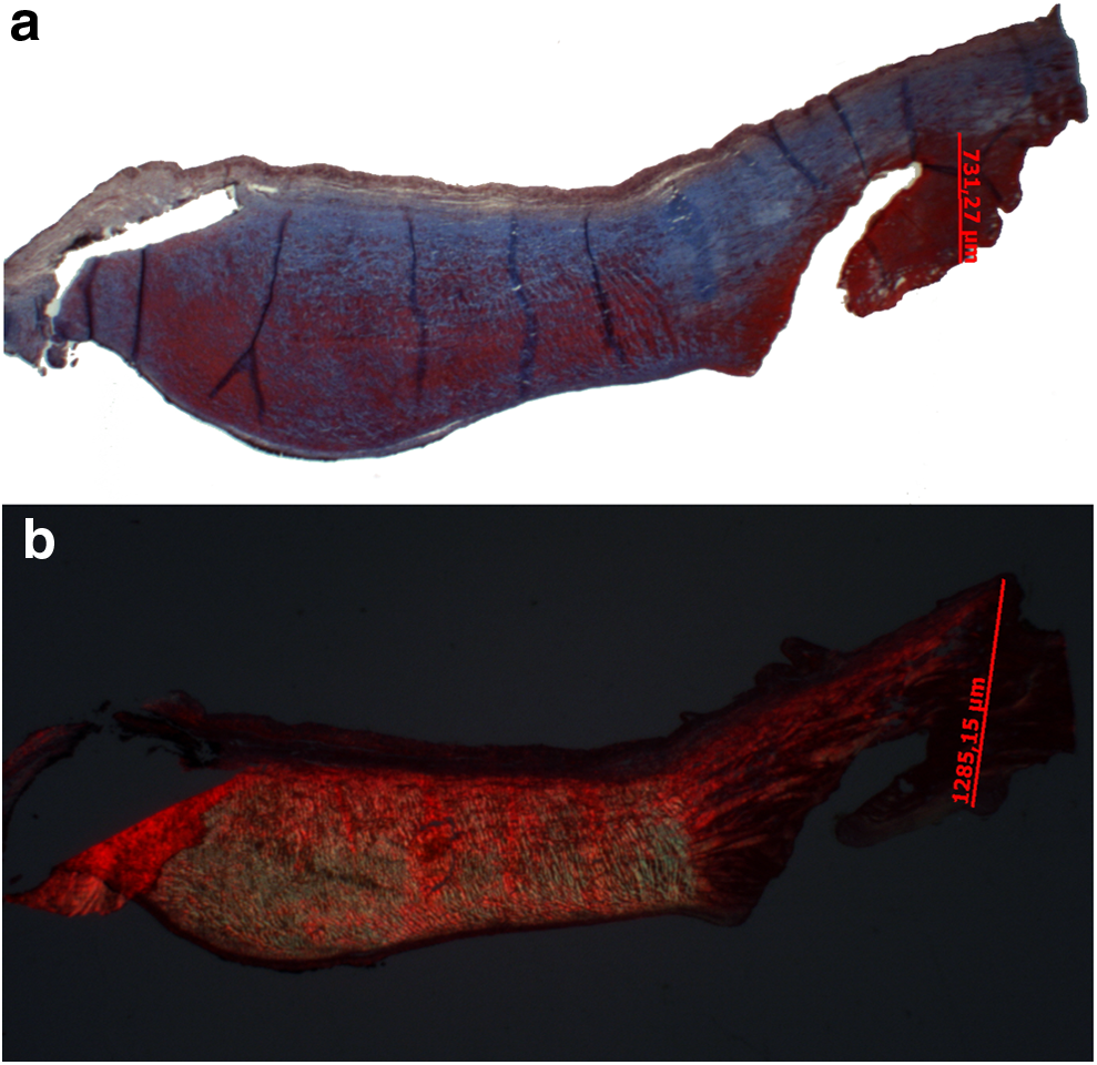
Histological sections of porcine cusps cut with laser, demonstrating the process of digital measurement of the distance between the free edge and the border of abnormal collagen.
The mean distances from the cutting edge to the end of the thermal lesion measured in the samples cut by the surgical laser and stained by Masson's trichrome and Sirius red were 416.7 and 778.6 μm, respectively; this difference was statistically significant (p = 0.044) (Fig. 3).
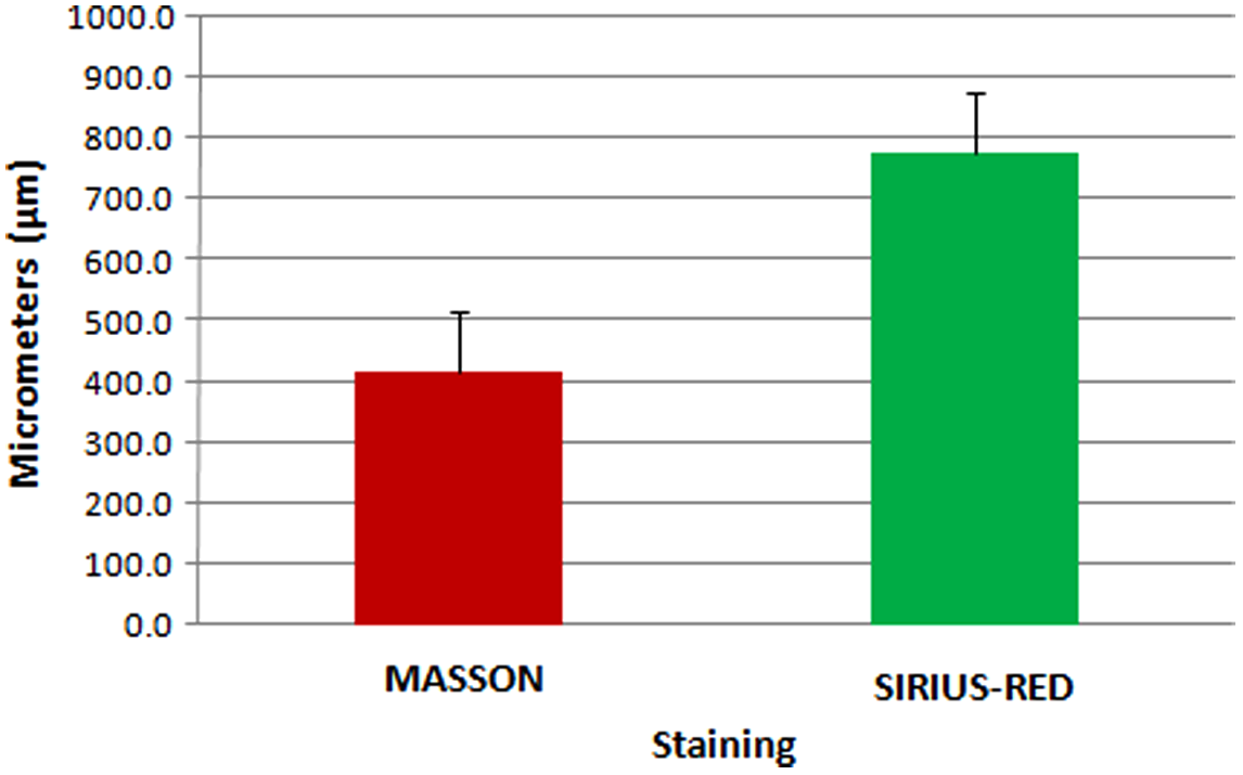
Means and standard deviations of the measurements of abnormal collagen in slides stained with Masson's trichrome and Sirius red. There was a significant difference between the measured distances (p = 0.044).
The ratio between the distances measured by Masson's trichrome and Sirius red was 0.58; in other words, the distance from the edge on the samples and the end of the thermal lesion in sections stained by Masson's trichrome was 42% shorter than in samples stained by Sirius red.
Discussion
Mitral stenosis is the most common chronic injury resulting from recurrent RF. Anatomically, it is the result of recurring postinflammatory crises that make the valve highly deformed and eventually obstructed. As the valvular vegetations of acute RF heal, they induce not only fibrous cusp thickening but also more important changes, including adherence between cusps at the level of the commissures and also fusion of the chordae tendineae. 14 Commissural fusion is the main feature of rheumatic mitral stenosis, and restenosis is prevalent. 15 Incorporation of technological innovations and improvements in surgical techniques are essential to improve outcomes.
In Azevedo's study, Wistar rats were submitted to tissue damage in the skin and tongue by scalpel, electrosurgery, and an HPL; less tissue damage (necrosis area) was observed in cases using a scalpel in both tissues. 16 The healing process proceeded similarly in all techniques by the end of the analysis period. Bernachio 17 found similar results in her study, which compared the healing process post-CO2 incision of diode lasers (2 and 4 W), electrosurgery, and scalpel incision into the skin of Wistar rats.
Additional studies have tested radiofrequency energy in a sheep model to improve mitral valve competence. One such study noted that radiofrequency energy applied for less than 4 min per case at subablative temperatures in four quadrants of the posterior mitral valve annulus reduced the anteroposterior and circumferential annular distances significantly, and eliminated nonischemic mitral regurgitation. 18 Likewise, the reaction to laser commissurotomy could vary from one individual to another, and the end result will depend on the quality and extent of this reaction.
In our study, we observed some differences in cusp structure (by HE, Masson's trichrome, and Picrosirius red staining) in the G1 and G2 groups; thermal damage resulting from the laser cut was more extensive than with the use of the conventional instrument (scalpel). We cannot establish with certainty whether the injury described histologically corresponds to irreversible tissue necrosis or whether it is the result of the structural changes of extracellular matrix proteins due to surgical laser application. We found that the distance of the tissue cutting edge when assessed by Masson's trichrome was 42% shorter than when assessed by Sirius red staining, indicating that the latter reveals more details and more accurately detects the extension engagement caused by laser cutting in our sample.
Further, the average distances of the areas with collagen injury from the laser cutting edge in the cusps stained by Masson's trichrome and Sirius red were less than 1 mm (416.7 and 778.6 μm, respectively). In other words, Sirius red staining revealed the highest precision in identifying the changes in mitral valve tissue. This indicates that even if such areas correspond to tissue necrosis, the distance from the edge (with the laser power and time period described in this study) was always less than 1 mm, suggesting a mild photothermal effect. This finding should guide future research on live animals; we also intend to study the local aspects of spontaneous or secondary regeneration of local sutures.
When an HPL focuses on tissue, we observe its central region and vaporization and carbonization zones. The next contiguous zones were the coagulation and edematous areas. As we move away from the central zone toward the periphery, the energy concentration decreases. In this more distant area, there is a residual effect of laser surgery tissue called photoactivation, with photophysical/chemical changes in that zone. This could incur photobiomolecular activation followed by a biomodulation response, which could aid the valvular repair effect and possibly decrease the restenosis process; the cusp edges postlaser commissurotomy may hinder the long-term fusion of these structures. 5
Thus, due to the effects of laser biomodulation in the tissue, the chances of restenosis could be smaller and the patient can get a longer time without the need for further surgical procedures.
Provided these data can be confirmed in experimental studies with appropriate follow-up in swine, this strategy could be used in the future as a possible novel therapeutic option for treating patients with mitral stenosis.
Conclusions
Optical microscopy demonstrated that the thermal changes found in the group cut with HPL were greater than in those treated with a scalpel blade. Nevertheless, these changes extension from the cutting edge were smaller than 1 mm, proving HPL to be a safe and feasible method of cutting. Sirius red staining is the most reliable technique to identify possible fine changes in mitral valve tissue after surgical laser application.
Footnotes
Acknowledgments
The authors thank Braile Biomédica for their contributions.
Author Disclosure Statement
No competing financial interests exist.