
Abstract
Introduction
T
Enterococcus faecalis is one of the most common bacteria associated with endodontic treatment failures. This species is Gram positive, facultatively anaerobic, commensal, 3,4 and resistant to many antimicrobial drugs used in endodontics, such as calcium hydroxide and some concentrations of sodium hypochlorite, and chlorhexidine. 5 Several techniques are being investigated in medicine and dentistry with the aim of further reducing these bacteria. One of these techniques is the photodynamic therapy (PDT) method, which has been used for the treatment of cancer 6 as well as oral infections 7 and caries. 8
The action mode of PDT is based on irradiation of a photosensitizer agent (PS), which is a non-toxic dye that is sensitive to light in the presence of oxygen. 9 The PS is irradiated with specific light, inducing the production of reactive species such as singlet oxygen-free radicals. 10 The triggered reactions cause bacterial cell wall rupture, leading to microbial death while also damaging essential cellular molecules, including proteins, membrane lipids, and nucleic acids. Gram-negative bacteria are less susceptible to photoinactivation than Gram-positive species. 11 This treatment is highly specific, because it only affects the bacterial cells that are impregnated by PS, constituting a safe procedure for periapical tissues. 12
The concentration of PS required for microbial death due to photodynamic activity depends on the dye, irradiation parameters, and bacterial genus. 13 Several dyes and light with different wavelengths are being researched in PDT. The most studied are the phenothiazine dyes such as toluidine blue and methylene blue (MB) dyes, which are associated with lasers at red wavelengths of irradiation (600–700 nm). 9 The reduction of E. faecalis may reach 99.89% 13 and 99.9%, 14 respectively. The important disadvantages of PDT using this type of PS are the staining of the dental structures 12,15 and the long-term consequences of exposure that have not been evaluated.
Rose bengal (RB) dye, a xanthene, is another dye that has been investigated and employed as a PS. RB dye is a derivative of tetrachloro-tetraiodo-fluoresceins, which are dark-red crystalline compounds, and it absorbs quite well in the visible light range of 500–800 nm. 16 These synthetic compounds are among the most active PSs and show good quantum yield of singlet oxygen production (80%). 17 Recent studies have shown the use of RB that is associated with green light (532 nm) in the reduction of cell counts of Candida albicans in biofilms 9,18 as well as in decreasing the cell count of Streptococcus mutans. 19
The aim of this in vitro study was to compare the PDT technique using RB (25 μmol/L) and MB (0.01% or 31.2 μmol/L) that are associated with visible light laser sources in the green (532 nm) or red (660 nm) wavelengths, respectively, on E. faecalis cell count reduction.
Materials and Methods
This study was conducted in the microbiology facilities at the University of the Region of Joinville in partnership with the Pontifical Catholic University of Paraná, Brazil, and the Federal University of Technology, Paraná, Brazil.
Preparation of E. faecalis
The strain E. faecalis ATCC 29212 was employed in all microbiological procedures. For long-term storage, it was initially inoculated in BHI (Brain Heart Infusion) broth (Prodimol Biotecnologia, Belo Horizonte, Brazil), which was prepared according to the manufacturer's recommendations plus 15% (v/v) glycerol, followed by incubation at 37°C for 48 h. Afterward, aliquots (1 mL) of bacterial suspensions were kept at −80°C.
An aliquot of the frozen strain was reactivated in 3 mL of BHI broth for 24 h at 37°C for each cell viability test performed. Afterward, a pre-inoculum containing 1 mL of reactivated bacterial culture and 9 mL of BHI broth was prepared under the same incubation conditions. These were also employed for inoculum preparation, which contained the complete volume of pre-inoculum culture plus 10 mL of fresh BHI broth. Finally, cell densities were visually estimated by comparison to the McFarland scale (Probac, São Paulo, Brazil), and the bacterial suspension was standardized to match 0.8 (∼3 × 10⁸ cells/mL) on the scale. The experiment was conducted with a planktonic culture.
PSs and lasers
MB (0.01% or 31.2 μmol/L), commercially available as Chimiolux 10 (DMC, São Carlos, Brazil), and RB (VETEC, Duque de Caxias, Brazil), which was formulated by a compounding pharmacist to reach 25 μmol/L in deionized water (pH 7.2), were employed as PSs. This concentration was obtained after preliminary studies in concentrations of 5, 10, 25, 50, and 100 μmol/L. In each experiment, all solutions were checked by spectrophotometry to maintain the standard concentrations of PSs, as shown in Fig. 1.

Absorption spectrum of methylene blue (0.01% or 31.2 μmol/L; MB) and rose bengal (25 μmol/L; RB) with peak absorptions around 660 and 540 nm, respectively.
Two different laser devices were used according to the PS peak absorption spectrum. Details and specific parameters regarding each protocol are indicated in Table 1.
MB, methylene blue; PDT, photodynamic therapy; RB, rose bengal.
The green laser device employed was a prototype made by the authors using a 2 W laser pen (Green Laser Pointer, JG, China), on which an optical fiber was added with adhesive tape. The power was reduced to 40 mW.
For both lasers, the power was tested at the tip of the fiber three times, both before and after each experiment, and the average obtained was used for the fluence calculation (Powermeter—Laser Check; Coherent, Inc., Alburn, CA).
Experimental design
Experiments were performed in a laminar flow chamber in the absence of light by a single operator. One milliliter of the bacterial suspension was gently mixed with 1 mL of PS (MB or RB; MB group and RBG groups, respectively) or 0.9% saline [control group (CG)] in a 2 mL cylindrical tube. The pre-irradiation time was set at 5 min. Thereafter, the laser fiber was immersed, and to guarantee uniform illumination of the samples, helical up-and-down movements were performed during the irradiation period of 3 min. For fluence calculations, we had to first consider the laser power delivered at the end of the optical fiber (300 μm diameter): 10.19 × 10³ J/cm2. However, the effective fluence has to take into account the Eppendorf tube dimensions (2 cm height, 1 cm diameter). Thus, we calculated the sample area that was actually irradiated by the laser. This was done by measuring the speed of the fiber tip during the helical up-and-down movements. We found that the effective fluence was ∼1/50 of the fluence at the fiber tip. It means that the effective energy density reaching the sample was 204 J/cm2.
The experiment was performed in triplicate for each tested condition.
The experimental groups were as shown in Table 2.
CG, control group; MBG, MB group/red laser; RBG, RB group/green laser.
After laser exposure, the bacterial suspension was serially diluted in 0.9% saline and plated (100 μL) in triplicate on BHI agar (90 × 15 mm), as shown schematically in Fig. 2. The plates were incubated at 37°C for 24 h, followed by colony-forming units (CFU) counting. All the cultivations were conducted in candle jars under a microaerophilic atmosphere and protected from light exposure. Only plates presenting counts between 30 and 300 CFU were considered for subsequent calculations of the CFU/mL.
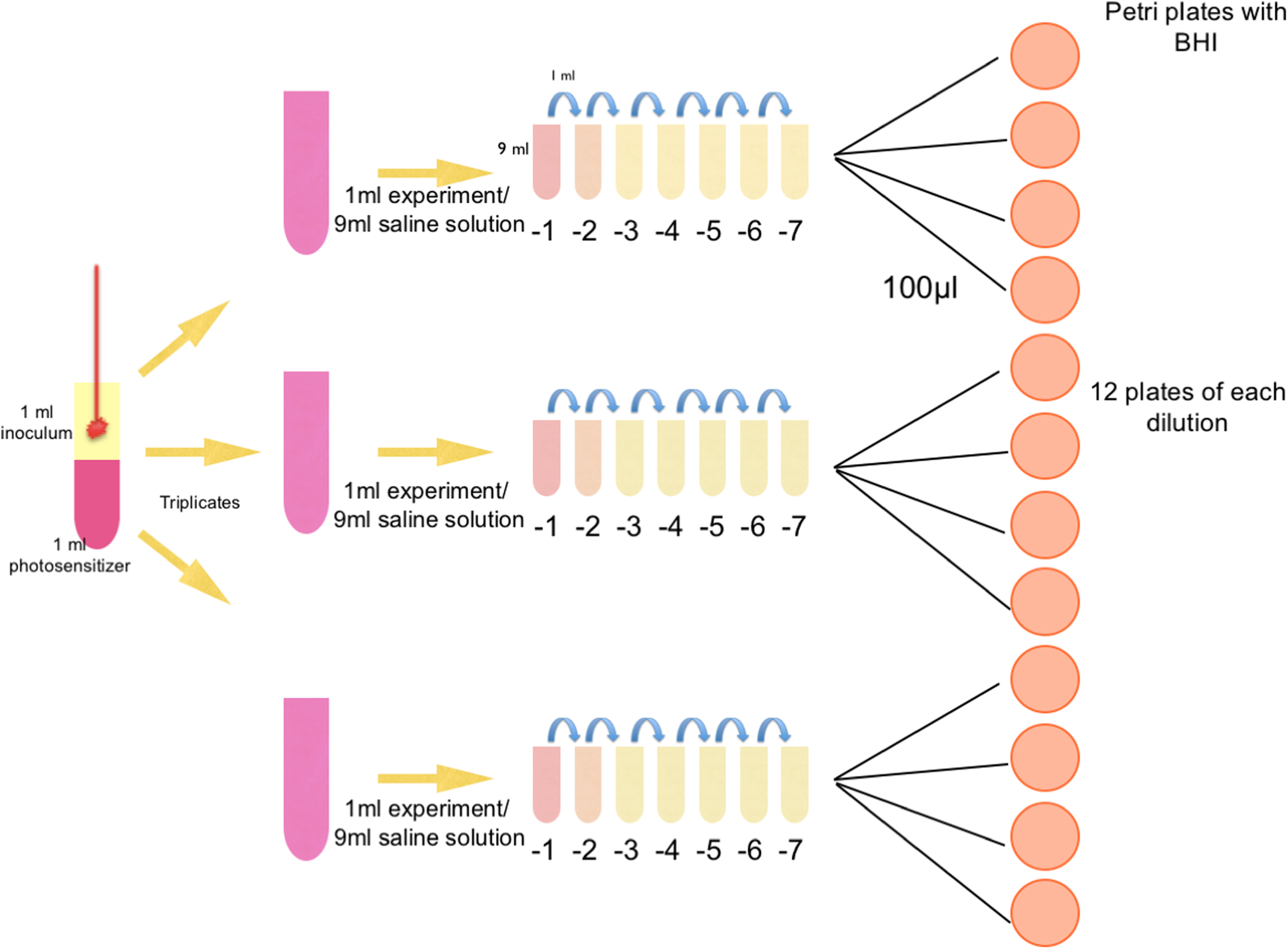
Research design from the irradiation of samples for plating.
The experimental protocol is shown by the diagram in Fig. 3.

Diagram of the experimental protocol.
Statistical analysis
The normality of data distribution was evaluated by the Kolmogorov–Smirnov test. The comparisons of the mean CFU values were performed using ANOVA, with p < 0.05 as the adopted level of significance.
Results
The results are summarized in Table 3. A reduction of 95.67% in viable cells was obtained in the RBG group. The mean number of the CFU/mL for the RBG group (0.12 × 10⁸) was significantly lower compared with the control (2.82 × 10⁸) and MBG (2.66 × 10⁸) groups. On the other hand, the decrease in viable cells observed in the MBG group (5.43%) was not significant relative to counts of the CG.
Significant statistical difference.
CFU, colony-forming units; MBG, MB group; SD, standard deviation.
Discussion
In this study, E. faecalis was selected, because it is one of the most resistant microorganisms found in infected root canals. 20 It is repeatedly found in endodontic cases, 21 and, further, it has been widely used as a valuable microbiological marker for in vitro studies, because it colonizes the root canal, forms a biofilm, invades the dentinal tubules, and is resistant to some endodontic treatment. 22
The negative CG, when saline solution was used for comparing it with the other experimental groups, showed an increasing and linear bacterial growth among the dilutions, thus demonstrating microbial viability.
This study compared two types of PS with the respective lasers (compatible with the absorption spectrum). In the first experimental group, MBG PS 0.01% (31.2 μmol/L) was used, because a vast amount of literature has results demonstrating its effectiveness in bacterial cell count reduction, 13,14,22 –24 but the results of this study showed a small reduction in the CFU/mL when compared with CG, with no statistically significant difference.
Phenothiazine dyes, such as MB PS, present a strong blue color when used in PDT. An adverse effect of using these dyes is that they stain the teeth. Only a few studies have addressed this drawback using methodologies with visual or even digital images. 12,15,25
To decrease the staining of the teeth, RB was selected as a PS. Although Hamblin and Hassan 26 reported that this PS did not bind to microorganisms, other research has shown that it is effective in reducing viruses, bacteria (Streptococcus mutans, E. faecalis, Porphyromonas gingivalis), and fungi (C. albicans). 4,11,17,19,27
In a pilot study, it was found that solely using the RB PS on microorganisms did not inhibit bacterial growth.
Pilot studies were performed with RB at different concentrations of 5, 10, 25, 50, and 100 μmol/L, and the most favorable reduction in the CFU/mL was at a concentration of 25 μmol/L. Below this value, the reduction of microorganisms was not significant, which differs from the study by Pileggi et al., 4 who showed a complete reduction of E. faecalis using a 10 μmol concentration of RB. However, Pillegi et al. used longer pre-irradiation (30 min.) and longer laser exposure times (4 min.), making this RB method clinically impractical. At concentrations higher than 25 μmol/L of RB, there was a total reduction in the CFU/mL, confirming their toxicity, as was also observed by Paulino et al. 17
In this study, an RB concentration of 25 μmol/L was used, and there was a significant reduction in the CFU/mL when compared with CG, in agreement with other studies 8 where the same concentration of PS and laser time were used, and they obtained a significant reduction in C. albicans.
Several studies 19,27 –29 have demonstrated reductions of Streptococcus mutans, P. gingivalis, Lactobacillus, and C. albicans using different concentrations of the PS RB with different light sources, which demonstrates their effectiveness.
In the studies by Kishen et al., 30 PSs such as MB and RB were used at a concentration of 100 μmol with 300–600 mW of power. This group obtained a greater reduction in the group using the MB PS that was irradiated with a red laser, differing from the results obtained in this study, because the energy used was lower.
To the best of our knowledge, no investigation has been conducted with the methodology employed in this study that compared a PDT technique using two PSs for the treatment of E. faecalis.
We concluded that the reduction in the CFU/mL of E. faecalis in the RBG group was higher than in the MBG and CG groups, demonstrating that the PDT technique with RB PS that was associated with a green light laser source, in the researched conditions, was effective in reducing these bacteria.
Footnotes
Author Disclosure Statement
No competing financial interests exist.