
Abstract
Introduction
D
Lasers have been recommended for dentin hypersensitivity treatment by a number of practitioners and scientists, including Kimura and Moritz. According to Kimura et al., 4 its effectiveness ranges from 5.2 to 100%, depending upon the type of laser and the parameters used. Four different kinds of lasers are used for dentin hypersensitivity treatment, including He-Ne (helium neon), GaAIAs (diode), Nd:YAG, and CO2. 4 Two mechanisms are involved in the treatment: low output lasers promote biomodulatory effects, minimizing pain and reducing inflammatory processes, 5 whereas middle output lasers increase surface temperature, which can result in the complete closure of dentinal tubules. 6
The energy of a 10,600 nm CO2 laser is easily absorbed by tissues with high water content. The effect of a CO2 laser is based on the closure or narrowing of the dentinal tubules and a reduction in dentin permeability, as it is a middle power laser. 7 –10 In vivo application of a CO2 laser in dogs and monkeys has been shown to cause no thermal damage to the pulpal tissue at an output power of 3 W. 10 However, morphologically, parameters >1 W lead to carbonization and cracks in human dentin, making it unfeasible for use in clinical procedures. 8 Therefore, the main concern regarding the use of CO2 lasers on dentin hypersensitivity treatment is their possible thermal effect/damage on the dental pulp. Whether the damage/effect on dental pulp is reversible or irreversible also needs to be clarified, as well as its efficacy in treating dentin hypersensitivity. Moreover, the dental pulp has its own reparative capacity. 11
The purpose of this study was to investigate the effects of CO2 lasers on the proliferation and differentiation of dental pulp cells, and their latent self-recovery in connection with their stemness assessed by reverse transcription polymerase chain reaction (RT-PCR) and by immunohistochemistry. We examined inflammatory marker heat shock protein 70 (Hsp70), mesenchymal stem cell markers, such as adenosine triphosphate (ATP)-binding cassette transporter G2 (ABCG2), CD34, and CD44, and cell differentiation markers, such as dentin sialophosphoprotein (DSPP) and dentin matrix protein 1 (DMP1).
Materials and Methods
Animal experiment protocol
Seventy male Sprague–Dawley rats, each weighing 150–200 g, were used in this study. There were a total of five groups in two categories: control and experimental. In the experimental groups, comprising immediate, day 1, day 3, and day 5, we applied a CO2 laser, whereas the control group was not treated with CO2 laser. Animals were anaesthetized using sodium thiopental (Ravonal; Tanabe, Japan) and were euethanized by cervical dislocation. Both control and experiment groups had been fed on standard diet. All animal studies were conducted in compliance with the Guidelines for the Treatment of Experimental Animals at the Tokyo Dental College (Approval Number 223206).
Laser irradiation protocol
A CO2 laser (Panalas CO5 Sigma; Panasonic Dental Co., Osaka, Japan) was used in this study. The experimental procedure was to irradiate the occlusal surface of the upper first molars of each rat given that there is no covering enamel because of the coarse abrasive character of the cariogenic diets and physiological abration (Fig. 1). 12 A Type 1A tip (defocus, diameter, 0.15 cm) was used in direct contact with the tooth surface and the laser irradiation time was 8.8 sec. The parameters for the CO2 laser were as follows: wavelength, 10.6 μm; power output, 2 W; super pulse mode; pulse, 0.6 m sec; and total laser energy of 4 J (density of 203.84 J/cm2) (Table 1). These laser conditions were based on clinical laser application standards, because the overall experimental purpose was focused on its possible clinical application. 13
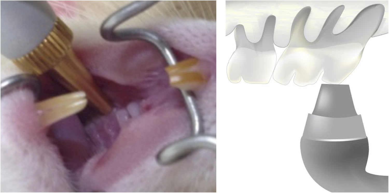
The scheme of the experiment. The experimental procedure is to irradiate the occlusal surface of the upper first molars, where there is no enamel covering.
Hematoxylin and eosin (HE) staining protocol
With respect to morphological analysis, all samples (n = 10) from five rats were stained with hematoxylin and eosin (HE). Experimental animals were euthanized immediately, or 1, 3, or 5 days after the laser irradiation with the control group. Each maxilla was fixed in 10% formalin buffer for 2 days and was then soaked in 10% formic acid solution for 4 days, resulting in decalcification. 14 Paraffin blocks were prepared and cut into thickness of 4 μm. The samples were then stained with HE, and were observed using an UPM Axiophot microscope (Carl Zeiss).
Quantitative RT-PCR
For quantitative RT-PCR analysis, 30 animals (n = 60) were euthanized by cervical dislocation immediately, or at 1, 3, or 5 days after the CO2 laser irradiation, with the control group. First, wash medium was prepared using alpha-Minimum Essential Medium (alpha-MEM, GIBCO, Carlsbad, CA) including 10% gentamycin and 1.2% fungizone. The upper molars were extracted and washed in wash medium for 5 min, after which the dental pulp was extracted mechanically. 14 Total RNA was obtained from each specimen using the acid guanidinium thiocyanate /phenol-chloroform method as follows. Trizol Reagent (Invitrogen, Carlsbad, CA) was used to homogenize the cells after rinsing the samples with phosphate-buffered saline (PBS).
Each sample was then mixed with chloroform and centrifuged at 14,000 rpm at 4°C for 20 min before incubation in 70% isopropanal at −20°C overnight. After centrifugation, the mRNA pellets were washed with 70% cold ethanol, and were then dissolved in RNase-free [diethylpyrocarbonate (DEPC)-treated] water. RT-PCR products were analyzed by quantitative real-time RT-PCR using TaqMan Gene Expression Assays for four target genes, Hsp70 to evaluate the stress level in cells, ABCG2 to evaluate the side population cell activation, DSPP and DMP1 as markers of cell differentiation, as shown in Table 2, and glyceraldehyde-3-phosphate dehydrogenase (GAPDH) as an endogenous control (Applied Biosystems). Gene expression quantitation using the Taqman Gene Expression Assay was performed in single-plex reactions containing Taqman Fast Universal PCR Master Mix, Taqman Gene Expression Assays, distilled water, and cDNA, according to the manufacturer's instructions (Applied Biosystems). The primary denaturation was performed at 95°C for 20 sec, followed by cycling for 40 cycles at 95°C for 3 sec and at 62°C for 30 sec. Experimental data were analyzed using one way ANOVA (p < 0.05) and Tukey's test was used for multiple comparison.
Immunohistochemical analysis
For immunohistochemistry, the streptavidin-biotin immunoperoxidase method was applied using a Histofine SAB-PO (MULTI) kit (Nichirei Co., Ltd. Tokyo, Japan).
Paraffin sections were deparaffinized with xylene, then dehydrated in 100% alcohol, and, finally, washed with distilled water. For the antigen retrieval step, heat-induced epitope retrieval (HIER) was used. Samples were microwaved for 30 min at 65°C in 0.01 M citrate buffer (pH 6.0), cooled at room temperature, and then rinsed in PBS three times for 3 min each. Endogenous peroxidase activity was blocked by incubating the sections with 3% H2O2 in methanol for 30 min. Before proceeding to application of the first antibody, 10% bovine serum albumin (BSA) was used to block the binding of nonspecific antibodies for 1 h.
The primary antibodies used in this study were: proliferating cell nuclear antigen (PCNA) as a cell proliferation marker (at a dilution of 1:100, DAKO, Glorstrup, Denmark), ABCG2 as a side-population cell marker (at a dilution of 1:100, Santa Cruz Biotechnology Inc., Santa Cruz, CA), CD44 as a mesenchymal stem cell marker (at a dilution of 1:100, Abcam, Nihonbashi, Tokyo, Japan), and CD34 as an early hematopoietic stem cell marker (at a dilution of 1:100, Santa Cruz Biotechnology Inc, Santa Cruz, CA).
Primary antibodies were incubated at 4°C overnight.
After primary antibody incubation, the samples were washed with PBS three times for 3 min each. The secondary antibody (biotinylated anti-mouse immunoglobulin G [lgG] or anti-rabbit lgG) was applied at room temperature for 30 min. 3,3′–diaminobenzidine-tetrahydochloride (DAB) was used to visualize the reaction after rinsing with PBS three times for 3 min each. Finally, each section (n = 70) from 35 rats (7 rats each group) was counterstained with Mayer's hematoxylin for 30 sec. Specimens were observed using a light microscope (BX41, Olympus, Shinjuku, Japan) and were photographed.
Results
HE Staining
A common finding after the CO2 laser irradiation in most of the groups was the disorganization of the odontoblast layer; however, the deep part of the pulp area was less affected. Along with the presence of small vacuoles as stated in Lee's experiment, 13 the displacement of odontoblastic nuclei into the dentinal tubules and degeneration in the pulp horn underneath the odontoblast layer was observed immediately after the laser irradiation in this experiment (Fig. 2a). However by day 5, the odontoblast layer was restored (Fig. 2b).
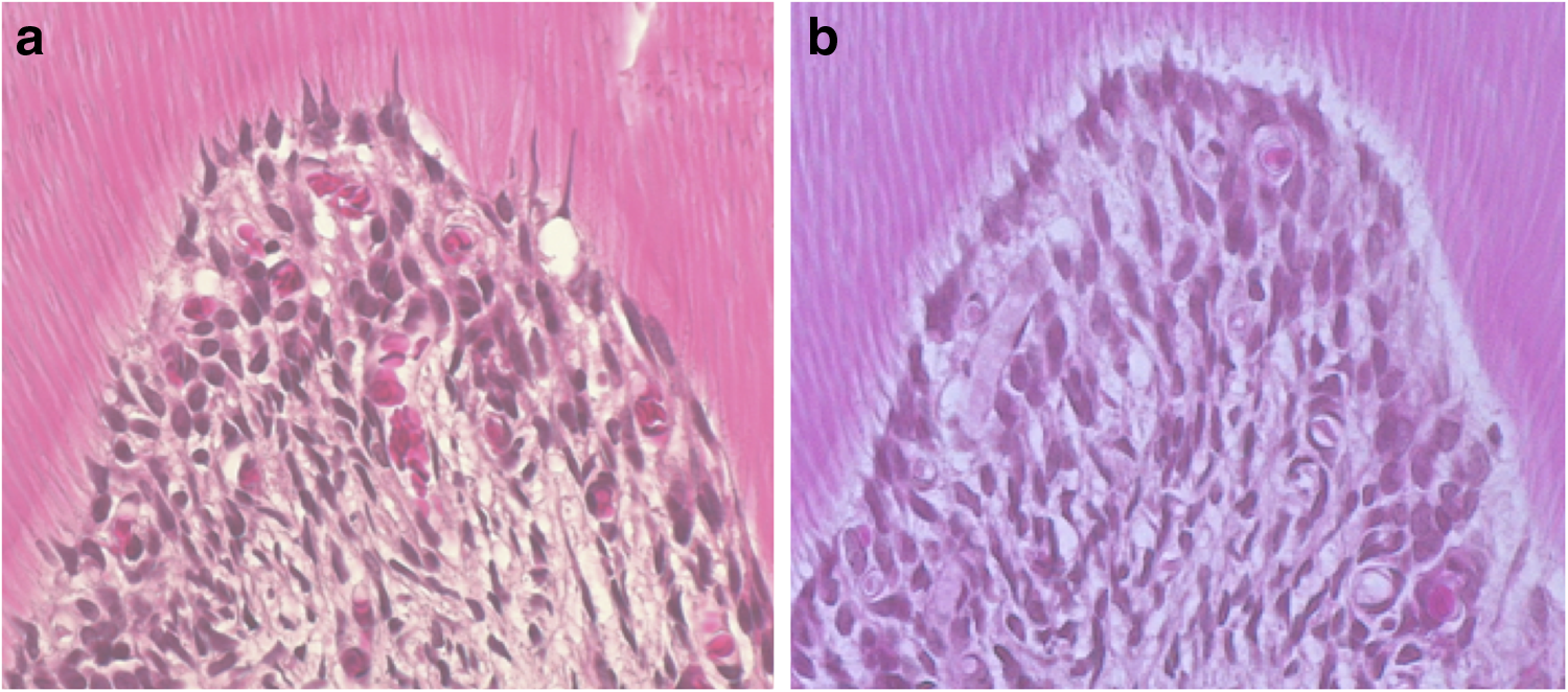
Hematoxylin and eosin staining. Immediately
RT-PCR analysis
The expression of HSP70 mRNA reached its highest level immediately after the laser irradiation, and then decreased gradually on days 1, 3, and 5 (Fig. 3a).
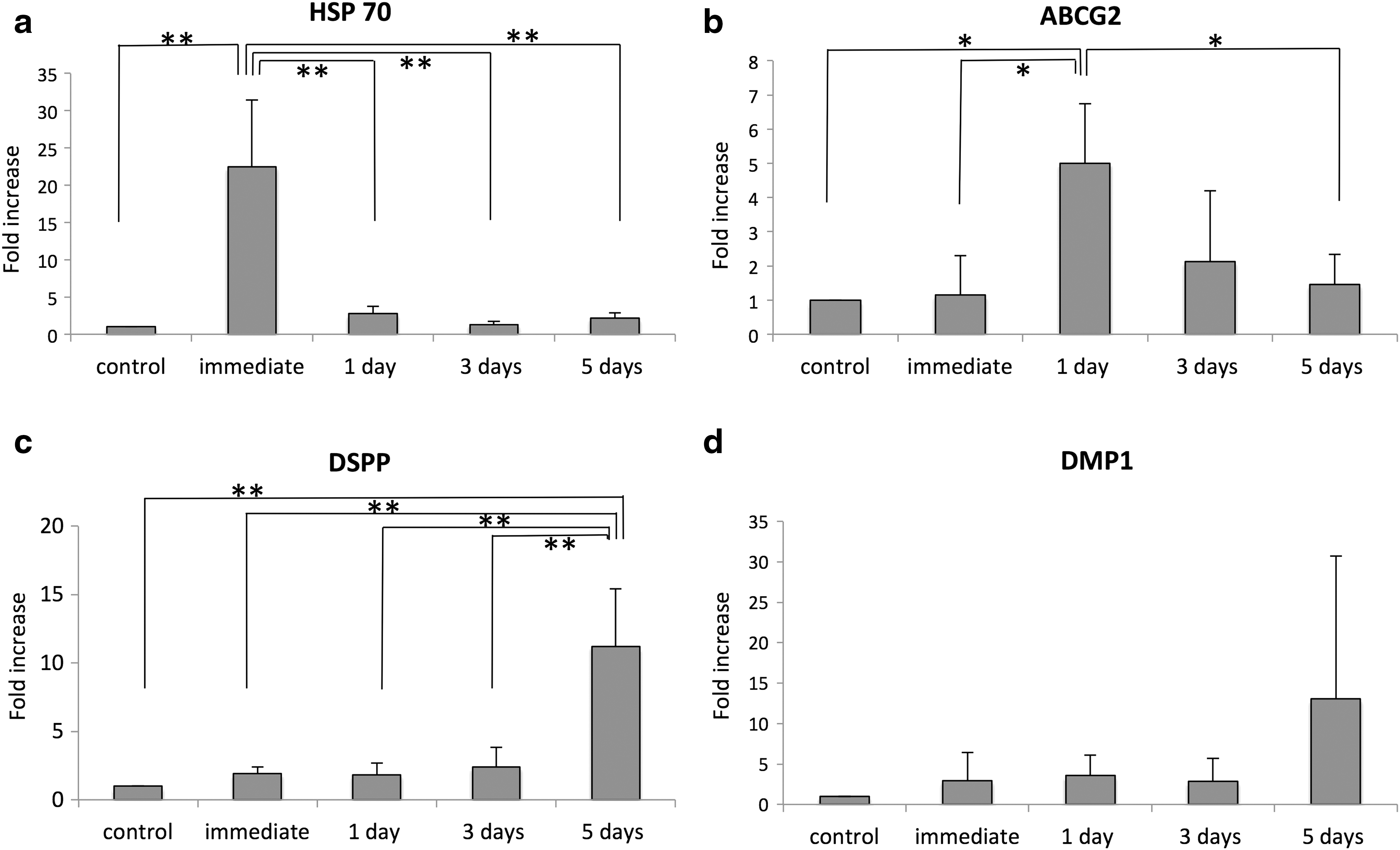
mRNA expressions after laser irradiation. The data are expressed as means+SD (n = 3). The p value for statistical analysis is *<0.05, **<0.01, ***<0.001.
The expression of ABCG2 mRNA reached its highest level on the 1st day after the laser irradiation, and then decreased gradually on days 3 and 5 (Fig. 3b).
DSPP mRNA expression was increased slightly after the laser irradiation, but then increased significantly, and the highest expression was found on day 5 (Fig. 3c).
The expression of DMP1 mRNA increased gradually from immediately after the laser irradiation and was highest on day 5. No significant difference was identified among the groups (Fig. 3d).
Immunohistochemical analysis
PCNA-positive cells were observed both in the control and the experimental groups (Fig. 4). PCNA-positive cells were mostly found in the odontoblastic layer, a cell-rich zone, and some were found in the deeper area of the dental pulp (Fig. 4b). Compared with the control group, there was no significant difference found in the immediate or day 1 groups. However, by day 3, PCNA-positive cells had increased significantly (Fig. 4c). By day 3 of the experiment, the number of ABCG2-positive cells was greater than in the other groups (Fig. 5). The presence of CD44-positive cells increased on day 1, and but these were found mostly on day 3, compared with the other groups (Fig. 6).
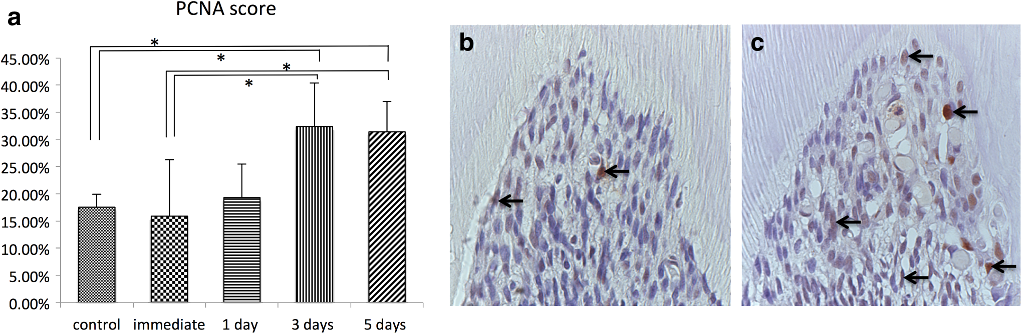
Proliferating cell nuclear antigen (PCNA)-positive cells after laser irradiation. The percentage of PCNA-positive cells
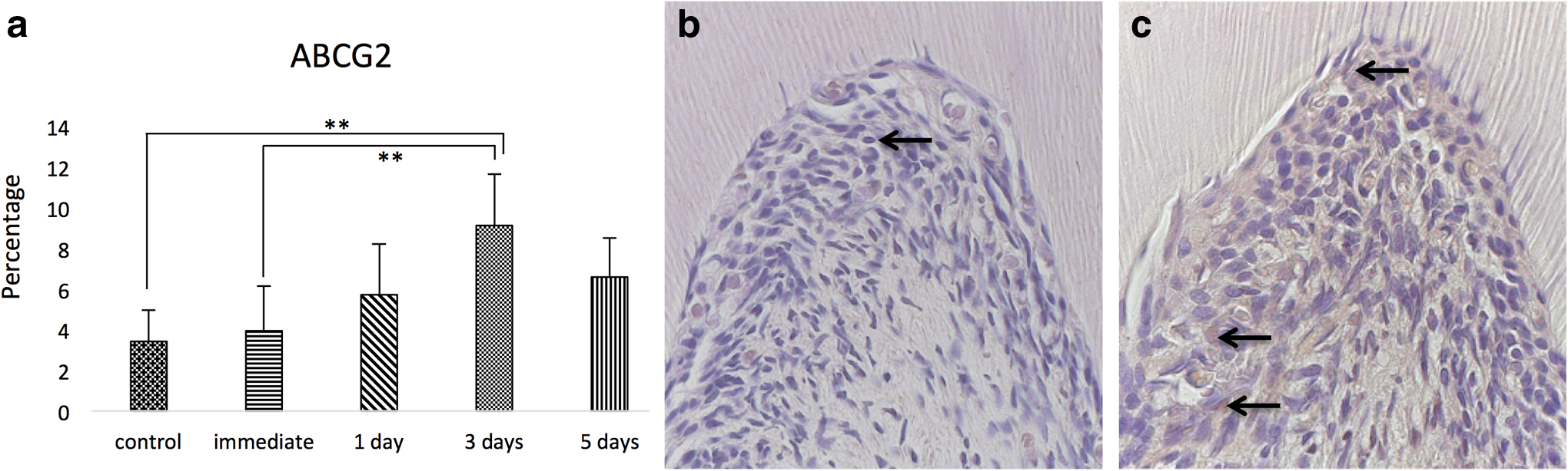
Immunohistochemical staining for ABCG2-positive cells. The percentage of ABCG2-positive cells
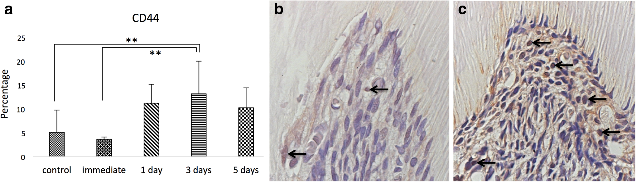
Immunohistochemical staining for CD44-positive cells. The percentage of CD44-positive cells
Expression of CD34, an early hematopoietic stem cell marker, was not observed in either the control or the experimental groups (not shown).
Discussion
The first use of a dental laser to treat dentin hypersensitivity was performed in 1985 by Matsumoto et al. 15 with an Nd: YAG laser, but it was not until 1996 that Moritz et al. used a CO2 laser in a dentin hypersensitivity study for the first time. This technique has been widely applied and recommended by scientists and clinicians since then. 15,16 According to Moritz et al, 16 its effect is based on the closure and narrowing of dentinal tubules, and the effectiveness of treatment ranged from 59.8% to 100%, depending upon the extent of the laser power used. According to Bonin et al., 17 a middle power CO2 laser was able to seal the dentinal tubules and reduce the permeability. Dentinal desiccation, yielding temporary clinical relief of dentin hypersensitivity, was also reported. 18 Therefore, we decided to use a middle power CO2 laser in this study.
The challenging part of this study was the unknown thermal effect on the dental pulp when sealing the dentin tubules. The ability of the dentin-pulp complex to respond to a variety of pathological conditions and injuries by deposition of reparative dentin by pulp “progenitors” has long been recognized. 19,20 Dental pulp has the potential ability to help repair itself at some level when injury is not fatal, because it contains a stem/progenitor cell population. 21 These stem/progenitor cell population can be the source of regenerative medicine as well. 22 Stem cells are nonspecialized cells that continuously divide, have the ability to self-renew, and are capable of generating complex tissues and organs. 23 Mesenchymal stem cells appear to both rely upon and generate a network that facilitates constant communication between normal and damaged cells in the body. 24 They migrate to the site of insult or injury in response to signals of cellular damage, known as homing signals. 25 –27 Lee et al. 13 reported that there was a slight degeneration of the dental pulp immediately under the odontoblast cell layer after the CO2 laser irradiation. However, the CO2 laser induced the inflammatory and pathological cytokine pathways to repair the damaged areas. 13
There were some degenerated areas observed in the pulp tissue in this study.
Hsp70 is an important factor for protein folding and plays a protective role against harmful factors. 28 The result of the experiment reveals a sharp increase in Hsp70 expression in the immediate group, although 1 day after the experiment it decreased substantially. Lee et al. 13 checked Hsp70 protein in three groups using immunohistochemistry, whereas in this experiment, two more groups, namely day 1 and day 3, were added to Lee's original experimental groups, and RT-PCR was performed. The result supports Lee's experiment, 13 which mentions the increased temperature stresses to the pulp cells causing slight morphological damage immediately after the laser irradiation, but dental pulp injury caused by laser irradiation is minimal, which subsides shortly and does not last long.
The highest level of ABCG2 mRNA expression was found on day 1 of the study compared with the other groups, and ABCG2-positive cells were observed mostly on day 3 by immunohistochemistry. This result suggests that the CO2 laser induces mesenchymal stem cells, which leads to their proliferation and recruitment to injured areas. It has been observed that dental pulp side-population cells can differentiate into and be a source of odontoblasts. Moreover, the majority of stem cells could be side-population cells, as the universal stem cell marker ABCG2 is expressed in side-population cells. 29 –31
The significant increase of CD44-positive cells on day 3 indicates the proliferation of mesenchymal stem cells. The dental pulp is extremely rich in stem cells capable of differentiating toward several stromal-derived differentiated cells. 32 The increased number of CD44-positive cells indicates that undifferentiated mesenchymal stem cells were induced by stimuli from the laser, as a result of which they regenerate and differentiate into odontoblast-like cells.
Dental pulp cells contain different kinds of cells, ranging from undifferentiated mesenchymal stem/progenitor cells to well-differentiated fibroblasts, as well as fully differentiated odontoblasts. 33 The number of PCNA-positive cells increased significantly on the 3rd day of the study, indicating an increase of the undifferentiated and differentiated dental pulp cell population. The increase of PCNA-positive cells overlaps the increase of CD44-, and ABCG2-positive cells, which are mesenchymal stem cell markers. The simplest scheme would assume that mitotic cells correspond to stem cells that are recruited to perform the repair process in response to injury. The nature of these proliferating cells might be odontogenic stem cells, pulpoblasts, and/or inflammatory cells. 34
No cells were positively stained for CD34, an early hematopoietic stem cell marker, in any of the control or experimental groups, suggesting that dental pulp stem cells are not hematopoietic in origin and that they contain pure mesenchymal stem cells. Dental pulp stem cells failed to react with hematopoietic markers, such as CD14, CD34, and CD45. 35
The expression of DSPP and DMP1 increased the most on day 5 of the study, which suggests that dentin-secreting odontoblast-like cells started to appear. Odontoblast differentiation, as revealed by DSPP and DMP1, occurs near the pulp exposure, leading to reparative dentin formation. 36,37 As for DMP1, mRNA expression was not significantly different among the groups.
Based on this experiment, it can be concluded that the clinical application of 203.84 J/cm2 CO2 laser on dentin hypersensitivity treatment will not be harmful to the dental pulp cells. Future experiments should focus on the application of CO2 laser on clinical cases.
Conclusions
The results of this study demonstrate that CO2 laser irradiation induces degeneration in the pulp tissue, which is then repaired by newly formed odontoblast-like cells.
Footnotes
Author Disclosure Statement
No competing financial interests exist.