
Abstract
Objective:
The aim of our study was to evaluate, from a histological point of view, the effect of photobiomodulation (PBM) with combined low-level laser therapy (LLLT)/light- emitting diode (LED) on porcine skin wound healing.
Materials and methods:
With the animals under general anesthesia, one full-thickness skin incision was performed on the back of each minipig (n = 10) and immediately closed using simple interrupted percutaneous sutures. The minipigs were randomly allocated into two groups: a PBM-treated group (LLLT λ = 685 nm, LED λ = 470 nm, both light sources producing power densities at 0.008 W/cm2; each light source delivering total daily doses of 3.36 J/cm2) and a sham-irradiated control group. Half of the animals in each group were killed on postoperative day 3, and the other half were killed on the postoperative day 7, and samples were removed for histological examination.
Introduction
D
It has been shown that low-level laser therapy (LLLT) at 670 and 685 nm is able to improve wound healing at energy densities of 5 and 10 J/cm2, respectively. 4,5 On the other hand, no differences in wound size were found following LLLT at 685 nm in patients with leg ulcers. 6 Moreover, light-emitting diode (LED) therapy at 525 and 425 nm produces significant bactericidal effects, and at 470 nm decreased proliferation of myoblasts, keratinocytes, and fibroblasts. 7,8 In contrast, LED of two separately tested wavelengths, 470 and 629 nm (30 J/cm2), improved ischemic wound healing by inducing angiogenesis. 9 From this point of view, photobiomodulation (PBM) with red/blue wavelengths could potentially accelerate healing and, possibly, prevent wound infection.
Materials and Methods
The experiment was approved by the State Veterinary and Food Administration of the Slovak Republic (Ro-1749/10-221).
Animal model
The present study was conducted in 10 1-year-old female minipigs weighing 30 ± 5 kg. Animals were allocated into two groups: a PBM group (PBM-G, n = 6) and a sham- irradiated control group (SIC-G, n = 4). Animals were housed in air-conditioned boxes with free access to tap water and were regularly fed twice a day.
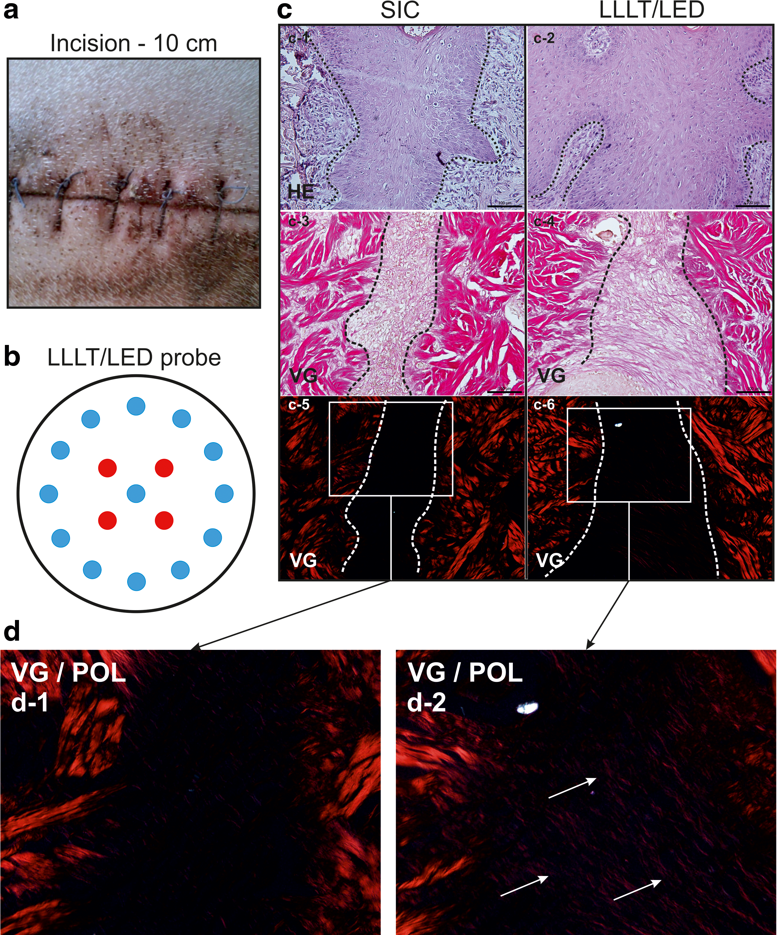
With the animals under general gas anesthesia [induced by a mixture of 50% N2O, 50% O2, and 1.5–2% Sevofluran (Abbvie, Campoverde, Italy)] under aseptic conditions, one incision, 10 cm in length, was created, and immediately closed using simple interrupted percutaneous sutures (Silon monofil 2/0, Chirmax, Prague, Czech Republic) on the back of each minipig (in the area between the processus spinosus of the third and fifth lumbar vertebra, Fig. 1a).
Half of the animals in each group were killed by anesthetic overdose (skin wounds were proceed for histology) on postoperative day 3 and the other half were killed on postoperative day 7.
PBM with red LLLT and blue LED
The wounds on the PBM-treated pigs were irradiated daily using a commercially available combined laser/LED device approved for clinical use: BTL-5000 (BTL Industries Ltd., Hertfordshire UK). The PBM probe (5.6 cm in diameter, S = 25 cm2, Fig. 1b) contained GaAs/GaAlAs diode laser with λ = 685 nm (4 laser diodes, output power per diode 0.05 W; shape of beam produced by each diode round, aperture 0.15 cm in diameter; laser light divergent; continuous mode) and blue LED with λ = 470 nm (13 diodes, output power per diode 0.016 W; shape of beam produced by each diode round; light divergent; continuous mode). For each of the red lasers and blue LEDs, the power density was 0.008 W/cm2; irradiation time per session was 420 sec; total energy density per session was 3.36 J/cm2; total daily dose was 84 J; and total cumulative dose for 3-day treated animals was 252 J and for 7-day treated animals it was 588 J). Minipigs from the SIC-group were sham irradiated.
Histological assessment
Wounds were processed routinely for light microscopy [fixation, dehydration, embedding, sectioning, and staining with hematoxylin-eosin (HE) and Van Gieson (VG) collagen staining]. The histological structures and processes [polymorphonuclear leukocytes (PMNL), re-epithelization, fibroblasts, and new collagen] were evaluated in blinded manner.
Results
Histological sections of the 3-day healed control wounds demonstrated that the incisions were not completely bridged by a layer of epithelial cells (not shown). The inflammatory phase was almost complete in both groups. The demarcation line, which consisted of PMNL, separated necrosis from vital tissue. In comparison with irradiated wounds, lower numbers of fibroblasts were present in control tissues. The incisional space at the layer of dermis was without a significant quantity of collagen in both groups (not shown).
On day 7, the epidermis completely covered the incisional gab with several layers of keratinocytes (Fig. 1c). The presence of a thin keratin layer was also recorded in both groups. In controls, mainly vertically oriented fibroblasts were present, whereas in irradiated wounds most fibroblasts were horizontally oriented (not shown). The predomination of cross-linked collagen fibers (Fig. 1c and d) indicated more progressive scar formation in irradiated wounds.
Discussion
The present study is the principal experimental work in which monochromatic coherent red light and monochromatic noncoherent blue light were used simultaneously to treat skin wounds in minipigs. Although the exact relationships between LLLT parameters and wound healing effect are still not fully understood, a wound-type-specific approach has already been encouraged by our group. 4 We have shown that LLLT effects depend upon dosing (5 J/cm2 delivered by 1, 5, 15, and 40 mW/cm2), wavelength (635 vs. 670 nm), and wound type (incision vs. excision). However, a literature review concluded that LLLT had a positive effect on the healing process of skin lesions in rats; however, the results in humans were inconclusive, 10 and justify our use of a pre-clinical porcine model to investigate how combined red/blue wavelengths of PBM may affect the incisional wound-healing process.
In addition, we observed no wound infection in either control or treated piglets. Therefore, the question of whether such combined PBM therapy is able to prevent occurrence of infected wounds remains to be answered in further research. The exact molecular mechanisms as to how this type of PBM modulates wound healing need to be clarified as well.
Conclusions
In this study, we have demonstrated that a daily wavelength-specific dose of 3.36 J/cm2 is able to accelerate the healing of sutured skin incisions when a combination of red/blue PBM has been used. Although the general molecular mechanisms in wound healing are similar, a direct extrapolation from this experiment to a clinical situation is not possible, because of interspecies variability. Therefore, further human research is warranted.
Footnotes
Acknowledgments
We thank Iveta Tomková, DVM for technical assistance. This study was conducted within the ITMS-26220220127 project supported by the Research & Development Operational Program funded by the European Research and Development Fund (ERDF). The experiment was also partially supported by the Slovak Grant Agency of Ministry of Education, Science, Research, and Sport (MESRS) (VEGA-1/0404/15 and VEGA-1/0048/15).
Author Disclosure Statement
No competing financial interests exist.