




Abstract
Introduction
P
We proposed an extracellular talaporfin sodium-induced photosensitization reaction with a short drug-light interval to induce photosensitization in interstitial spaces. Using this specific reaction, we intended to induce immediate blockade of electrical conduction by membrane damage and/or dysfunction of ion channels, and permanent blockade of electrical conduction by cell necrosis by irradiation through a laser catheter. 3 In our catheter ablation treatment methodology, we intend to insert the newly developed laser catheter from the femoral vein and achieve direct contact between the catheter tip and the myocardium for irradiation. The developed laser catheter is composed of a line irradiation diffusion fiber made from a plastic optical fiber, and shape memory alloys that allow fitting with any ablation lines. This electrical conduction blockade mechanism would be performable in a maze procedure for atrial fibrillation, 10,11 by using, for example, a linear irradiation device.
Talaporfin sodium (2.5 mg/kg, intravenous) binds with proteins present in serum, especially albumin (65–70%), high-density lipoprotein (HDL) (29–33%), and low-density lipoprotein (LDL) (1–2%). 9 It has been reported that uptake of talaporfin sodium and photocytotoxicity induced by absorbed talaporfin sodium decrease with increasing albumin concentration. 12 We previously reported that binding of talaporfin sodium with albumin reduces the photocytotoxicity of the extracellular talaporfin sodium-induced photosensitization reaction. 13
The drug-protein binding ratio changes with temperature. 14 Dubbelman et al. 15 reported that the photodynamic cross-linking of red cell membranes was temperature-dependent because of a reaction they termed a “secondary reaction.” The cell killing effect of the photosensitization reaction was dependent on temperature in the case of the intracellular photosensitizer-induced photosensitization reaction. 16 The target-tissue temperature should be close to body temperature during interventions, but may be lower during surgical ablation or open surgery 17 and the temperature would differ between a blood-filled heart and an arrested heart. In the case of photodynamic therapy, the temperature differs between normal tissue and tumor tissue. 18 There are no previous reports on the temperature dependence of photocytotoxicity in the extracellular photosensitizer-induced photosensitization reaction. Understanding the temperature characteristics of photocytotoxicity in myocardial cells using the extracellular talaporfin sodium-induced photosensitization reaction with a short drug-light interval is important because the target-tissue temperature will differ according to the treatment procedure. The cell killing effect would be excessive with a temperature increase by laser irradiation under catheter intervention, and insufficient under open surgery. Binding of talaporfin sodium with albumin (and the resultant photocytotoxic behavior) must be ascertained quantitatively to ensure that the treatment is safe. The aim of this study was to understand how temperature affects the extracellular talaporfin sodium-induced photosensitization reaction in a catheter-based procedure (around 37°C) or open surgery (lower temperature setting).
We investigated the temperature dependence of the binding of talaporfin sodium by measuring its absorbance spectrum. We also investigated the temperature dependence of the death of myocardial cells using a water-soluble tetrazolium-8 assay.
Materials and Methods
Solution preparation
For measurement of the binding ratio, three flowing solutions were prepared: Solution 1, saline with human serum albumin (molecular weight, 66,437–66,600; A9731-10G; Sigma-Aldrich, St. Louis, MO) 0–20 mg/mL; Solution 2, saline with HDL (RP-037; 20 mg/mL; Intracel Resource; Cosmo Bio, Tokyo, Japan) 0–0.04 mg/mL; and Solution 3, saline with LDL (RP-031; 5 mg/mL; Intracel Resource, Cosmo Bio) 0–0.14 mg/mL. These solutions were designed to cover the estimated human serum albumin, HDL, and LDL concentrations in interstitial spaces. 19, 20
Talaporfin sodium was dissolved in the dark. The concentration was set to 20 μg/mL in the three solutions containing the serum proteins described above. For measurement of cell lethality, talaporfin sodium concentration varied at ≤30 μg/mL in a solution comprising Dulbecco's modified Eagle's medium/nutrient mixture F-12 (DMEM/F-12; Invitrogen, Carlsbad, CA) with 10% fetal bovine serum (Invitrogen) that corresponded to an albumin concentration of 2.1 mg/mL.
Measurement of binding of talaporfin sodium
To investigate the temperature dependence of the binding ratio of talaporfin sodium, talaporfin sodium solutions containing serum proteins were set at 17°C, 27°C, and 37°C. The talaporfin sodium solutions were passed through ultrafiltration devices (Centrifree; Merck Millipore, Bedford, MA) containing a membrane with a nominal molecular-weight cutoff of 30 kDa, and centrifuged (200 × g, 10 min) at each temperature. The extracted filtrates were placed in optical cuvettes (optical path length, 1 cm). The absorbance spectrum of the Q band of talaporfin sodium was measured using a spectrophotometer (UV-3600; Shimadzu, Kyoto, Japan) from 645 to 665 nm at a resolution of 0.05 nm. The same solutions were filtered without any proteins as a reference. We obtained the differences between the reference solutions and the corresponding filtrates. The photosensitizer molecules that passed through the ultrafiltration devices were regarded as the unbound part, and the binding ratio of talaporfin sodium was calculated with the measured wavelength of the absorbance peak.
Evaluation of photocytotoxicity
Rat primary myocardial cells (Cardiomyocyte Culture Kit CMC02; Cosmo Bio) were seeded (2.0 × 10⁵ cells/mL in 0.1 mL of culture medium) and cultured for 6–7 days in a collagen-coated black 96-microwell plate under 5% CO2 at 37°C. A red diode laser (wavelength, 663 ± 2 nm) was used to excite talaporfin sodium. The optical setup was designed to irradiate the bottom of the microwell plate homogeneously at 0.29 W/cm2, as shown in Fig. 1. A prototype of a temperature-controlled chamber (Blast, Tokyo, Japan) with a glass heater, Peltier element, and proportional-integral-derivative controller was set on the irradiation table.

Optical setup for a photosensitization reaction under temperature control.
The microwell plate containing myocardial cells was placed in the temperature-controlled chamber and the solution temperature was set at 17°C or 37°C. Each microwell was filled with a prepared talaporfin sodium solution in DMEM/F-12 and regulated at 17°C or 37°C, thereby replacing the culture medium. The radiation exposure was varied from 0 to 40 J/cm2, with constant irradiance of 0.29 W/cm2 and changing irradiation duration from 0 to 137 sec, within 15 min after the beginning of the drug contact with the cells.
The morphology of the myocardial cells was observed before and immediately after laser irradiation by bright field microscopy (IX70; Olympus, Tokyo, Japan). Each microwell was filled with the culture medium (0.1 mL) and WST-8 assay solution (10 μL; Cell Counting Kit-8; Doujinkagaku, Kumamoto, Japan), replacing the talaporfin sodium solution just after the photosensitization reaction. The myocardial cells were cultured in the dark with the water-soluble tetrazolium-8 solution in an atmosphere of 5% CO2 for 2 h at 37°C. The absorbance at 450 nm of the solution in the microwell was measured using a microplate reader (Sunrise™; Tecan Group, Mannedorf, Switzerland). Cell lethality was defined as described in our previous report. 5,13
Statistical analysis
Data are presented as mean ± standard deviation from three or six independent experiments. SPSS version 22 (IBM, Armonk, New York, NY) was used to carry out Student's t-tests to evaluate the significance of differences in comparisons. p values of less than 0.05 were considered to indicate significant differences and are shown in the figures.
Results
Changes in binding of talaporfin sodium according to temperature
The binding ratios of talaporfin sodium with human serum albumin, HDL, and LDL are shown in Fig. 2a–c. The binding ratio of talaporfin sodium with human serum albumin was not associated with increasing temperature of the talaporfin sodium solution (Fig. 2a). By increasing temperature from 17°C to 37°C, the mean binding ratio of talaporfin sodium with HDL decreased by 6.3% at an HDL concentration of 0.04 mg/mL, and that with LDL decreased by 12.8% at an LDL concentration of 0.14 mg/mL (Fig. 2b, c). The plots represent the mean values of three repeated measurements.
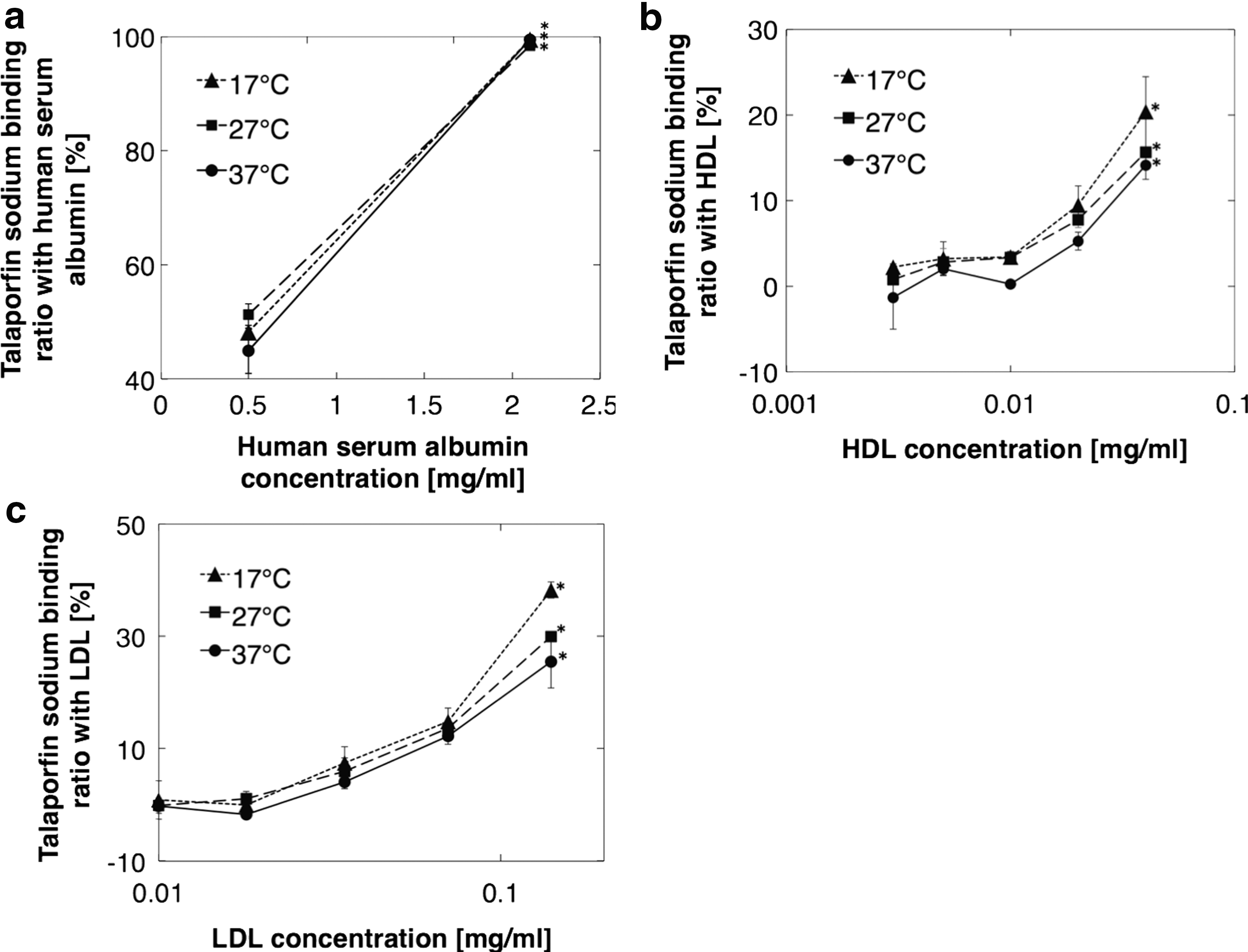
Obtained binding ratios for talaporfin sodium with human serum albumin
Changes in photocytotoxicity according to temperature
The morphologies of the myocardial cells before and after the photosensitization reaction are shown in Fig. 3. Many bleb formations were observed after the extracellular talaporfin sodium-induced photosensitization reaction under the 37°C condition (Fig. 3, black arrows). Figure 4 shows the dependence of cell lethality on photosensitization at different temperatures. Cell lethality increased depending on the concentration of talaporfin sodium and irradiation exposure. Cell lethality increased significantly with increasing temperature at irradiation exposures of 20 and 40 J/cm2 and a talaporfin sodium concentration of 20 μg/mL (Fig. 4c, d).
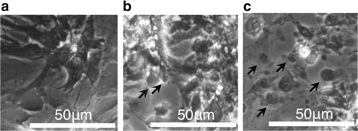
Cell morphology before
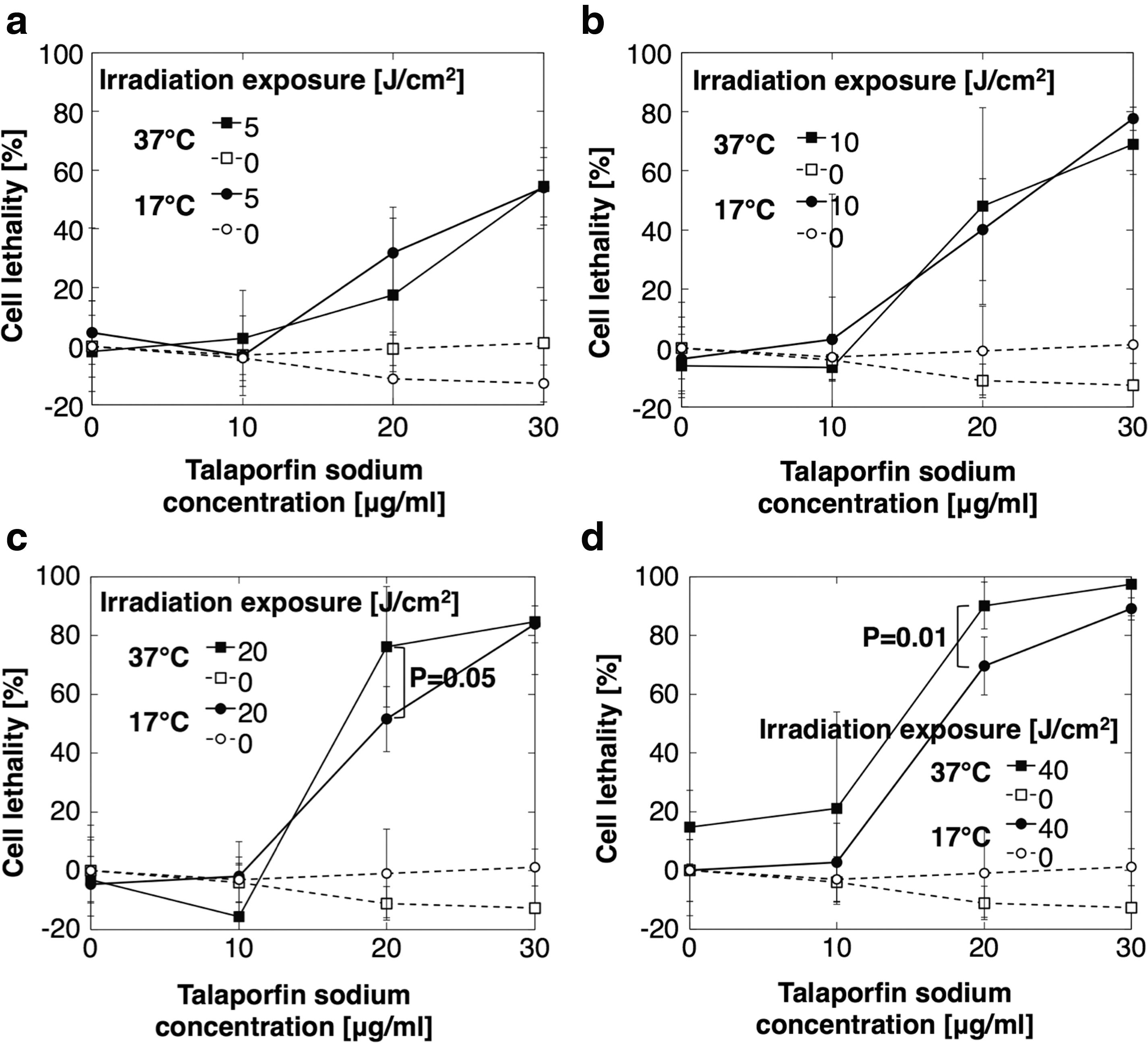
Dependence of cell lethality on photosensitizer concentration at 17°C and 37°C. White characters with dotted lines indicate reference lines. Irradiation exposure was 5
Discussion
The increases in cell lethality of 20–30% shown in Fig. 4c and d may have been derived from decreases in the binding ratios of talaporfin sodium with HDL and LDL.
In our proposed method for ablation, 4 we use a red diode laser to irradiate the myocardium. It might be expected that the temperature of the myocardium would be increased through absorption of the laser irradiation by the myocardium and/or blood. In our proposed method for photodynamic ablation, we intended to use a maximum input power of a few 100 mW/cm2. We can calculate the temperature increase caused by absorption of laser irradiation to a myocardial tissue depth of 1 mm and surface area of 1 cm2 to be 0.08°C/sec using the following parameters 21 : adiabatic 0.12 mm−1 as the absorption coefficient of the myocardial tissue; 4.2 as the specific heat of the myocardium; 1.0 as the specific weight of the myocardium; and 300 mW/cm2 of irradiance at the tissue surface.
In surgical ablation or open surgery, the temperature of the tissue surface can decrease to around 30°C. 16 A cell lethality decrease of around 10% would be suggested in the case of open surgery that has a lower temperature setting, compared with catheter-based treatment at 37°C, assuming that the photocytotoxicity changes linearly with temperature changes. The temperature of the target organ should be maintained for an efficient photosensitization reaction because photocytotoxicity decreases with decreasing temperature in these situations. Alternatively, it could be necessary to input extra energy in the case of open surgery to complement the efficiency decrease caused by the temperature decrease.
As it is suggested that these photocytotoxicity differences arise from binding ratio changes with serum proteins, it is possible that there may be photocytotoxicity differences among animal species because their serum albumin structures are different. 22 A limitation of this study was that we only used rat primary myocardial cells to investigate the therapeutic efficiency in humans.
Footnotes
Acknowledgments
This work was supported in part by the Adaptable and Seamless Technology Transfer Program (#AS2415004P) of the Japan Science and Technology Agency and Japan Agency for Medical Research and Development. We greatly appreciate the clinical consultation by Dr. Seiji Takatsuki and Dr. Takehiro Kimura from Keio University School of Medicine.
Author Disclosure Statement
This work has been supported in part by Acceleration of Transformative Research for Medical Innovation of the Japan Agency for Medical Research and Development.