
Abstract
Introduction
M
The first medical application of lasers was reported by Dr. Goldman regarded as the father of lasers in medicine. Ten years later, high-energy CO2 lasers were utilized for myocardial acupuncture. These lasers are high energy for their high-energy densities, which range from 1 to 100 W/cm2. The CO2 lasers are able to produce microchannels, without debris or fibrosis formation, allowing blood to flow directly from the ventricular cavity into the ischemic myocardium. 2 This technology is known as transmyocardial laser revascularization (TMR). TMR provides a possible treatment for patients suffering from diffuse coronary artery disease (CAD) that is not amenable to percutaneous coronary interventions or coronary artery bypass grafting.
Low-level lasers, on the other hand, emit nonthermal beams that do not deliver power to damaged tissue, but do deliver sufficient energy for heat-independent biostimulation effects. 3,4 Low-level laser irradiation (LLLI) has been applied in clinical practice for more than 40 years. 5,6 Initial studies have reported that LLLI has an effect on various biological processes, which accelerate wound healing and promote the regeneration of muscle. 7 –9 It could stimulate the photoreceptors that transfer light energy into chemical energy in the form of ATP, which further promotes cell proliferation. 10
In 2000, Dr. Uri from Tel Aviv University began to apply LLLI to treat MI in the rat and dog. He found that infarct size was reduced by 52% in the group that received LLLI treatment after induced MI. 11 Later studies indicated that LLLI could stimulate proliferation and differentiation of bone marrow-derived mesenchymal stem cells (BMSCs) and attenuate the hostile pathological microenvironment of damaged myocardium. 4,12 Further, another study reported that LLLI has a positive effect on proliferation of cardiac stem cells (CSCs) in vitro. 13 Most laser studies use BMSCs as cell sources because it is easy to get through aspiration procedures.
In this review, we summarized published studies on molecular mechanisms of LLLI preconditioning for cardiac cell-based therapy.
Using LLLI to Precondition Stem Cells Before Implantation
Although the beneficial effect of BMSC implantation on infarcted myocardium has been described, 14,15 other studies have failed to find improved cardiac function after implantation. 16 This might be a consequence of the high apoptotic percentage and limited myogenesis ability of BMSCs, so sufficient numbers of BMSCs with improved differentiation ability are necessary before transplantation. 17 On the other hand, when it comes to CSCs, we also encounter the same problem: few autologous CSCs and long preparation time.
In 2005, Abramovitch-Gottlib et al. observed that combining a 3D biomatrix with LLLI biostimulates the proliferation of BMSCs. 18 Another study reported similar results, where LLLI promoted proliferation of BMSCs and CSCs in vitro. 13 Our studies have also shown that LLLI facilitates proliferation and myogenic differentiation of BMSCs. 12 These findings indicate the positive stimulating effect of LLLI as a potential solution to the abovementioned problem.
LLLI preconditioning promotes stem cell proliferation
Increased proliferation after LLLI has been found in other cell types, such as lymphocytes, fibroblasts, and osteoblasts. 19 However, stem cells are different from these cells due to their multi-lineage differentiation potential. To date, the mechanism of LLLI proliferation effects on stem cells remains poorly understood.
Laser parameters
Laser parameters play a role in the proliferation-stimulating effect of LLLI. Table 1 summarizes laser parameters used in studies from 2006 to 2015, looking at the effect of LLLI on BMSC proliferation. Most studies have observed a positive effect of LLLI on the proliferation of BMSCs.
ADSCs, adipose-derived stem cells; BMSC, bone marrow-derived mesenchymal stem cell; CSC, cardiac stem cell; LLLI, low-level laser irradiation.
LLLI stimulates proliferation of BMSCs without cytotoxicity at a wavelength of 635 nm and a power of 60 mW, which is in agreement with later studies. 12 However, Bouvet-Gerbettaz et al. reported that LLLI fails to promote proliferation of BMSCs at a wavelength of 808 nm and a power of 800 mW. 20 This indicates that the laser effect on cells depends on optimal wavelength and power. Moreover, Soleimani et al. found that LLLI significantly stimulates BMSC proliferation at 2, 3, or 4 J/cm2, but not at 6 J/cm2, which indicates that energy density also plays a part in this process. 21
It has been found that different wavelengths and energy densities lead to different gene expression profiles. Studies have shown that the transforming growth factor (TGF)-β pathway switches from canonical to noncanonical signaling as wavelength increases. Meanwhile, receptor activator of nuclear factor kappa-B ligand and matrix metalloproteinase-10 increase in dose–response curves as energy density increases. 22 These results could help to explain paradoxical results in the above studies when wavelengths and energy densities differ. Nevertheless, the optimal wave lengths and energy densities for stimulating cell proliferation are still under debate.
LLLI preconditioning activates cell proliferation signaling pathways
As a downstream effector of PI3K/Akt signaling pathways, Akt is a serine/threonine kinase, which has a positive effect on the proliferation of cells by inhibiting apoptotic processes. 23,24 It has been reported that many growth factors modulate angiogenesis, migration, and cell proliferation through PI3K/Akt signaling pathways. 19,25,26 Microarray technology was applied to investigate the gene expression profiles of BMSCs after LLLI. It was found that 119 genes were differentially expressed between control and irradiated groups. 7 Among them, five genes (PI3Kca, Akt1, Ptpn6, StK17b, and Ccnd1) were confirmed by real-time polymerase chain reaction (real-time PCR). In the irradiated group, levels of PI3Kca, Akt1, and Ccnd1 were increased, while Ptpn6 and Stk17b were decreased. PI3Kca is the catalytic subunit of PI3K; Akt1 is a family member of Akt; and Ccnd1 is a downstream effector of PI3K/Akt and plays an important role in proliferation. Ptpn6 is able to decrease the level of p-Akt, which increases cell apoptosis, but inhibits proliferation. Stk17b is a kinase, which promotes cell apoptosis as well. Another study also reported that the expression of Akt1 pathway components is increased in BMSCs exposed to infrared radiation wavelengths at a wavelength of 830 nm. 22 Interestingly, PI3Kca, Akt1, Ptpn6, and Ccnd1 are all involved in the PI3K/Akt/mTOR/eIF4E signaling pathway. According to the results above, PI3K in BMSCs could be activated after LLLI, which induces phosphatidylinositol 4, 5-bisphosphate (PIP2) phosphorylation and converts it to phosphatidylinositol 3, 4, 5-triphosphate (PIP3). PIP3 facilitates Akt activation by 3-phosphoinositide-dependent protein kinase-1 (PDK1). Activated Akt then activates rapamycin (mTOR), which further phosphorylates eukaryotic initiation factor 4E-binding protein 1 (eIF4EBP1). The phosphorylated eIF4EBP1 dissociates from eukaryotic initiation factor 4E (eIF4E), allowing it to form the initiation complex. This complex triggers Ccnd1 gene translation, which stimulates the proliferation of BMSCs 7 (Fig. 1).
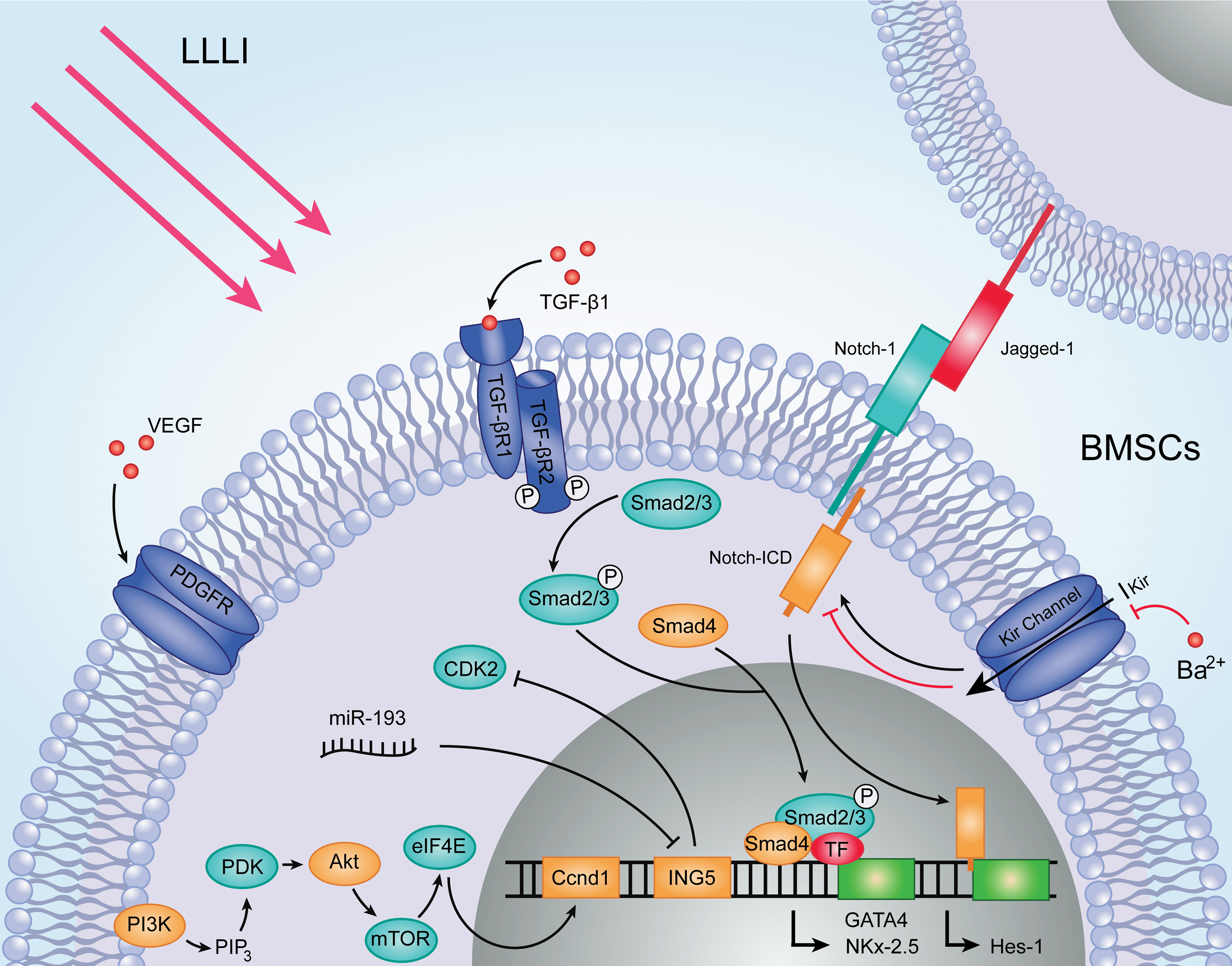
Molecular profiling of LLLI preconditioning in cardiac regenerative therapy. LLLI preconditioning activates the PI3K/Akt signaling pathway and notch-1 signaling pathway, upregulates the expression of miR-193, and the secretion of VEGF and TGF-β1, which stimulates the proliferation and differentiation of stem cells. LLLI, low-level laser irradiation; TGF-β1, transforming growth factor-β1; VEGF, vascular endothelial growth factor.
Notch-1 is a transmembrane receptor whose ligands are located on the surface of neighboring cells. 27 It has been reported that Notch signaling plays an important role in the proliferation of mesenchymal stem cells (MSCs). 28 Dr. Giannelli and his group found that the expression of Notch-1 and its intracellular domain and ligands were significantly upregulated in BMSCs after LLLI. 27 Hes-1, a downstream effector of canonical Notch-1 signaling, was also increased. Moreover, they performed electrophysiological whole cell patch-clamp analysis on BMSCs after LLLI and found that inward rectifier K+ current (IKir) and L-type Ca2+ current (ICa, L), as well as T-type Ca2+ current (ICa, T), were increased compared with the control group. As a result, suppressed Kir currents significantly reduced MSC proliferation and Notch-1 expression. This suggests the Kir channel plays a central role in regulating Notch-1 activation and controlling LLLI-induced BMSC proliferation (Fig. 1). However, how Kir channels influence the expression of Notch-1 is still unknown, which would be worth investigating in the future.
LLLI preconditioning alters microRNA expression
MicroRNA (miRNA) has an effect on almost every aspect of cellular activity. 29 It has been found that a subset of miRNA plays a crucial role in the proliferation of stem cells. 30 Global changes in miRNA expression in BMSCs following LLLI treatment were investigated. 31 As a result, 34 miRNAs were found to be differentially regulated, in which 19 miRNAs were upregulated, while 15 miRNAs were downregulated. Among them, miR-193 was the most significantly upregulated. Further, we found the expression of growth family member 5 (ING5) proteins is 50% lower than the control group in miR-193-overexpressed BMSCs. Anti-miR-193 can increase the expression of ING5. It was also shown that cyclin-dependent kinase 2 (CDK2), a downstream effector of ING5, is upregulated by overexpression of miR-193 and downregulated by inhibiting miR-193. Applying siRNA of ING5 also increases CDK2. Previous studies have demonstrated that CDK2 plays an important role in cell proliferation because of its effect on G1/S transition during mitosis. 32 In conclusion, we proposed a possible model of miR-193 regulation of BMSC proliferation promoted by LLLI (Fig. 1).
Moreover, we reported that another miRNA, miR-133b, can be induced by LLLI. It has been reported that miR-133b is able to promote cell proliferation by repressing serum response factor. 32 Our results showed that miR-133b can influence the expression of cyclin D1 (CCND1) and CDK2. They both are cell cycle-specific proteins, which indicate that miR-133b may regulate BMSC proliferation through a complicated signaling pathway.
LLLI precondition promotes differentiation of stem cells
When irradiated and nonirradiated BMSCs are treated with myogenic differentiation inductor, 5-azacytidine, we found the expression of myogenic-specific proteins, sarcomeric α-actin, and desmin, is dramatically higher in the irradiated group, which confirms the positive effects of LLLI on BMSC myogenic differentiation. 12
On the other hand, the therapeutic potential of implanted BMSCs is partly attributed to their potential endothelial cell differentiation, important for angiogenesis in infarcted myocardium. 33 A study has shown that BMSCs are able to enhance vascular density at 5 days after implantation. 34
Although a myriad of studies have proposed many molecular mechanisms underlying cardiomyogenesis of BMSCs, 35,36 little is known about the molecular mechanisms behind the myogenic and endothelial conversion of BMSCs by LLLI.
LLLI preconditioning stimulates paracrine secretion
BMSCs can secrete various antiapoptotic as well as angiogenic factors, such as vascular endothelial growth factor (VEGF), insulin-like growth factor 1 (IGF1), and TGF-β1. Different studies have established that these factors mediate an improvement of cardiac function and reduce the infarct scar. 37,38
VEGF is commonly known as a signal protein playing an essential role in angiogenesis and vasculogenesis. Although VEGF can be secreted by BMSCs under standard culture conditions, LLLI preconditioning could significantly increase the production of VEGF. 12 Moreover, studies have reported that VEGF is able to induce BMSC proliferation through binding with platelet-derived growth factor receptors, suggesting that VEGF may contribute to the LLLI proliferation effect on stem cells. 39
IGF1 is a multi-functional peptide that regulates cell growth and differentiation. 40 From the analysis of the mRNA of growth factors in LLLI-treated BMSCs, it was found that the expression level of IGF1 was significantly increased. Further, after cells were treated with IGF1-neutralizing antibodies, proliferation was significantly decreased. The combination of these results provides support for the importance of IGF1 in the biostimulation effect of LLLI. 27
TGF-β1 is a multi-functional cytokine involved in the growth and differentiation of various cells. 41 Previous studies have demonstrated that TGF-β1 can promote embryonic stem cell differentiation into cardiomyocytes. 42 In a recent study, LLLI significantly stimulated the expression of TGF-β1 in BMSCs. They found upregulation of TGF-β1, TGF-β receptor 1 and 2, and Smad1 and 2, components of the canonical TGF-β1 pathway. Further, their TGF array also detected increased phosphoserine 187 of Smad1, phosphothreonine 220 of Smad2, and phosphoserine 179 of Smad3. 22 Consistently, another study reported a similar result of increased expression of phosphorylated Smad2 and Smad3 in BMSCs at 24 h after TGF-β1 stimulation. 43 Given these facts, it indicates that TGF-β1 may play an important role in the LLLI stimulating effect on myogenic differentiation of BMSCs (Fig. 1).
LLLI Preconditioning Creates a Friendly Milieu in Infarcted Myocardium for Implanted Cells
Although many studies have reported that BMSC transplantation can significantly improve cardiac function, the low survival rate of BMSCs after transplantation limits the efficacy of cell therapy. Numerous strategies have been used in cell preconditioning to improve cell survival. 44,45 However, these direct stem cell interventions are complicated and may lead to undesired effects generating safety problems for clinical application.
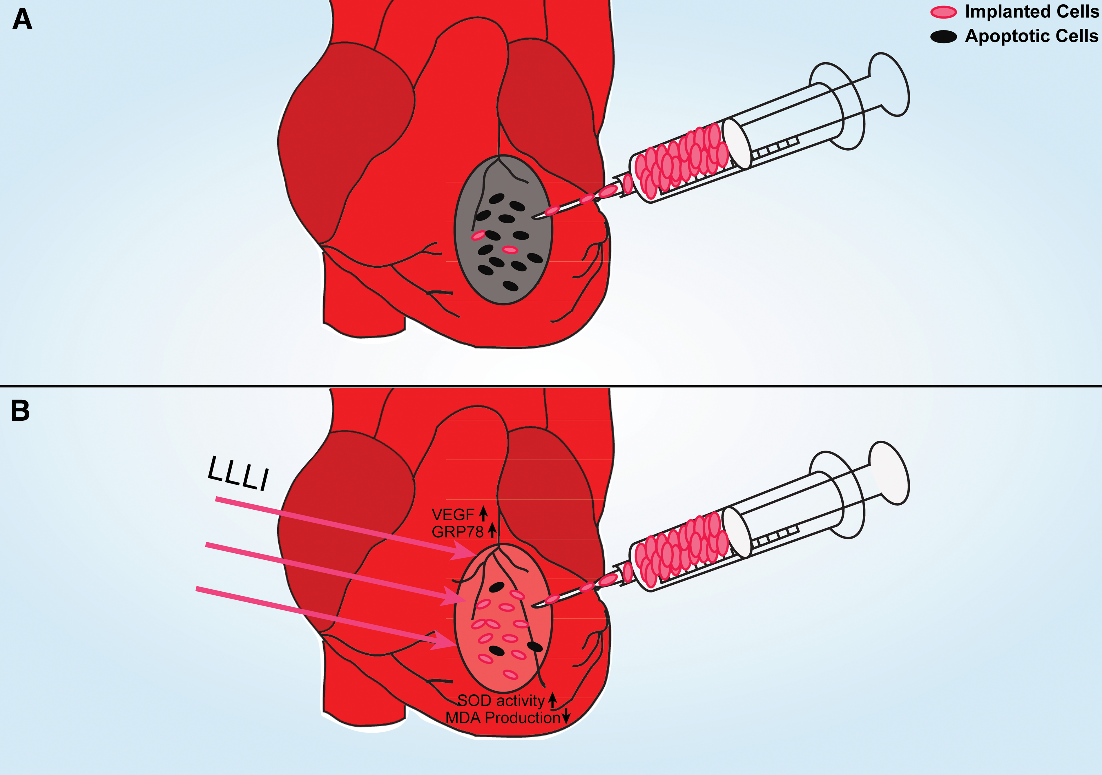
Our group applied a preconditioning of 635 nm diode laser with an energy density of 0.96 J/cm2 on infarcted myocardium before cell transplantation. 4 We found that LLLI significantly increased early survival rate of implanted cells by twofold. LLLI also decreased their apoptotic percentage and enhanced vascular density in the myocardium compared with the nonirradiated group, indicating LLLI preconditioning might remodel the hostile milieu of infarcted myocardium.
Our further studies showed that LLLI significantly stimulates the expression of VEGF and glucose-regulated protein (GRP78) in myocardium. Thus, increased VEGF and GRP78 may mediate the remodeling process triggered by LLLI preconditioning. Further, we found that LLLI enhances the activity of superoxide dismutase (SOD) and inhibits the production of malondialdehyde (MDA), which decreases the level of superoxide in infarcted myocardium. It may also contribute to improved survival rate of implanted cells. Taken together, we speculate that LLLI myocardial preconditioning creates a friendly milieu in infarcted myocardium for implanted cells. This is accomplished through increased expression of VEGF and GRP78, as well as enhanced SOD activity and decreased MDA production (Fig. 2).
LLLI Preconditioning Alters Cardiac Cytokine Expression in Infarcted Myocardium
Cytokines released from injured myocardium play an important role in the inflammatory response during cardiac repair. Previous studies have shown that LLLI is able to protect tissues through regulating cytokine expression and inhibiting inflammation responses. 46 –49 A cytokine protein array system was applied to screen cardiac cytokines following LLLI preconditioning to explicate the altered cytokine expression profile in damaged myocardium. 50 The results showed that LLLI preconditioning stimulates the expression of interleukin (IL)-4 and granulocyte/marcrophage colony-stimulating factor (GM-CSF) and decreases the production of chemoattractant-3 (CINC-3) and fractalkine.
It has been reported that GM-CSF can mobilize BMSCs to regenerate the infarcted myocardium. 51 Additionally, a study reported that intracoronary or subcutaneous administration of GM-CSF is able to improve collateral flow in a patient suffering from CAD. 52 These results indicate that LLLI promotes stem cell homing and stimulates collateral formation by increased expression of GM-CSF. On the other hand, fractalkine plays a role in leukocyte accumulation and modulates local inflammatory activity. 53 Decreased fractalkine can inhibit leukocyte accumulation in the infarcted myocardium and alleviate inflammatory responses, which may improve the survival rate of implanted cells. Above all, LLLI preconditioning exerts a protective effect on infarcted myocardium by stimulating the expression of GM-CSF and inhibiting the production of fractalkine, which may benefit implantation.
Conclusions and Summary
In the past, LLLI has been applied for wound healing, inflammation reduction, and pain relief. Recent years have witnessed a growing interest for the application of LLLI in cardiac regenerative therapy. It has been well demonstrated that LLLI preconditioning stimulates proliferation and differentiation of stem cells. Moreover, LLLI could remodel the hostile milieu in infarcted myocardium, but this beneficial effect vanishes rapidly within 1 week. It suggests that repeated irradiation is necessary to amplify its positive effect. Further, a myriad of studies only concentrated on LLLI preconditioning before cell transplantation. In fact, LLLI after transplantation may also increase the survival rate of implanted cells, but evidence is still lacking.
At last, although lots of studies have reported the possible molecular mechanisms involved in LLLI preconditioning, the exact mechanisms of this biological effect are still not clearly understood. In future studies, it will be necessary to use new platforms, such as proteomics and metabolomics, to gain a clear understanding of the molecular profiling of LLLI preconditioning effects in cardiac regenerative therapy.
Footnotes
Acknowledgments
This study was supported by the Program for New Century Excellent Talents in University, Foundation for Young Investigator at Peking Union Medical College (XHQN09 and XR01-YP), the Program for Distinguished professor in PUMC, and National Natural Science Foundation of China (81525002 and 81270225).
Author Disclosure Statement
No competing financial interests exist.