
Abstract
Introduction
I
In previous INS studies, infrared lasers in the 1800–2200 nm wavelength range have been mainly used because of the relatively high water absorption coefficient. 5 –7 Recently, pulsed short-wavelength near-infrared (SWNIR) laser was proposed for auditory neural stimulation because of its deeper penetration depth in fluids and tissues. 8,9 Because the cochlea is a hollow chamber of bone filled with lymph fluid, the laser irradiation has to penetrate the fluids before it acts on the target neurons within the modiolus. 10 Especially when the SWNIR laser with deeper tissue penetration depth is used, 11 laser divergence in the lymph fluid should be considered and controlled properly to ensure accurate stimulation. In our study, for the first time, the fiberoptic collimation technique was developed, using the 808 nm wavelength pulsed near-infrared laser to stimulate cochlear neurons. The auditory neurons were stimulated with collimated and uncollimated laser irradiation and the neural responses to the laser modes were compared by recording the optically evoked auditory brainstem responses (OABRs). The study aimed to investigate the effect of the laser collimation technique on activation of auditory neurons in the cochlea with 808 nm wavelength SWNIR lasers.
Materials and Methods
Ethics statement
All procedures performed in studies involving animals were in accordance with protocols approved by the Laboratory Animal Ethics Management Regulations instituted by the State Science and Technology Commission.
Animal anesthesia and surgery
Hartley guinea pigs (specific pathogen free, 3–4 months of age) of either sex were anesthetized by urethane solution (1200 mg/kg body weight), through an intraperitoneal injection. Depth of anesthesia was evaluated every 15 min by a paw withdrawal reflex, and 0.1–0.2 mL urethane solution was injected when necessary. The animal was then positioned on a thermostatically controlled heating pad to maintain its body temperature at 38°C. Stereotaxic equipment (DW-2000, Taimeng bio-instruments Co., Ltd., Chengdu, China) was used to stabilize the animal's head for surgical preparation. A “C” shaped retroauricular incision behind the right pinna of animal was done after shaving the hair around the ear. Then the muscle tissue and soft tissue were dissected and a hole (∼5 mm2) was opened on the posterior-lateral part of the bulla to get access to the round window niche (Fig. 1).
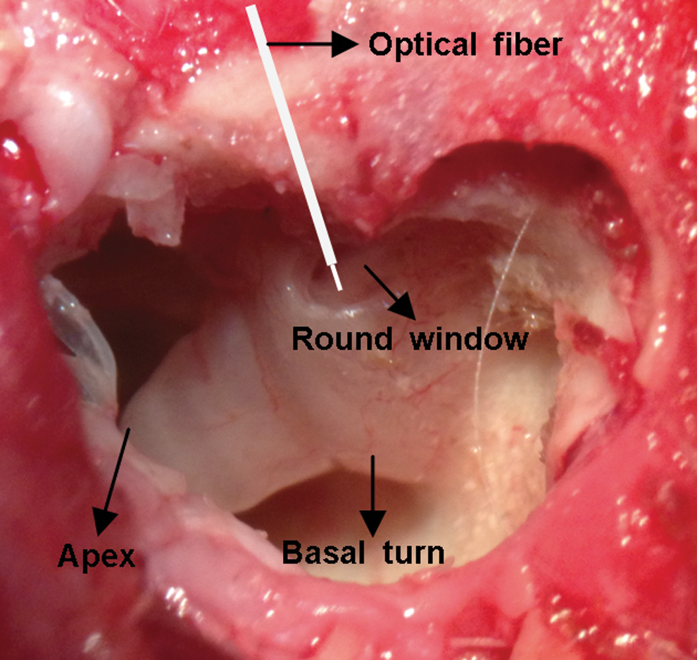
The access to the cochlea and the optical fiber placement.
Animal deafening procedure
At present, the mechanism of INS in the auditory system is still under investigation. Most cochlear INS studies suggest that infrared radiation activates the spiral ganglion neurons directly via the photothermal effect. 12,13 However, some researchers reported that the pulsed laser induced optoacoustic events, and that pressure waves could also evoke the neural responses when the cochlear hair cells are still functional. 14,15 Therefore, strong experimental controls are indispensable to eliminate the hair cell function before the optical stimulation. In our study, we deafened the cochlea with an additional surgery. The cochlear osseous spiral lamina was exposed and the basilar membrane of the basal cochlea was destroyed using a medical electrical drill (XY-ND3A, Xiyi Medical Instrument Co., Ltd., Tianjin, China). The surgery eliminated the hair cell function and caused hearing loss.
Acoustic test and ABR recording
Cochlear function was evaluated before and after the deafening procedure by utilizing the evoked potentials system (SmartEP USB, Intelligent Hearing Systems, USA) to record the acoustically evoked ABRs (AABRs). Three subdermal needle electrodes were located at the vertex (record), the ipsilateral mastoid (reference), and the neck (ground). The ABR signals were filtered between 100 Hz and 3 kHz and averaged across 512 trials. The sound stimuli were generated with an acoustic transducer (ER-3A Insert Earphones, Intelligent Hearing Systems, USA). A silicone tube was connected to the transducer and coupled the sound stimuli to the inner ear canals. A series of 100 dB sound pressure level (SPL) click-sound stimuli (11 Hz repetitions, 200 μs-duration, alternating polarity) was transmitted into the cochlea before and after the deafening process, to evaluate the hearing function. The entire experiment was conducted in an electromagnetic isolation soundproofed room.
Optical stimulation protocols
Two 808 nm wavelength fiber-coupled diode laser systems (105 μm core diameter fiber with 0.22 NA, TTL modulation, Beijing STONE Laser Co., Ltd., China) were applied in the optical stimulation process, one with an uncollimated fiberoptic output (Fig. 2a) and the other with a C-lens collimating element (silica glass, 1 mm diameter, sz-ark optical device Co., Ltd., Shenzhen, China) on the fiber tip (Fig. 2b). For the uncollimated laser output, the full divergence angle was 19.1 degrees measured in fluids. The collimated laser output has a full divergence angle of 0.3 degrees in fluids. The laser beam profile is Gaussian, and the spot size was defined as the diameter over which the laser intensity decreased to 1/e 2 of its peak intensity. During the optical stimulation, the laser pulse duration and repetition rate were set to 200 μs and 11 Hz, and the distance from optical fiber tip to neurons was kept at 0.5 mm. The radiant exposures at the surface of tissue for both the divergent and collimated outputs were maintained at the same level, ranging from 0.1 to 1.5 J/cm2, by calculating and adjusting the laser peak power according to the spot sizes at the tissue surface of each laser output. The laser power was measured at a 0.5 mm distance from fiber tip to probe surface by a power meter (Nova II meter, PE10BF-C probe, Ophir Photonics, Israel).
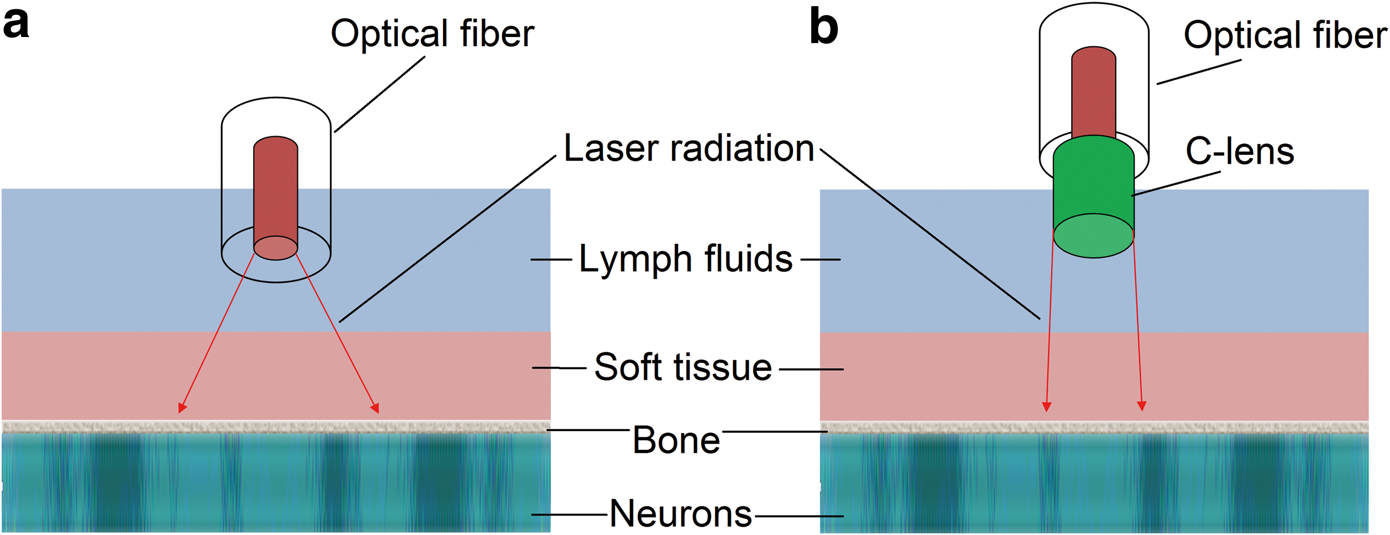
Diagram of the simplified laser neuron stimulation in the cochlea. The graphs are not to scale.
Optical stimulation was performed with deafened animals. The collimation fiberoptic was carefully placed into the surgical access using a three-axis micromanipulator (TSD-40 XYZ, SIGMA KOKI, Japan). The optical fiber was inserted into the round window and oriented toward the modiolus (Fig. 1). Next, the radiant exposure at the tissue surface was adjusted from 0.1 to 1.5 J/cm2 in 0.1 J/cm2 steps, and the OABR at each radiant exposure was recorded accordingly. In addition, we made the OABR amplitudes increase from 0.5 to 4 μV in 0.5 μV steps by adjusting the stimulating laser energy. The energy value at each OABR amplitude step was measured correspondingly for the energy consumption analysis. An identical operation process was then repeated with the uncollimated laser output setup.
Data acquisition and statistical analysis
In this study, the ABR wave III peak-to-peak amplitude, measured from the third positive peak to the following negative peak, was utilized to quantify the neural response intensity. The ABR data was exported to OriginPro8.6 software for data analysis and curves drawing. The one way analysis of variance (ANOVA) statistical model was implemented in results analysis.
Results
A total of five guinea pigs were used, and ABR was recorded when stimulating the auditory neurons acoustically and optically. The results showed classical Jewett waves evoked by 100 dB SPL sound clicks with normal hearing animals, and the AABR wave III amplitude was 5.6 μV (upper waveform, Fig. 3a). While stimulating the same animal after deafening, the absence of ABR confirmed that the animal hair cells function was eliminated (lower waveform in Fig. 3a).
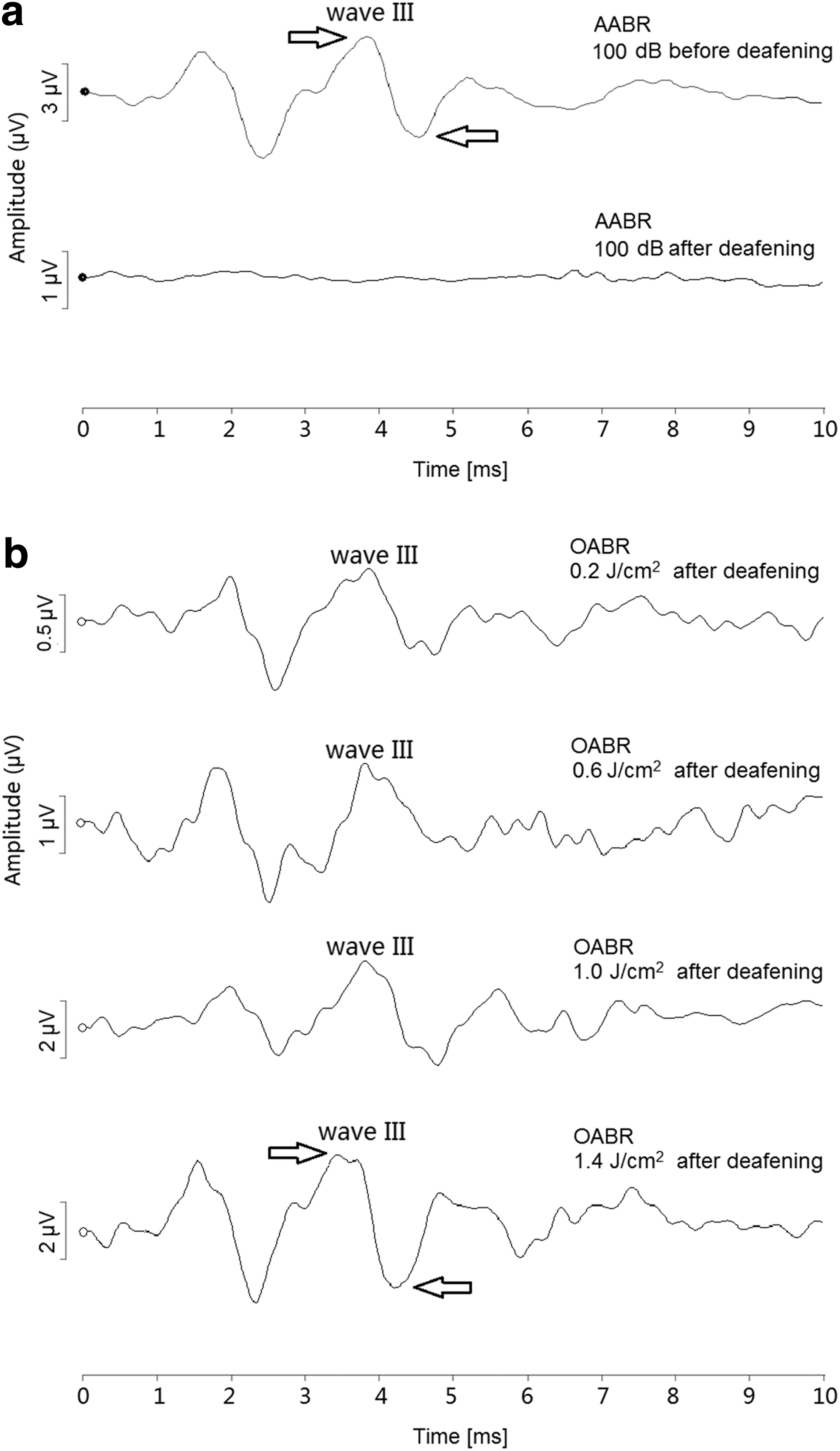
Acoustically and optically evoked auditory brainstem response (ABR) waveforms with animals.
In response to optical stimuli, the OABR was successfully recorded with deafened animals. The OABR showed a similar wave shape compared with the AABR waveform, and its intensity increased with the increased radiant exposures (Fig. 3b). In order to contrast the different effects with collimated and uncollimated laser output, the OABR wave III amplitudes under the two output approaches were measured as a function of the increasing radiant exposure levels at the tissue surface (Fig. 4a,b). Input-output (I/O) curves demonstrated that each animal (n = 5) followed a similar trend with ABR amplitude increase along with the increasing radiant exposure levels. ANOVA showed no statistically significant differences among the individual animals (p > 0.05). The averaged I/O curves across the animals further revealed the performance difference between the divergent and collimated laser stimulations (Fig. 4c). OABRs with the laser collimation showed nearly double amplitude (0.78 vs. 0.38 μV) than the uncollimated output with the same radiant exposure of 0.2 J/cm2. Moreover, the OABR intensity disparity between the two output modes increased to 1.51 μV (5.27 vs. 3.76 μV) at the 1.5 J/cm2 radiant exposure.
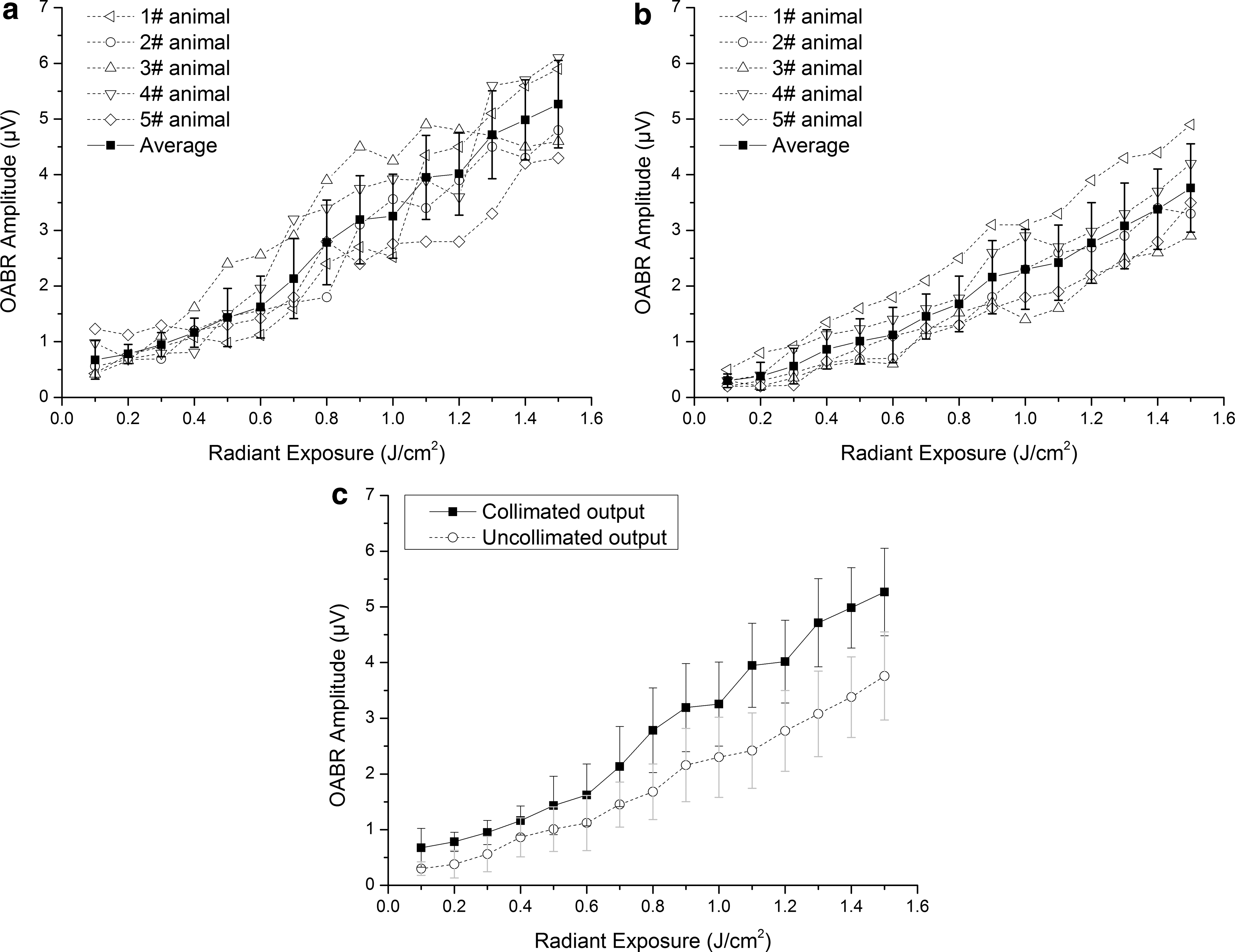
Input/output (I/O) functions relating the optically evoked auditory brainstem response (OABR) amplitude to radiant exposure at tissue surface with the collimated output
Whether the fiberoptic collimation technique could reduce the energy demand of the INS was also investigated. The I/O curves demonstrated the energy consumption difference of the two output modes while evoking the same OABR intensities from 0.5 to 4 μV in 0.5-μV steps (Fig. 5). Averaged data (Fig. 5c) showed that the collimated laser configuration consumed only 43.1% laser energy (0.22 vs. 0.51 mJ/pulse) compared with the uncollimated mode when evoking 1.5 μV OABR amplitude, and this percentage was reduced to 35.6% (0.42 vs. 1.18 mJ/pulse) when the evoked OABR amplitude reached 4 μV.
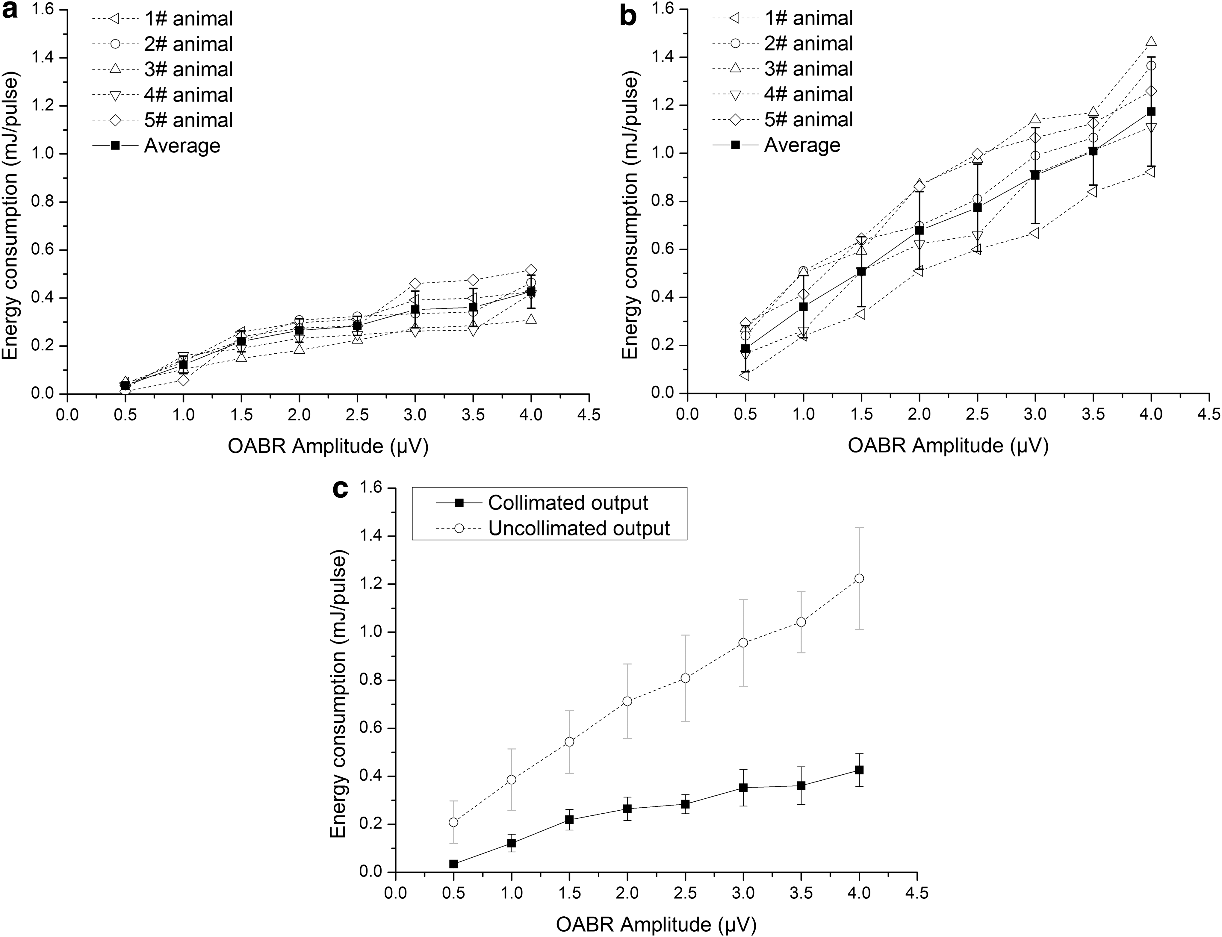
Input/output (I/O) functions relating the optically evoked auditory brainstem response (OABR) amplitude to the stimulating energy consumption with the collimated output
Discussion
In this study, the pulsed SWIR laser with the collimation technique was used to activate the auditory neurons in the cochlea. It was suggested that the laser wavelength in the range of 800–1000 nm might have less lymph or tissue obstruction of light delivery to the target neurons because of its lower water absorption. However, the divergent laser out from the optical fiber could be one of the negative effects on the INS performance, which enlarged the irradiation area and reduced the stimulation selectivity. 16 Our data confirmed that the laser collimation approach could have a positive effect on auditory neural stimulation in two main aspects. First, the collimated laser output could evoke a slightly higher OABR intensity than the uncollimated output in the average data (shown in Fig. 4c), with the same radiant exposure level at the tissue surface. We consider that this phenomenon could be relevant to the laser irradiation diffusion in tissue. Because the spiral ganglion neurons are housed beneath the tissue surface, the collimated, approximately parallel laser has less diffusion in tissue than the divergent laser output before it acts on the neurons. Therefore, the actual radiant exposure at the neuron surface with the uncollimated output was lower than the level with the collimated output, which evoked a weaker neural response than the collimated laser stimulation.
In addition, the collimated laser setup consumed less than one third of the energy compared with the uncollimated setup. This economization of stimulating energy consumption could bring several advantages to the engineering applications of optical neural activation. First, the system power consumption is always one of the key considerations in electrical cochlear implants. Similarly, in optical cochlear implants, the reduction of laser energy could save the limited system energy sources, which are usually powered by batteries. Additionally, with the lower laser energy demand, the optical stimulation system could be further miniaturized and integrated. Finally, in the multichannel optical neural stimulation configuration, the laser site spacing and heat distribution have an effect on the stimulation performance. 16,17 The collimated output approach with lower beam divergence could reduce the crosstalk between the channels and make the heat distribution more focused, which could improve the spatial selectivity of optical neural stimulation.
Although this study provided some evidence that the fiberoptic collimation technique could have a positive effect on cochlear neuron stimulation with near-infrared laser, some limitations still exist. In our study, the C-lens was selected as the collimating optics for its relatively low cost and longer working distance compared with the Gradient Index (GRIN) lens, but the size of the collimator is still overlarge for the multichannel optical neural stimulation. To overcome this limitation, ways to optimize the collimation structure, such as using the self-collimated fiberoptics or machining microstructure at the fiber tip for collimation will be explored. Moreover, the collimated laser neural stimulation requires higher precision in the placement and orientation of the optical fiber, which could be solved by designing a special waveguide for the laser irradiation delivery.
Conclusions
This study examined a method to stimulate cochlear neurons with a laser collimation technique and successfully activated the auditory neural response with deafened guinea pigs in vivo. The fiberoptic collimation technique improved the stimulation efficiency and reduced the stimulating energy demand in 808 nm wavelength near-infrared neural stimulation in the cochlea. The positive effects of this laser collimation technique could bring benefits to further research in optically based cochlear implants.
Footnotes
Acknowledgments
This work was supported by the National Natural Science Foundation of China (11474185), the Shandong Provincial Science Foundation for Distinguished Young Scholars of China (2013JQE27056), the Shandong Provincial Natural Science Foundation of China (ZR2012FM030), and the Fundamental Cross-decipline Research Foundation of Shandong University (2015JC029).
Author Disclosure Statement
No competing financial interests exist.