
Abstract
Introduction
D
Despite the advantages of Er:YAG lasers, the absorption of the laser energy in the “cloud” of debris formed during ablation reduces the cutting ability and redirects the beam from its original path, resulting in cavity preparations that are not well defined. 13 –15 In order to resolve this problem, more recently the quantum square pulse (QSP) technology was introduced, extending the treatment options available for Er:YAG lasers. In the QSP mode, a longer laser pulse is divided into several short pulses that follow each other at an optimally fast rate. 14 Short, low-energy pulses in the QSP mode have the efficiency of long duration pulses but also the precision provided by short duration pulses. 14 The main advantage of the QSP mode is a significant reduction of the undesirable effects of the laser beam scattering and absorption in the debris cloud during hard tissue ablation. 13
Another innovation in dental laser technology is the digitally controlled “X-Runner” handpiece. Conventional laser handpieces project laser beam diameters ≤1 mm; therefore, treatments of larger areas need extremely precise laser handpiece movements by the operator. The new X-Runner provides the possibility of automatic laser beam guidance in the required shape (circular, square, rectangular) and of setting the size of the selected shape, with areas of up to 6 mm in diameter, width, or length.
The available literature has limited data on the efficacy of different operating modes, pulse durations, and the new digitally controlled handpiece of the Er:YAG laser on human dentin. Therefore, the purpose of this in vitro study is to compare ablated volume and ablation rate of human dentin with different Er:YAG laser modes: QSP and digitally controlled dental laser handpiece (X-Runner) versus VSP, and to examine the dentin surface after ablation using scanning electron microscopy (SEM).
Materials and Methods
Seventy-two human molar teeth, freshly extracted for periodontal reasons, were used in this study. The teeth were obtained from the Department of Oral Surgery, School of Dental Medicine, University of Zagreb. After extraction, all teeth were thoroughly cleaned using brushes and curettes and sterilized in an autoclave at 121°C and 2.1 atm for 30 min, then stored in normal saline solution. The teeth were randomly divided into two experimental groups (n = 36): Group 1) QSP group, and Group 2) VSP group (Table 1). Specimens in each group were randomly divided into three subgroups (n = 12/subgroup). In the QSP subgroups, preparations in dentin were performed using 250 and 500 mJ of pulse energy and with the X-Runner handpiece using 250 mJ of pulse energy. In the VSP group, cavity preparations were performed using three pulse variables: super short pulse (SSP), micro short pulse (MSP), and short pulse (SP) (Table 1).
QSP, quantum square pulse; VSP, variable square pulse; SSP, super short pulse; MSP, micro short pulse; SP, short pulse.
Cavity preparation in dentin
Specimens in all groups underwent the following procedures: Teeth were sectioned with a fissure diamond drill (GV878K.314.014, Diacut, Edenta, Switzerland) at the cementoenamel junction, perpendicular to the long axis of the tooth, to create a flat dentin surface. In each specimen, three cavities were prepared by one operator on the exposed dentin surface using the Er:YAG lasers for 1, 2, and 5 sec of irradiation time (Fig. 1). A stopwatch (Motorola C 139, Schaumburg, IL) was used to ensure precise time intervals. The cavities in the QSP group were prepared using the LightWalker AT Er:YAG laser (Fotona, Ljubljana, Slovenia). In the first two subgroups, cavities were prepared using the QSP with pulse energies of 250 and 500 mJ and pulse rates of 10 and 12 Hz, respectively. A HO2 handpiece was used, with a spot size of 0.9 mm in a noncontact mode using continuous water spray with a 3/6 water/air ratio for a pulse energy of 250 mJ and a 4/6 water/air ratio for a pulse energy of 500 mJ. In the third subgroup, the X-Runner handpiece was used in noncontact mode. For treatment with the X-Runner, the circular shape with a 2 mm diameter was selected, and the cavities were prepared using a QSP with a pulse energy of 250 mJ, a 10 Hz repetition rate, and water cooling with a 4/6 water/air ratio.
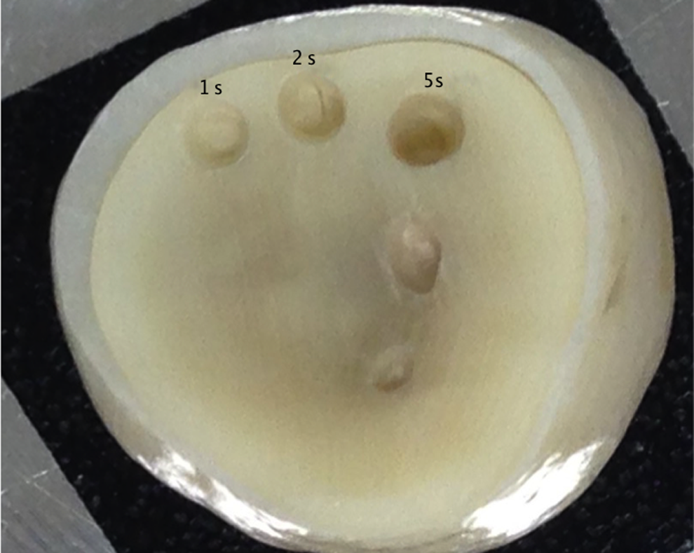
Three cavities prepared on the exposed dentin surfaces using the Er:YAG lasers for 1, 2, and 5 sec of irradiation time.
The cavities in the VSP group were prepared using the Fidelis Plus II Er:YAG laser (Fotona, Ljubljana, Slovenia) with a wavelength of 2940 nm, a 10 Hz pulse rate and 250 mJ of pulse energy. The laser energy was delivered by a RO2-C handpiece with a spot size of 0.9 mm in diameter, in a noncontact mode under continuous water spray (40–60 mL/min).
Two specimens were randomly selected from each subgroup for SEM analysis. In all other specimens, cavity volumes were measured using a laser triangulation profilometer (constructed at the Faculty of Mechanical Engineering, University of Ljubljana, Slovenia).
Volume measurement
For cavity volume measurement, the prepared surfaces were coated with a special spray (Met-l-Chek, Metallfinish Gmbh, Siegen, Germany). Each specimen was inserted into a plastic base and placed onto the movable table. The laser projector was turned on and the camera scanned the sample. Cross-sections of all cavities were scanned (80 cross-sections/sec with an accuracy of 0.02 mm) and the cavity volumes were determined using a specially designed computer program. A nonuniform, rational B-spline (NURBS) surface, representing the tooth surface prior to cavity formation, was approximated onto already measured cavity cross-sections. The computer program determines cavities volumes by comparing NURBS and cross-section surfaces (Fig. 2). The accuracy of this measurement method is ±5%. The results are presented by a computer program in three-dimensional (3D) image form. Measured cavities are differently colored depending upon their depth. Negative and positive volumes are given next to the 3D image of the tooth and the prepared cavity. The negative volume is the missing volume of the tooth after laser preparation, taking into consideration the NURBS surface. The positive volume is necessarily the least possible, because this is the remaining volume of the NURBS surface after cavity preparation. The final cavity volume was calculated by subtraction of the two volumes already mentioned. The ablation rate was also calculated, by dividing the volume of ablated hard dental tissue by the time interval in which the dentin was ablated. The ablation rate was measured in mm3/sec.
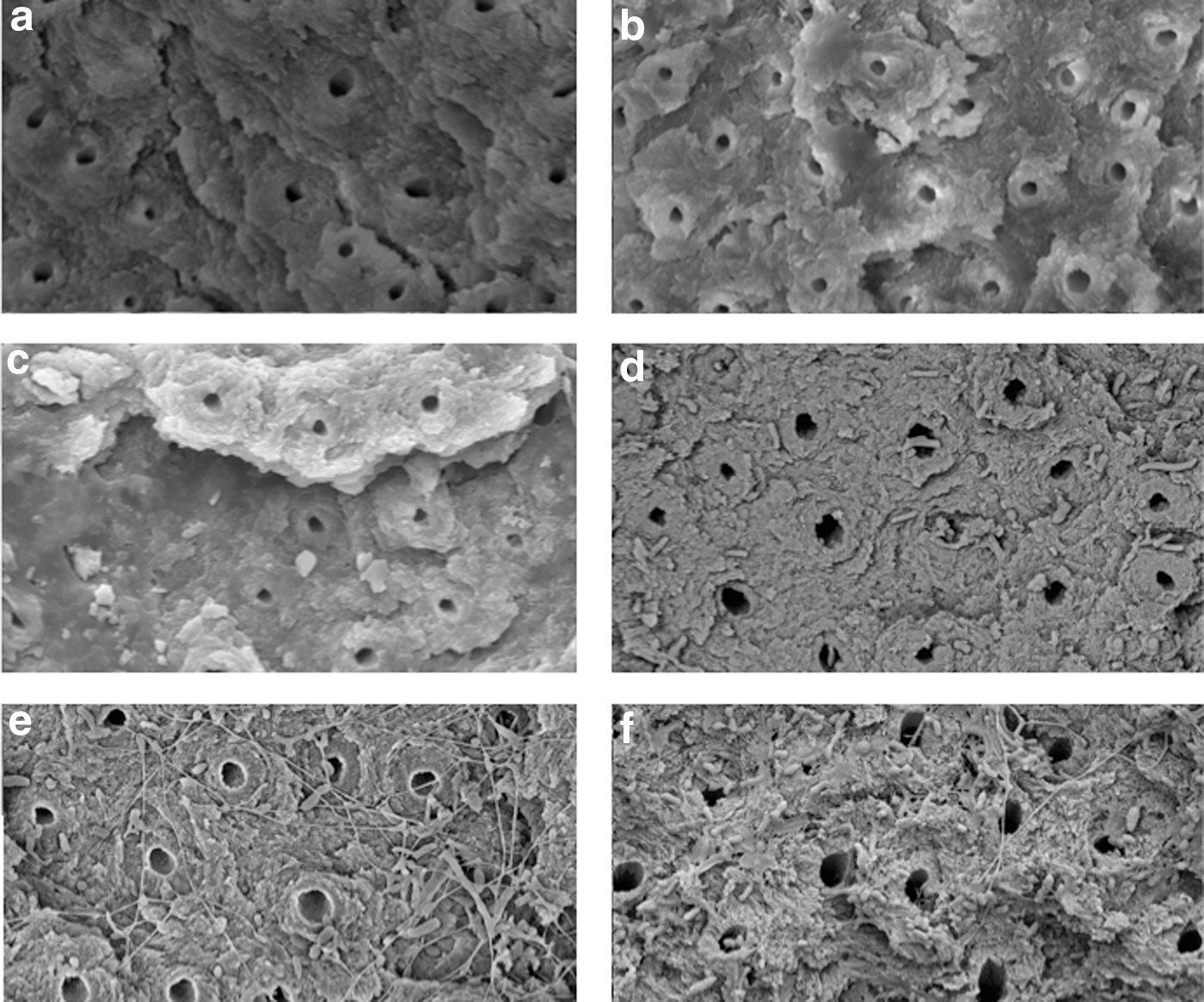
Scanning electron microscopy (SEM) showing dentin surface after cavity preparation in
SEM evaluation
For the SEM analysis, specimens were washed, air dried, dehydrated in an ascending ethyl alcohol series (70%, 80%, 90%, and absolute alcohol) for a total of 8 h, critical-point dried, and sputter-coated with a gold layer 10–15 nm thick. Specimens were observed under an SEM, and micrographs of lased surfaces were taken.
Data analysis
For statistical analysis of dentin ablation rates, the data were compared with the ANOVA test. Multiple comparisons between the experimental groups were performed using the Tukey test. The level of significance was set at 5%. The statistical analysis was performed using the SAS system for Windows (release 8.02).
Results
For the time intervals of 1 and 2 sec, ablated volume and ablation rate for QSP with 500 mJ was significantly higher than for all other groups (p < 0.0001), (Tables 2 and 3). A significant difference was also found between the ablated volumes and ablation rates of QSP 250 mJ and SSP and MSP (p < 0.0001) for a 1-sec time interval (Tables 1 and 2). For the time interval of 2 sec, the ablated volume and ablation rate for the X-Runner group was found to be significantly higher than the ablated volumes of VSP and QSP-250 mJ (p < 0.0001), (Tables 2 and 3), whereas ablated volume and ablation rate for QSP 250 mJ were found to be significantly lower than for the VSP modes (p < 0.0001), (Tables 2 and 3). For the 5 sec time interval, the ablated volumes and ablation rates of the X-Runner and QSP 500 mJ groups were significantly higher than for other experimental groups (p < 0.0001), (Tables 2 and 3).
p value for ANOVA test.
Means with the same letter are not significantly different according to Tukey multiple comparison test at 0.05 significance level.
SSP, super short pulse; MSP, micro short pulse; SP, short pulse; QSP, quantum square pulse.
*p value for ANOVA test.
Means with the same letter are not significantly different according to Tukey multiple comparison test at 0.05 significance level.
SSP, super short pulse; MSP, micro short pulse; SP, short pulse; QSP, quantum square pulse.
SEM analysis
In all experimental groups, the ablated dentin surfaces were free of smear layer, with open dentinal tubules (Fig. 2). The dentin surfaces were irregular because of intertubular dentin, which was selectivly ablated more than the peritubular dentin, making the tubules protrude.
Discussion
This in vitro study demonstrated the influence of different pulse geometries, pulse durations, and laser handpieces, on the efficacy of dentin ablation with Er:YAG lasers. Although different methods have been reported for measuring the ablation rate of hard dental tissues, 16 –22 in this study, the laser profilometry was selected to compare the volume loss of ablated dentin. This measuring method has high accuracy, repeatability, and is far less time-consuming than other methods. 12
Results showed that after a 1- and 2-sec time interval, the largest volume and the highest ablation rate were recorded for QSP with 500 mJ of energy and pulse rate 12 Hz. For the same time intervals, ablated volume and ablation rate of dentin with X-Runner were lower than for the QSP 500 mJ group. This can be explained by the difference in energy settings, of 250 mJ for X-Runner and 500 mJ for the QSP group, plus the difference in the frequency of 10 Hz versus 12 Hz, respectively. This result is in accordance with previous studies on human enamel 23 and bovine teeth. 24 Mironov et al. 24 confirmed that the QSP 500 mJ/12 Hz is the fastest method for hard dental tissue removal, even in comparison with high-speed drills. What supports the findings of Mironov et al. 24 and the present study is the fact that power density and frequency are important in the ablation of mineralized tissues. 25 –27 If the frequency is increased during laser ablation, more hard dental tissue is removed during the same time interval, although higher frequencies (40–50 Hz) offer no further gain in laser efficacy. 28 On the other hand, increasing energy also seems to have an influence on Er:YAG laser's efficiency, but to a lesser degree than the frequency. 26,29 After a 5-sec time interval, QSP 500 mJ/12 Hz and X-Runner 250 mJ/10 Hz were both the most efficient for dentin ablation. Equal efficiency, even with the lower energy and frequency for the X-Runner, could be attributed to the selected circular shape of 2 mm in diameter for dentin ablation in comparison with 0.9 mm diameter of the handpiece used for the QSP 500 mJ group.
This study showed no difference in ablated volume or ablation rate with different pulses of the VSPt Er:YAG laser. Knowing that the shape of the pulse influences the pulse length and energy, 12 the square-shaped pulse that is characteristic for VSP technology enables a constant pulse energy, which does not change during pulse duration. Therefore, the entire energy of the pulse is used up for ablation, and pulses of 50–300 μm length, similar to the ones used in this study, are equally efficient in dentin ablation. Similar results were published by Brugnera et al., 30 who investigated the efficiency of dentin ablation with MSP and SP of Er:YAG laser and found no difference between the two pulses. Also, Nishimoto et al. 31 concluded that there was no difference in ablated volume of dentin for pulses of Er:YAG lasers between 100 and 500 μm, as increasing the pulse duration resulted in increased depth of ablated dentin, but decreased diameter. The better performance of QSP in comparison with VSP can be explained by the absorption of the VSP laser beam in the “cloud” made of particles of the tissue removed, which lowers the ablation rate of hard dental tissues. 9,32 According to Gutknecht et al., 13 QSP has reduced laser beam scattering and absorption in the debris cloud.
In all the experimental groups in this study, the SEM analysis of the dentin showed that the smear layer was completely removed leaving open dentinal tubules and irregular surfaces. The smear layer is a 1 μm thick layer composed of inorganic and organic material, which is a result of conventional methods of dentin preparation with rotary and/or hand instruments. Er:YAG lasers cause vaporization of water within hard dental tissues resulting in volume expansion and disruption of tissues by micro-explosions, 3 removing inorganic and organic tissues and the smear layer as well. Similar findings were found in other published SEM studies. 33 –35 Regarding the influence of VSPt Er:YAG laser on dentin surface morphology, a study by Baraba et al. 36 showed that the use of different pulses resulted in irregular surfaces without smear layer, which is in accordance with the results of the present study.
As micromehanical attachement is one of the main mechanisams for successful adhesion of composite resin materials, 37 it is possible that the effect of Er:YAG laser irradiation of dentin and removal of the smear layer may be beneficial for bonded restorations.
Conclusions
The results of this study suggest that the QSP in combination with the new digitally controlled handpiece (X-Runner) is highly efficient, with a significantly high ablation rate. The X-Runner modality enables efficiency with 250 mJ similar to that of the QSP 500 mJ for a 5-sec time interval. All tested treatment modalities in this study produced cavity preparations with dentin surfaces clean and smear layer free.
Footnotes
Acknowledgments
The authors thank Fotona for providing the laser equipment, and Tadej Perhavec and Tomaz Suhovrsnik for support with the cavity volume measurements.
Author Disclosure Statement
No competing financial interests exist.