
Abstract
Introduction
P
PDT induces cell death in the targeted tissue based on the combined effects of three nontoxic components as follows: a photosensitizer (PS), light, and oxygen. 5 Subsequent to administration, the PS accumulates in tumor tissues. 6 On illumination by light of the appropriate wavelength, the PS undergoes photochemical reactions that result in the production and accumulation of reactive oxygen species. 7 Photodamage of tumor cells then occurs and leads to cell death through activation of apoptosis and/or necrosis. 8 Currently, PDT is clinically approved in several countries for the treatment of bladder, lung, esophageal, head and neck cancers, as well as nonmalignant growths such as actinic keratosis, Barrett's esophagus, and nonmelanoma skin cancers. 4
Osteosarcoma is the most common type of malignant bone tumor in the United States, especially among adolescents and children. 9 Conventional treatments for osteosarcoma include surgical resection, radiotherapy, and chemotherapy, all of which could lead to severe limb function impairment and risk of local recurrence. 10 PDT holds promise as a novel, less invasive treatment for osteosarcoma for chemoresistant tumors, for tumors that cannot be removed by surgery, or for retention of excellent limb function subsequent to surgery. 11
The 5-aminolevulinic acid (5-ALA) is an endogenous metabolite found as an early intermediate in the heme biosynthesis pathway. The 5-ALA is a PS precursor as it is converted by cellular enzymes of the heme pathway to the natural PS protoporphyrin IX (PpIX). 12 –14 5-ALA uptake and metabolism occur in most human cells and tissues; however, ALA is preferentially taken up by cells with increased metabolic activity, leading to increased intracellular accumulation of PpIX. 15 Therefore, in addition, selectivity can be achieved by administration of this prodrug.
ALA is currently used in clinical applications in relation to bladder cancer, actinic keratosis, and basal cell carcinoma. 4 Further, numerous studies implicate ALA-mediated PDT as a promising treatment for breast, bladder, prostate, ovarian, renal, and lung cancers. 16 However, data on the use of ALA-mediated PDT for osteosarcoma are much more limited. In this study, we determined the ability of ALA-PDT to induce cell death in the human osteosarcoma cell line MG-63. The results of our study not only support the ability of ALA-PDT to kill human osteosarcoma cells but also provide optimal in vitro parameters for ALA-PDT, which could be useful for clinical applications.
Materials and Methods
Chemicals
Aminolevulinic acid hydrochloride (ALA) was purchased from Sigma-Aldrich as a powder. It was reconstituted in phosphate-buffered saline (PBS) at a concentration of 100 mM and was kept at −20°C until use. Storage, dilution, and incubation were performed under conditions that prevented the exposure of ALA to light. All chemicals and reagents for the cell culture were purchased from Gibco® Life Technologies, unless otherwise specified.
Cells and cell culture
MG-63 human osteosarcoma cells were derived from a tumor that developed in the bone of a 14-year-old Caucasian boy and were obtained from the American Type Culture Collection (ATCC). Cells were cultured in 75-cm2 flasks in Dulbecco's modified Eagle's medium (DMEM) supplemented with 10% fetal bovine serum (Atlas Biologicals) and 100 IU/mL penicillin and 100 μg/mL streptomycin. Cells were incubated at 37°C in a humidified atmosphere containing 5% CO2 and 95% atmospheric air.
Cytotoxicity assay
Experiments were carried out in black, clear bottom 96-well plates (VWR). MG-63 cells were plated at a density of 4 × 10³ cells per well. After 24 h, the medium was removed and replaced with serum-free DMEM containing either vehicle alone (1 × PBS) or varying concentrations of ALA. Cells were incubated in ALA-containing medium for either 4 or 24 h. Subsequent to the incubation time, the medium was replaced with complete DMEM, and cells were allowed to recover for 24 h. Experiments were performed in the dark to minimize light exposure. Cells were then fixed in ethanol, stained with crystal violet (Sigma-Aldrich), and solubilized with deoxycholate (Sigma-Aldrich). Cell density was measured by detecting absorbance at 595 nm in a BioTek Synergy Ht Multi-Mode Reader (BioTek). Cell viability was calculated as the ratio of cells treated of vehicle-treated (0 mM ALA) to ALA-treated cells.
Measurement of intracellular accumulation of PpIX
MG-63 cells were plated in 25-cm2 flasks at a density of 200,000 cells per flask in a complete medium. After 48 h, the medium was removed and replaced with serum-free, phenol-red minus DMEM containing either vehicle alone (1 × PBS) or varying concentrations of ALA. Cells were incubated in ALA for either 4 or 24 h. Subsequent to ALA incubation, cells were lysed in SDS/NaOH buffer, and lysates were cleared by centrifugation. Experiments were performed in the dark to prevent light exposure. One milliliter of the lysate was collected for fluorometry; the remaining supernatant was subjected to a modified Bradford protein assay (Bio-Rad) to ensure equal loading. Briefly, protein concentration of each lysate was calculated using a bovine serum albumin (Sigma-Aldrich) standard curve. Protein content of each lysate was then equalized by dilution with lysis buffer before being subjected to fluorometry to measure PpIX accumulation. PpIX content in the cell lysates was determined using a HORIBA Scientific/Jobin-Yvon FluoroMax-4 spectrofluorometer (HORIBA Scientific). The fluorescence signal was measured for each sample using 405 nm excitation and 650 nm emission wavelengths. PpIX concentration was calculated from a standard fluorescence curve of PpIX (Sigma-Aldrich).
Light source for PDT
An 8 × 12 light emitting diode (LED) array of 5.0 mm red LEDs (Part No. 1586189; James Electronics) was built to control illumination in individual wells of black 96-well plates (VWR). Grouped in columns of 8, the LEDs were controlled by a microcontroller board (Arduino Duemilanove) and the associated Arduino open source software. Each of the 12 LED columns was connected to one of the digital output pins through a Darlington configuration of two transistors to amplify the output of the digital pins. All of the transistors' collectors were connected in parallel to the constant 5 V output pin. The LEDs were rated at a maximum forward voltage of 2.5 V and forward currents of 20 mA. Given the parallel connection of each group of eight LEDs, an individual LED was supplied by an average forward voltage of 1.96 and 6.9 mA of forward current. The LED array was calibrated accordingly to account for attenuation. Spectra were collected from each of the individual LEDs in the array using a fiber optic cable (USB650 Red Tide Spectrometer; Ocean Optics, Inc.). The peak wavelength of the LED emission spectrum was 636 nm (FWHM = 16 nm). In addition, the output power was detected with a power meter calibrated to a 635 nm wavelength (PM100D Power Meter; Thorlabs). To measure power incident on the cells, measurements were taken at the same height above the LEDs as the plate. Reflection at the bottom of the plastic well was also accounted for.
Measurement of the cytotoxic effects of ALA-mediated PDT
Experiments were carried out in black, clear bottom 96-well plates (VWR). MG-63 cells were plated at a density of 4 × 10³ cells per well in the complete medium. After 24 h, the medium was removed and replaced with serum-free DMEM containing either vehicle alone (1 × PBS) or varying concentrations of ALA. Cells were incubated in ALA-containing medium for either 4 or 24 h. Subsequent to ALA incubation, cells were exposed to varying light fluencies using the custom-built LED array. The LED array was measured to have a power density of 14 mW/cm2. To achieve a 0.3 J/cm2 energy density, a duration of 21.5 s was used; to achieve a 0.6 J/cm2 energy density, a duration of 42.9 s was used; to achieve a duration of 3 J/cm2, a duration of 214.3 s was used; to achieve a duration of 6 J/cm2, a duration of 428.6 s was used (Table 1). Treatments were performed one time for each energy density. Cells were allowed to recover for 24 h in complete medium before viability measurement.
To assess viability, cells were subjected to an MTT (3-[4,5-dimethylthiazol-2-yl]-2–5-diphenyltetrazolium bromide) assay (Sigma-Aldrich) as described in Mosmann. 17 Briefly, MTT solution was added to each well after recovery. After 3 h, cells were solubilized in HCl, and the resulting absorbance was measured at 595 nm with a BioTek Synergy Ht Multi-Mode Reader (BioTek).
Statistical methods
All experiments in this study were performed at least in triplicate, and the data were statistically analyzed using the Student's t-test. Statistical significance is defined as a calculated p-value <0.05.
Results
Dark toxicity of MG-63 cells induced by ALA
To identify ALA toxicity in the absence of light, MG-63 human osteosarcoma cells were treated with various concentrations of ALA for 4 or 24 h without illumination (Fig. 1). There was no significant cell death with respect to those cells treated with up to 1 mM ALA (>90% viability) for both the 4- and 24-h time points. Cell viability significantly decreased at ALA concentrations of 2, 5, and 10 mM ALA for both time points. There were no observed significant differences in cell viability between the 4- and 24-h time points.
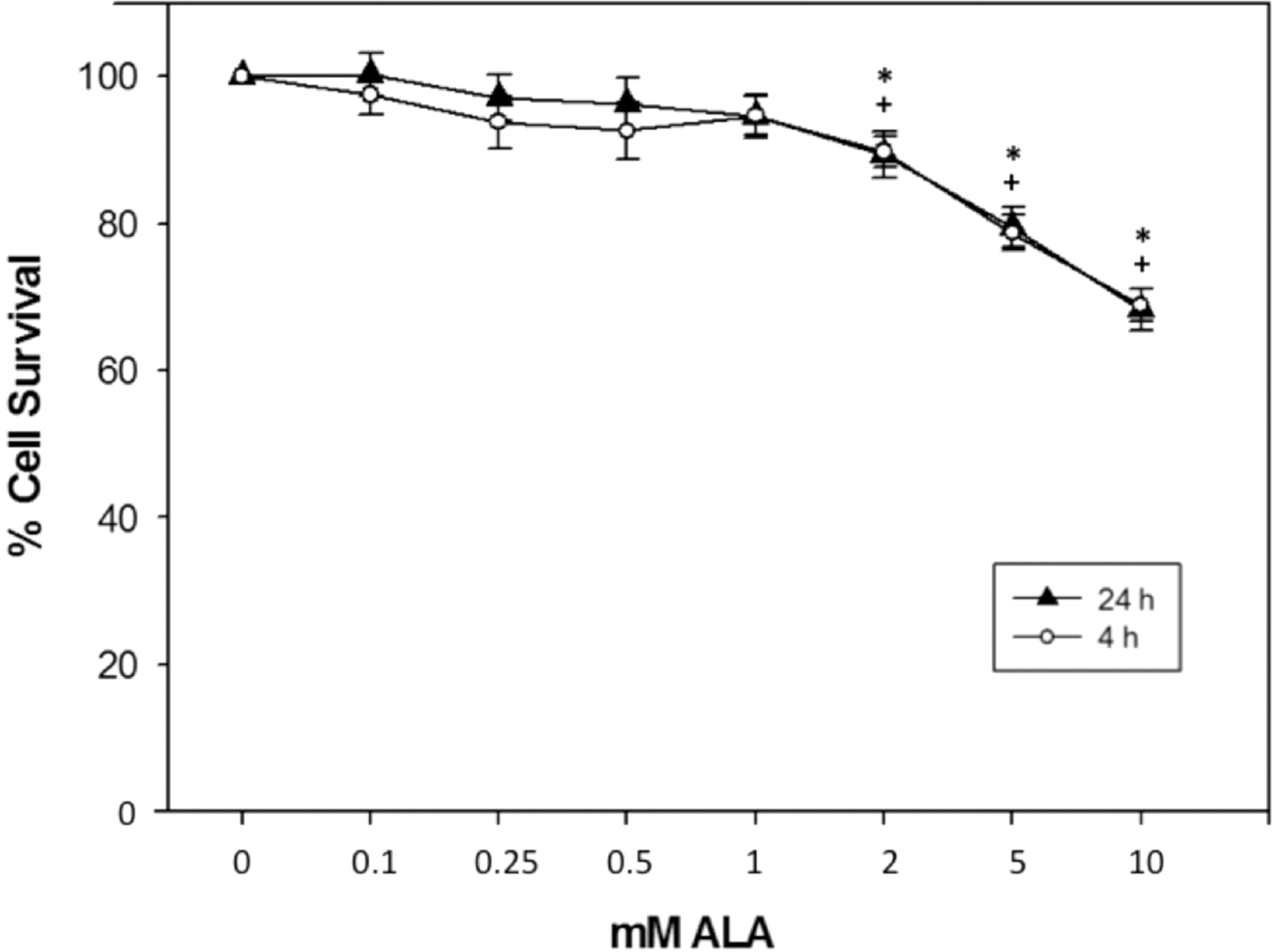
ALA-induced cell dark toxicity in MG-63 cells. MG-63 cells were treated with ALA (0, 0.1, 0.25, 0.5, 1, 2, 5, or 10 mM) in serum-free media for either 4 or 24 h. Subsequent to ALA incubation for indicated times, media were changed to cDMEM for 24 h before analysis of cell survival. Cell survival was determined using a crystal violet assay. ALA concentrations of up to 1 mM did not cause significant decreases in cell survival. Error bars represent ±SEM; *p < 0.05 compared to 4-h vehicle-treated control; + p < 0.05 compared to 24-h vehicle-treated control.
PpIX accumulation in MG-63 cells subsequent to ALA incubation
PpIX accumulation in MG-63 cells incubated with various concentrations of ALA was assessed subsequent to either 4- or 24-h treatment (Fig. 2). With respect to the 4-h time point, PpIX accumulation was significantly increased compared to vehicle-alone control at 0.5, 1, and 2 mM ALA. With respect to the 24-h time point, PpIX accumulation was significantly increased compared to the vehicle-alone control at 0.25, 0.5, 1, and 2 mM ALA. For either time point, maximal PpIX accumulation occurred at 1 mM ALA. PpIX accumulation differed significantly between time points at 0.5, 1, and 2 mM ALA. Maximal PpIX accumulation occurred at 1 mM ALA, 24 h post-ALA incubation.
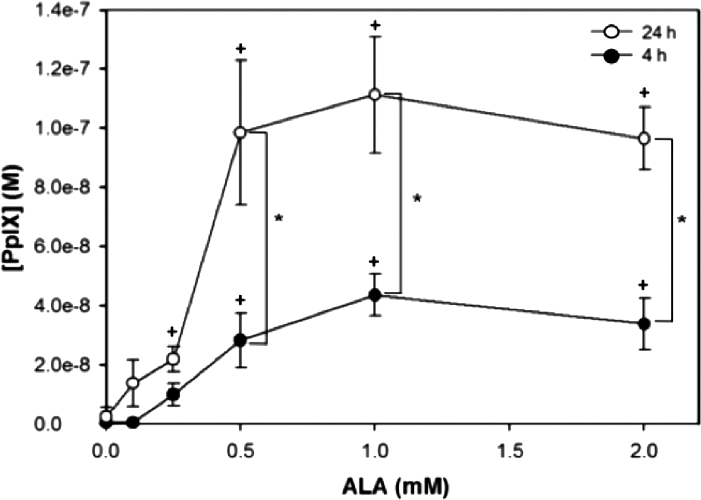
PpIX accumulation in MG-63 cells subsequent to incubation with ALA. MG-63 cells were treated with indicated concentrations of ALA in serum-free media for 4 or 24 h. Subsequent to ALA treatment, cells were lysed and PpIX concentration was spectrofluorometrically determined based on 405 nm excitation and 650 nm emission wavelengths. PpIX concentration was calculated using a standard fluorescence curve of known PpIX concentrations. Error bars represent ±SEM; *p < 0.05 compared between 4- and 24-h time points; + p < 0.05 compared to vehicle control. PpIX, protoporphyrin IX.
Cell death in MG-63 cells subjected to ALA-mediated PDT
Cell death subsequent to ALA-mediated PDT was assessed after the 24-h incubation with ALA by MTT assay.
Cells were incubated with 0 (vehicle alone), 0.5, or 1 mM ALA for 24 h to achieve maximal PpIX accumulation and then subjected to illumination with 0, 0.6, 3, or 6 J/cm2 light (Fig. 3). No significant difference in cell viability was observed in nonilluminated cell populations, even in the presence of ALA. Cell viability of vehicle-treated cells appeared to decrease with increasing light fluence, but no statistical significance was observed. However, all light fluencies significantly decreased cell viability subsequent to 0.5 mM ALA treatment compared to the nonilluminated control, with maximal death occurring at 6 J/cm2. Similarly, all light fluencies significantly decreased cell viability subsequent to treatment with 1 mM ALA compared to the nonilluminated control, with the maximal death occurring at 6 J/cm2. Subsequent to illumination at 0.6 J/cm2, both 0.5 and 1 mM ALA treatments significantly increased cell death compared to the vehicle-alone (0 mM ALA) control. Subsequent to illumination at 3 J/cm2, both 0.5 and 1 mM ALA treatments significantly increased cell death compared to the vehicle-alone (0 mM ALA) control. Subsequent to illumination at 6 J/cm2, both 0.5 and 1 mM ALA treatments significantly increased cell death compared to the vehicle-alone (0 mM ALA) control. Maximal cell death with minimal light toxicity was achieved with either 0.5 or 1 mM ALA treatment and illumination with 3 J/cm2.

Effect of ALA-mediated photodynamic therapy in MG-63 cells. Cells were treated with vehicle alone (0 mM ALA), 0.5, or 1 mM ALA for 24 h in serum-free media. Cells were then exposed to varying light fluencies (0, 0.6, and 3 J/cm2) and allowed to recover for 24 h. Subsequent to recovery, cell viability was measured using a standard MTT assay. Formazan crystal formation was measured at 595 nm. Error bars represent ±SEM; *p < 0.05 compared to 0 J/cm2 control; + p < 0.05 compared to 0 mM ALA control.
Discussion
PDT is a promising therapy for the treatment of many tumors and is currently clinically approved for the treatment of several types of neoplasms, including cutaneous lesions, nonsmall cell lung carcinomas, and esophageal cancers. 4 Clinical and preclinical studies focused on expanding the use of this therapy to other oncologic conditions, such as head and neck cancers, neurological tumors, and gynecological neoplasms, are promising. 14 However, studies evaluating the use of PDT for the treatment of human osteosarcomas are more limited. Studies using ALA-PDT for the treatment of osteosarcoma are limited only to the preclinical setting. 11 Because this therapy has numerous advantages over traditional treatments for human osteosarcoma, more research is necessary to ascertain the potential efficacy of ALA-mediated PDT in this context. Therefore, we have used the human osteosarcoma cell line MG-63 to evaluate the potential of ALA-mediated PDT to kill osteosarcoma cells and to determine the most effective light and PS dose to achieve maximal effect.
Several studies have reported the efficacy of PDT to kill osteosarcoma cells in vitro. 11 However, only a very limited number of these studies investigate the use of ALA as the PS. 18 –21 Further, these studies use the cell lines HOSM-1, HOSM-2, and U2OS and report varied results. 18 –21 These results suggest that the efficacy of ALA-PDT in vitro is likely cell type and treatment parameter specific. It is important then to understand the intricacies of this treatment on many types of cells to further determine potential efficacy in translational and clinical trials. To date, no study has reported the effects of ALA-mediated PDT on human MG-63 osteosarcoma cells. Therefore, this study aimed to understand the most efficient and effective parameters for ALA-PDT on this particular cell line.
Our study found that ALA alone is toxic to MG-63 cells at concentrations only above 1 mM ALA. This finding is consistent with the ALA dose used in similar studies with other human osteosarcoma cell lines. 19,21 However, another study conducted by Yanase et al. found significant toxicity in HOSM-1 human osteosarcoma cells at concentrations of 0.5 mM ALA and above. 18 Because it is thought that ALA can penetrate nonselectively into all cells through the cell membrane, 22 it is most likely the activity of the enzymes involved in the metabolism of ALA that determines ALA uptake levels. 15 Further, recent studies show that PpIX accumulation is generally less in normal human cells subsequent to ALA incubation. 19,23,24 Therefore, it is possible that MG-63 cells express differential metabolic activities and profiles compared to other human osteosarcoma cell lines and normal human cell lines. Our study found that ALA conversion to PpIX was maximal at 24 h postincubation. Other studies have found maximal PpIX accumulation at 9–12 h post-ALA treatment. 18,20 This result could suggest differences among osteosarcoma cell lines in the amount of ferrochelatase or other enzymes associated with the heme pathway. 25
Our study found that ALA-mediated PDT was most effective at 3 and 6 J/cm2. This fluency is notably less intense than other reports in the literature, both pertaining to the death of other osteosarcoma cell lines as well as normal cells. 18 –21,23,24,26 –28 This finding is significant because it has been reported that high rates of light fluency have been correlated with necrotic cell death, while low-fluency rates have been associated with apoptotic cell death. 29 It has been reported that necrotic cell death can result in the release of immunogenic proteins and cytokines that can lead to immune system activation, including local acute inflammatory reaction. 30 However, recent evidence suggests that the proinflammatory effects of PDT might increase the immunogenicity of dead tumor cells and increase immune cell activation, thus leading to increased immune response against the tumor. 30 –33 Therefore, it is essential to understand the molecular and cellular differences associated with ALA-mediated cell death among cell types.
Consistent with reports using other osteosarcoma cell lines, we found that ALA-PDT is effective at killing MG-63 cells. However, our study did report notable differences with respect to ALA toxicity, PpIX accumulation, and effective light doses among MG-63 cells and other human osteosarcoma cell lines. These data are useful in determining translational and preclinical parameters for ALA-PDT of osteosarcoma, as well as characterizing the molecular mechanisms of this specific therapy.
Summary and Conclusions
We have shown that ALA alone is not toxic to MG-63 human osteosarcoma cells up to a concentration of 1 mM when cells were treated for either 4 or 24 h. This result established a range of nontoxic working concentrations to use for further studies. Working within these nontoxic concentrations of ALA, we also showed that ALA is maximally converted to PpIX at 0.5 and 1 mM ALA 24 h postincubation. This result provided a range of the most effective ALA concentrations, with which to carry out further studies. Finally, we showed that ALA-mediated PDT can cause significant cell death with minimal light toxicity in MG-63 cells by incubating cells with 0.5 or 1 mM ALA for 24 h, followed by illumination with light at 3 J/cm2. Taken together, these results strongly suggest that ALA-PDT can induce cell death in human osteosarcoma cells.
Footnotes
Acknowledgments
The authors acknowledge the M.J. Murdock Charitable Trust for project funding (Scientific Research Start-up Grant No. 2007164:JVZ:11/15/07). With deep appreciation, they acknowledge Dean Dr. John Hayes and the College of Arts and Sciences at Pacific University for project funding (Pacific University Start-up Grant). They also thank the Pacific Research Institute for Science and Mathematics for project funding (PRISM Summer Research Grant). Finally, the authors gratefully acknowledge the technical assistance of Mariko Newton, Scott Loring, and Lane Higa.
Author Disclosure Statement
No competing financial interests exist.