
Abstract
Introduction
H
The traditional use of herbal components in UVB-mediated oxidative damage is a common practice in the domestic medicine of many countries. Rhodiola rosea L. is a Chinese traditional herbal medicine used for protection against UV radiation, 18 and salidroside is the major active component of Rhodiola rosea. 19 Figure 1 shows the structure of salidroside. Previous studies have shown that salidroside has protective effects against oxidative stress. 20 –24 Recently, salidroside has been found to increase the survival rate against UVB-induced cell death 25 and to decrease the hyperpigmentation of UVB-stimulated guinea pig skin. 26 However, whether salidroside can reduce UVB-mediated oxidative damage remains unclear.
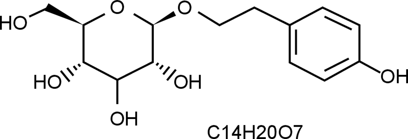
Chemical structure of salidroside.
Therefore, in this study, we tested the effects of salidroside on photo-oxidative stress in a UVB-induced sun damage model. Using HaCaT cells, the oxidative damage and possible regulatory factors (including Nrf2, NQO1, and HO-1) were examined. In addition, the regulatory effects of salidroside on apoptotic sunburn cells (SBCs) and 8-OHdG-positive epidermal cells in UVB-exposed guinea pig skin were also investigated.
Materials and Methods
Treatment of cells
The immortalized HaCaT keratinocytes, gifted by Prof. Lu CR, were cultured in Dulbecco's modified Eagle's medium (DMEM) supplemented with 10% fetal bovine serum and 1% penicillin/streptomycin and maintained at 95% humidity in a 5% CO2 environment at 37°C. Salidroside (99.9%; Biopharmaceutical Co., Ltd.) dissolved in DMEM was used to treat the cells. The cells (70–80% confluent) were treated with salidroside (0–80 μg/mL) for 24 h in DMEM, after which the media were removed, the cells were washed with phosphate-buffered saline (PBS), and fresh PBS was added. The salidroside-pretreated cells were irradiated with UVB at a dose of 50–100 mJ/cm2 with emission at 280–320 nm and a peak at 315 nm (Shanghai SIGMA High-Tech Co. Ltd.). The irradiation doses were monitored using a calibrated SUN5 digital spectroradiometer (National Institute of Measurement, Beijing, China).
MTT cell viability
The effects of UVB or salidroside (or both) on the viability of the HaCaT cells were determined using the MTT assay (Sigma). The cells were seeded at a density of 1000 cells per well in a culture medium containing 0, 10, 20, 40, or 80 μg/mL salidroside in 96-well plates at 37°C for 24 h in a humidified chamber. Each concentration of salidroside was repeated in eight wells. After incubation for 24 h at 37°C in a humidified incubator, the cells were washed with PBS and irradiated with UVB (50–100 mJ/cm2). Twenty-four hours after UVB exposure, 10 μL of MTT/100 μL of culture media was added, and the plate was incubated for 4 h. The plate containing the cells was centrifuged. The MTT solution was then removed from the wells, and the formazan crystals were dissolved in DMSO. The absorbance was recorded on a microplate reader at a 505-nm wavelength. The cell viability was calculated as the percentage absorbance relative to the control cultures.
Immunocytochemistry for Nrf2
Twenty-four hours after salidroside (20–40 μg/mL) and UVB (100 mJ/cm2) treatments, the cells were fixed in methanol for 5 min at −20°C and then incubated with rabbit anti-Nrf2 1:100 (Abcam), followed by secondary antibodies conjugated with horseradish peroxidase. The cells were then stained with 3-amino-9-ethyl-carbazole (Maxin Company) until satisfactory staining was obtained (the procedure was monitored under an inverted microscope) and the translocation of Nrf2 to the nucleus was observed and analyzed. The results were expressed as the amount of Nrf2 translocated to the nucleus per 10 high-power fields, as previously described. 27
Western blot analysis for Nrf2, NQO1, and HO-1
Twenty-four hours after UVB exposure (100 mJ/cm2) for cells that were pretreated with salidroside (20 and 40 μg/mL), the cells were harvested, and cell lysates were prepared. A Western blot analysis was used to detect Nrf2 (Abcam), NQO1 (Abcam), and HO-1 (Abcam). Briefly, cell protein extract was resolved over 8–12% polyacrylamide gels and transferred into a nitrocellulose membrane. The membranes were incubated with primary antibodies, followed by horseradish peroxidase-conjugated secondary antibodies, and developed with enhanced chemiluminescence reagents (Amersham) and autoradiography. Densitometric measurements of the bands on the Western blot were performed using the digitalized scientific software program UN-SCAN-IT (Silk Scientific Corporation).
Nrf2, NQO1, and HO-1 expression by real-time reverse transcription polymerase chain reaction (RT-PCR)
HaCaT cells were treated with salidroside (20 and 40 μg/mL) for 24 h and were then exposed to UVB irradiation (100 mJ/cm2). Total RNA was isolated from cultured cells with the TRIzol reagent (Invitrogen), and cDNA was prepared (Bio-Rad). Real-time RT-PCR was performed using the one-step RT-PCR system (SYBR Green). Relative mRNA expression levels were calculated using the ΔΔCt method 28 and were normalized to β-actin. The primers were designed with Primer3 software (version 4); the primer pairs used are listed in Table 1.
Measurement of intracellular ROS with flow cytometry
UVB irradiation-mediated intracellular ROS and their suppression by salidroside were analyzed by flow cytometry. Intracellular ROS were assessed by the fluorescent probe 2′, 7′-dichlorodihydrofluorescein diacetate (H2DCFDA; Invitrogen Ltd.), 29 24 h after cell irradiation by UVB (100 mJ/cm2) and salidroside treatment (20 and 40 μg/mL, respectively). Then, HaCaT cells were trypsinized, collected, incubated with 5 μg/mL H2DCFDA for 30 min at the temperature of 37°C, and washed with PBS. Flow cytometric analysis (FACScan; Becton, Dickinson, and Company) using Cell Quest 3.2 software (Becton, Dickinson, and Company) was then followed. The excitation wavelength was 480 nm, and the emission wavelength was 525 nm. Mean fluorescence intensity was used to express the results of flow cytometry.
Animals and treatment
Female guinea pigs were purchased from Fangshan Guandao Shunli Laboratories (Beijing, China). All of the procedures were performed in accordance with animal care guidelines and were approved by the Institution Ethics Board of the Air Force General Hospital. All of the guinea pigs used in this study were maintained in laboratory animal facilities under specific pathogen-free conditions. Thirty female guinea pigs were divided into three groups of ten animals in each group as follows: (a) animals fed 0.1% (w/w) salidroside in the diet for 2 weeks (Sal); (b) animals irradiated once a day for three consecutive days with UVB at doses of 200 mJ/cm2 (UVB); and (c) animals fed 0.1% (w/w) salidroside in the diet for 2 weeks; the back skin was shaved and was followed 24 h later by UVB irradiation (200 mJ/cm2) once a day for three consecutive days (Sal+UVB). The selection of the dietary salidroside dose in the present study was based on our previous study, which showed that 0.5% (w/w) salidroside in the diet for two months is safe and nontoxic (data not shown). The guinea pigs were then killed 2 or 24 h after UVB exposure, and the skin biopsy specimens were fixed in formalin.
Histological examination and apoptotic SBC counts
Twenty-four hours after UVB exposure, the guinea pigs were killed. The fixed skin biopsy specimens were processed for routine histology. Apoptotic SBCs were identified by a round shape with loss of connections with surrounding keratinocytes, typical eosinophilic cytoplasm with dense and contracted nuclei, or by severely swollen nuclei with a vacuolated cytoplasm. 30 The results were expressed as SBCs per 10 high-power fields. 27 All of the histological specimens were read in a blinded manner.
Immunohistochemical detection of 8-OHdG
Two hours post-UVB exposure, the guinea pigs were killed. The skin biopsy specimens were fixed in formalin. The immunohistochemical detection of 8-OHdG was performed as described previously. 27 In brief, formalin-fixed tissue slides were deparaffinized, dehydrated, and blocked with 10% goat serum in 2% bovine serum albumin/Tris-buffered saline (BSA/TBS) for 30 min at room temperature. The tissue sections were incubated with a primary antibody against 8-OHdG (Sigma) overnight at 4°C in 2% BSA/TBS. After the sections were washed, they were incubated with horseradish peroxidase-conjugated streptavidin (NeoMarkers); immunoreactivity was developed by adding 3,3′-diaminobenzidine (DAB; Maxin Company), and counterstaining was performed using hematoxylin.
Statistical analyses
The results are expressed as mean ± standard deviation. Statistical analyses of all the data between the groups were carried out using Student's t-test. p < 0.05 was considered statistically significant.
Results
Salidroside treatment protects HaCaT cells from UVB-mediated decreases in cell viability
We investigated the effects of salidroside (0–80 μg/mL) against UVB-mediated decreases in cell viability. The salidroside treatment (0–80 μg/mL) showed no cytotoxic effects. As expected, the UVB (50–100 mJ/cm2) irradiation of the HaCaT cells resulted in a decrease in cell viability. This UVB-induced inhibition of cell growth was significantly prevented by the pretreatment of cells with salidroside (0–80 μg/mL) in a dose-dependent manner as determined by the MTT (3-[4, 5-dimethylthiazol-2-yl]-2,5-diphenyl tetrazolium bromide) assay. The results are shown in Fig. 2.
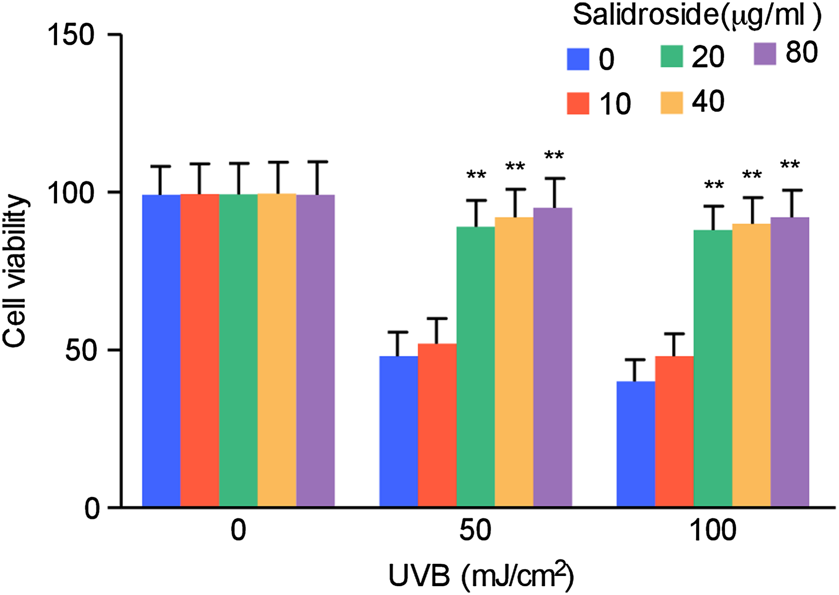
Salidroside treatment protects HaCaT cells against UVB-mediated decreases in cell viability. HaCaT cells were treated with different doses of salidroside (0, 10, 20, 40, or 80 μg/mL) for 24 h, after which the medium was removed and the cells were washed with PBS. Then, fresh PBS was added, and the cells were exposed to UVB irradiation (50–100 mJ/cm2). Twenty-four hours after UVB irradiation, the percent cell viability was assessed using the MTT assay. The data shown are the mean ± SD of three separate experiments, in which each treatment was repeated in eight wells. **p < 0.01 compared to UVB. PBS, phosphate-buffered saline; SD, standard deviation.
Salidroside treatment enhances the nuclear translocation of Nrf2 in HaCaT cells
In this study, we found Nrf2 in the cytoplasm of HaCaT cells. However, UVB irradiation (100 mJ/cm2) caused Nrf2 translocation to the nucleus in only a few HaCaT cells. This suggests that epidermal cells can provide some natural protection against UVB-induced damage by activating the Nrf2/ARE pathway. However, this response is limited and is not sufficient to prevent damage. Salidroside treatment (20–40 μg/mL) significantly increased Nrf2 translocation to the nucleus in cells (Fig. 3A, B).
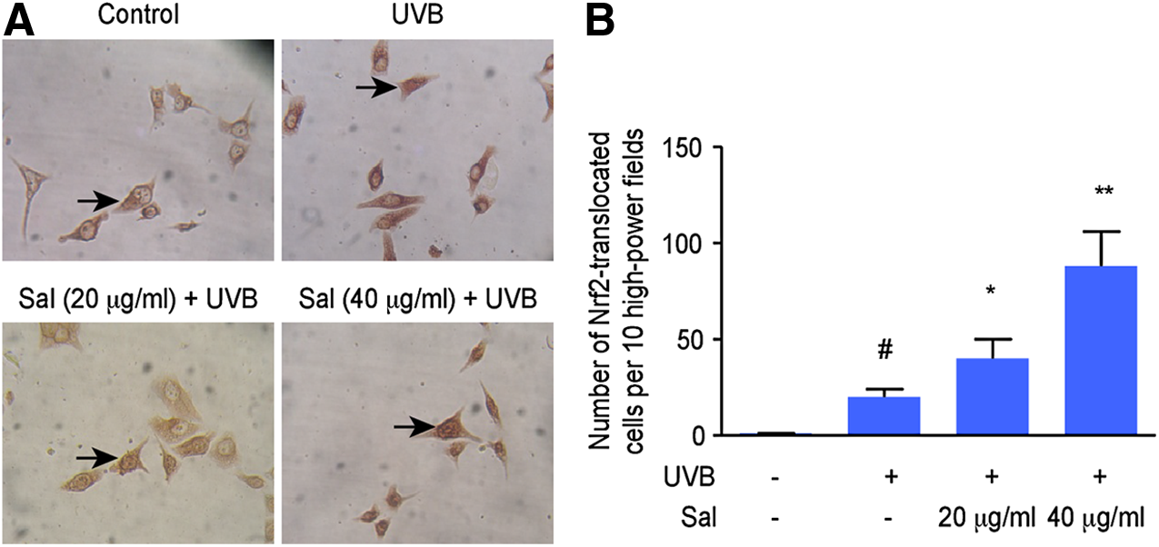
The effects of salidroside treatment on Nrf2 translocation to the nucleus. HaCaT cells were treated with salidroside (20 or 40 μg/mL) for 24 h, then the medium was removed, after which the cells were washed with PBS and fresh PBS was added three times, and then the cells were exposed to UVB (100 mJ/cm2) irradiation. Twenty-four hours after UVB irradiation, the cells were washed with PBS and fixed, and Nrf2 was detected by immunocytochemistry.
Upregulation of Nrf2 and its target genes by salidroside treatment in HaCaT cells with UVB irradiation
We showed that salidroside treatment can enhance Nrf2 translocation to the nucleus in immortalized human skin HaCaT keratinocytes that are exposed to UVB radiation (Fig. 2A, B). Further, we detected the expression of Nrf2 and its target genes NQO1 and HO-1 in HaCaT cells exposed to UVB radiation following salidroside treatment. First, we found that salidroside treatment significantly enhanced protein levels of Nrf2 and its target genes NQO1 and HO-1 in irradiated HaCaT cells in a dose-dependent manner (Fig. 4A, B). Using RT-PCR, we also demonstrated that salidroside (20 and 40 μg/mL) treatment significantly upregulated the expression of Nrf2 and its target genes NQO1 and HO-1 (Fig. 4B).
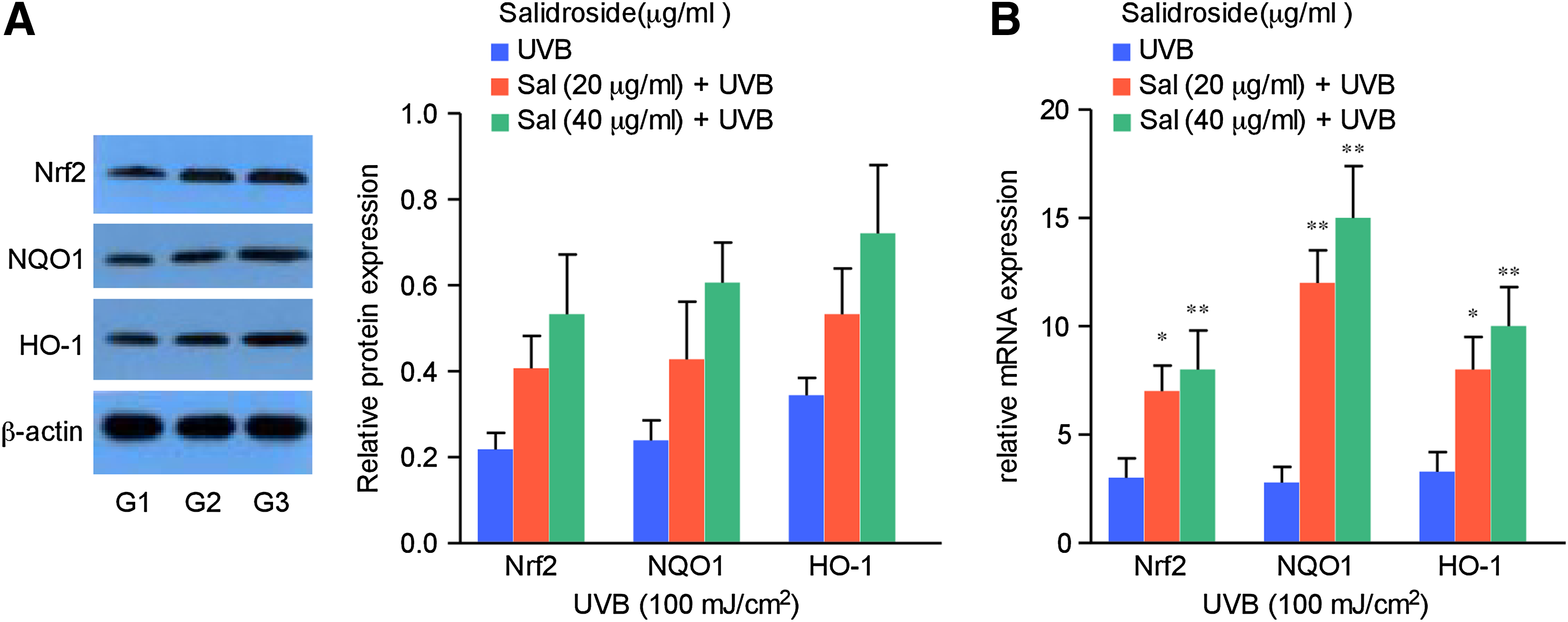
Salidroside treatment enhances the gene and protein expression of Nrf2, NQO1, and HO-1 in HaCaT cells.
Salidroside treatment decreases intracellular ROS in irradiated HaCaT cells as detected by flow cytometry
We further investigated the effects of salidroside (20 and 40 μg/mL) on UVB-mediated changes in ROS. After UVB irradiation (100 mJ/cm2), ROS in HaCaT cells increased. This UVB-induced increase in ROS was significantly prevented by salidroside pretreatment (20 and 40 μg/mL) according to flow cytometry. These results are shown in Fig. 5A, B.

Effects of salidroside treatment on UVB-mediated ROS as detected by flow cytometry. Intracellular ROS levels were assessed according to the intensity of H2DCFDA fluorescence as determined by FACS analysis.
Salidroside treatment decreases the UVB-induced sunburn reaction and inhibits the formation of apoptotic SBCs and 8-OHdG-positive epidermal cells
We further observed the effects of salidroside in the diet (0.1% w/w) of guinea pigs on UVB-induced damage. We found that salidroside treatment decreased the sunburn reaction, including congestion and telangiectasia of the skin, in guinea pigs that were exposed to UVB irradiation at a dose of 200 mJ/cm2. Biopsy specimens were then taken from the skin 2 and 24 h after irradiation with UVB. Histologically, the skin samples showed more significant edema, thickening, and the elimination of obvious rete ridges of the epidermis after irradiation with UVB, whereas the salidroside treatment decreased these changes. Moreover, we found that UVB irradiation (200 mJ/cm2) significantly increased the formation of apoptotic SBCs in the epidermis of guinea pigs. However, we found that the administration of salidroside inhibited UVB-induced apoptosis. These results are shown in Fig. 6A-a. 8-OHdG is considered to be an important biomarker of DNA damage. 4 Therefore, using immunohistochemical analysis, we also assessed the effects of salidroside (0.1% w/w) in the diet on UVB-mediated 8-OHdG formation in the epidermis of guinea pigs 2 h after UVB irradiation. As shown in Fig. 6B, UVB irradiation on the skin of guinea pigs induced the formation of 8-OHdG.
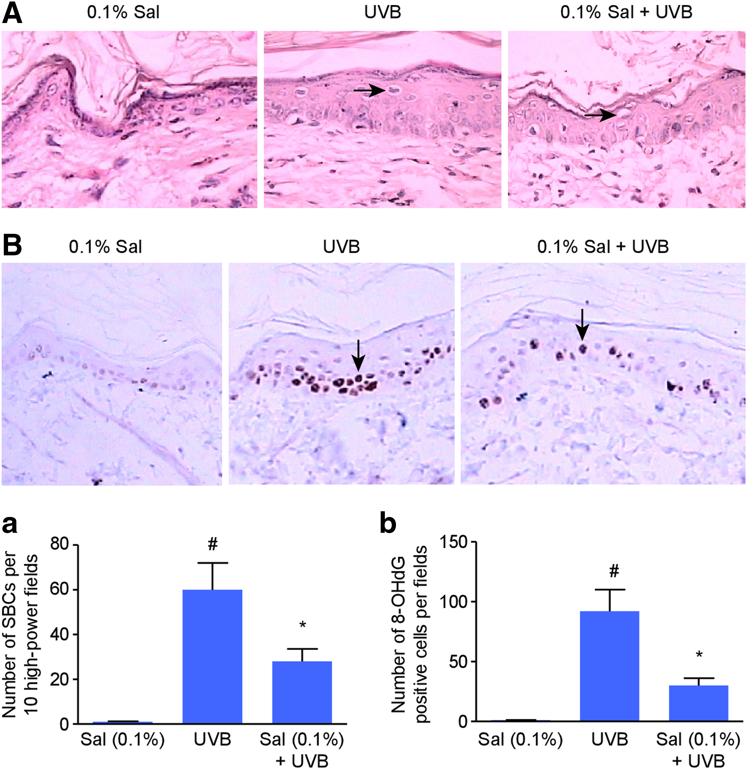
Oral salidroside inhibits UVB-mediated damage in the skin of guinea pigs.
Our data demonstrate that oral salidroside resulted in obvious inhibition in the number of 8-OHdG-positive epidermal cells (Fig. 6B-b).
Discussion
Solar UV radiation is associated with many skin disorders in humans and induces a number of harmful responses, including erythema, edema, photoaging, and skin cancer, which creates an urgent need for better agents for skin photoprotection. 3,31 The use of existing agents is rather limited because of poor skin penetration, low stability, high toxicity, and insufficient activity. 32 In recent years, there has been an increase in the use of natural Nrf2 activators as skin photoprotective agents. 33
Rhodiola rosea L., an adaptogenic herb, is a Chinese traditional herbal medicine that is grown in regions with an altitude of 3500–5000 m, such as the Tibetan Plateau and the Yunnan-Guizhou Plateau, where the air is thin and the sunshine is strong. The ancient Chinese people used it as a remedy for fatigue or to improve pulmonary/cardiac function. 34 Early in “Ben Cao Gang Mu,” one of the earliest Chinese medical records, the therapeutic effects of Rhodiola rosea included the improvement of blood circulation, antifatigue, the reduction of chest tightness, enhanced oxygenation, and adaptogenic effects. Modern research has shown that it has many pharmacological effects, such as antioxidant effects, cardioprotective effects, hepatoprotective effects, adaptogenic and stress-protective effects, and stimulating effects on the central nervous system such as attention and antifatigue effects. Although it has been used to protect against UV irradiation in China for a long time, its mechanisms remain elusive. Salidroside, the main component of Rhodiola, a phenylpropanoid glycoside extracted from Rhodiola rosea L., exhibits potent antioxidative effects. 22,23 Therefore, in this study, the in vitro and in vivo photoprotective effects of salidroside were investigated. Our results first showed that salidroside protected HaCaT cells from UVB-mediated decreases in cell viability in a dose-dependent manner. We also found that the pretreatment of cells with salidroside upregulated Nrf2 translocation to the nucleus according to immunocytochemistry, increased the gene and protein expression of Nrf2 and its targeting genes NQO1 and HO-1, and inhibited UVB-mediated ROS. In addition, salidroside inhibited the UVB-mediated formation of apoptotic SBCs and 8-OHdG-positive epidermal cells in the skin of guinea pigs.
Exposure of the skin to UV light irradiation initiates a photo-oxidative reaction that impairs the antioxidant status and increases the cellular ROS with the activation of many ROS-sensitive signaling pathways. 35 The increased ROS level further impairs the skin to protect itself from the excessive generation of ROS and thus results in increased oxidative stress. 36 Studies have shown that the involvement of photo-oxidative stress mediated by ROS is a crucial mechanism of skin damage relevant to the photoaging and photocarcinogenesis of human skin. 37 In this study, we found that the UVB-mediated increase in the ROS level and salidroside treatment significantly decreased ROS levels.
The upregulation of Nrf2 expression can reduce oxidative stress, and Nrf2-activators can serve as potentially photochemopreventive ingredients. 38 Under physiological conditions, Nrf2 is bound by Kelch-like ECH-associated protein 1 (Keap1) and resides in the cytoplasm before it is targeted for proteosomal degradation. During oxidative stress, Nrf2 that liberated from Keap1 enters the nucleus and induces the expression of genes encoding antioxidant modulators. Nrf2 plays an important cytoprotective role in UVB-induced cell damage. 11 In the present study, the salidroside-treated group had a significantly higher level of nuclear Nrf2 compared to the UVB group. This result indicates that salidroside induced the nuclear translocation of Nrf2 in UVB-induced cell damage. Given that Nrf2 nuclear translocation can induce the expression of multiple cytoprotective genes, this Nrf2 nuclear translocation might mediate the protective effects of salidroside that were observed in this study. For the first time, we also observed increased transcription and expression of the HO-1 and NQO1 genes in HaCaT cells that were treated with salidroside. HO-1 and NQO1 are preferentially inducted by Nrf2 because of the large number of ARE sequences to which Nrf2 can bind in its promoter. The induction of HO-1 and NQO1 renders cells more resistant to the potential subsequent challenges of greater stress. 39 Salidroside treatment also significantly increased the mRNA and protein expressions of HO-1 and NQO1 in the HaCaT cells. Therefore, HO-1 and NQO1 could be important targets of Nrf2 nuclear translocation induced by salidroside and may mediate the protection of salidroside.
SBCs can be found in skin areas of acute sun injury and are often signs of apoptosis. 40,41 8-OHdG is a sensitive marker of oxidative DNA damage; when produced by UVB irradiation, 8-OHdG results in DNA damage via ROS. 7,42 In the present study, we examined the effects of salidroside against UVB-mediated DNA damage. Our results suggest an attenuated sunburn reaction with the reduced formation of UVB-induced apoptotic SBCs in the skin of guinea pigs with salidroside in diets for 2 weeks compared to the UVB-alone group. The oral administration of salidroside (0.1% w/w) inhibited the UVB-induced formation of 8-OHdG. This indicates that the attenuation of the sunburn reaction and the reduced formation of apoptotic SBCs and 8-OHdG can be attributed to the scavenging activity of salidroside against ROS and the activation of the Nrf2/ARE pathway and downstream antioxidative systems, such as NQO1 and HO-1.
In summary, using immortalized HaCaT keratinocytes and a guinea pig skin model, we found that the Nrf2 activator salidroside can protect against UVB-induced damage. The results suggest that treatment with salidroside inhibits UVB-mediated ROS formation, thereby protecting the cells from UVB-induced DNA damage. Moreover, our results suggest that the potential of salidroside against the adverse effects of UVB radiation is associated with the upregulation of Nrf2 nuclear translocation and transcription activity, which in turn may protect skin cells against UVB-induced oxidative damage. In addition, salidroside inhibits the UVB-mediated formation of apoptotic SBCs and 8-OHdG-positive epidermal cells in the skin of guinea pigs. Our results show the photochemopreventive effect of salidroside and suggest that it may be a protective agent against UVB-induced damage in human skin.
Footnotes
Acknowledgments
This work was supported by a grant from the National Natural Science Foundation of China (81000706), a project from the Key Laboratory of Mental Health, Institute of Psychology, Chinese Academy of Sciences. We thank Prof. G.J.Y. for the critical review of the article.
Author Disclosure Statement
No competing financial interests exist.