
Abstract
Introduction
T
Technological advances have encouraged studies of Photodynamic Therapy (PDT) as a potential alternative to combat the formation of dental caries. PDT requires the application of a photoactive drug that can be activated upon exposure to the appropriate light source. Photon absorption leads to an electronically excited state, which in the presence of oxygen, catalyzes a cytotoxic photodynamic reaction that produces reactive oxygen species (ROS); these sequential oxidative reactions lead to cell death. 5
A large number of studies have shown promising results for photodynamic inactivation of microorganisms related to dental caries; extensive investigations utilizing various photosensitizers (PS) and light combinations have been performed. 6 –9 In addition, several publications have shown that when curcumin is utilized as PS, PDT is able to kill cariogenic bacteria, 10 –14 but few studies analyzed the best PDT dose–response for bacterial photodestruction in dentinal tubules using curcumin as a photosensitizer.
An ideal PS should be nontoxic and should display local toxicity only after activation by light. 15 Curcumin is a dye extracted from the rhizomes of the plant Curcuma longa and effectuates antitumor, anticancer, anti-inflammatory, antioxidant, and antimicrobial medicinal activities. 16,17 A number of cell culture and whole animal studies have demonstrated the essentially nontoxic effects of curcumin, which can be detected through a rather broad absorption peak in the range of 300–500 nm (maximum 430 nm). 18,19 Moreover, with the capability of producing high quantities at a reasonable cost, this PS could provide an economically enticing solution.
Common clinical PDT light sources include LASER, light-emitting diode (LED), and halogen lamps. LASER provides a monochromatic and highly efficient (>90%) radiation source that can be transmitted through optical fibers in endoscopic and interstitial light-delivery devices. However, their high cost can discourage their use. However, a low priced system, such as the Diode LASER, is both convenient and reliable. This light source is limited to a single wavelength and each photosensitizer does require a separate unit due to their different absorption wavelengths. Therefore, light sources such as LED have become an attractive alternative for PDT, particularly for irradiating superficial tissue surfaces. The easy configuration of LED arrays into different irradiation geometries and their low cost provide this system with a unique advantage over LASER or diode LASER sources. Although LED does have a fixed output wavelength as well, the cost per watt is significantly lower, thus diminishing the drawback of requiring a separate unit for each photosensitizer. 20
The simple PDT mechanism that activates photosensitizers such as curcumin could provide an exceptional alternative to eradicate the pernicious microorganisms that populate dental cavities. The objective of this study was to assess the photodynamic effects of curcumin activated with an LED light source (central wavelength of 450 nm) on Streptococcus mutans and Lactobacillus acidophilus grown concurrently in dentine carious lesions.
Materials and Methods
Dentin caries-like lesion formation
One hundred eighty structurally normal human third molars were used, after scaling the periodontal tissue. The teeth were rigid, without the presence of caries, and there were no visible structural abnormalities. The teeth used in this study were extracted by orthodontic indication in the surgery department and Buco-Maxillofacial, Faculty of Dentistry, University of Pernambuco. A titanium trephine drill with a 4.0 mm internal diameter was utilized to obtain identically sized dentine slabs from the cervical third of the root. Each slab was then air-dried for 1 min, and an average weight of 23 mg was calculated. After an autoclave cycle at 121°C for 20 min, 6 the slabs were deposited into a 24-well plate. The wells contained 2 mL of a BHI (brain-heart infusion) culture medium augmented with 1 g/100 mL glucose and 2 g/100 mL sucrose Pro-analysis. Each 50 mL of medium solution was inoculated with 5 mL of 108 CFU/mL L. acidophilus (ATCC #ITAL-523) and 5 mL of 108 CFU/mL S. mutans (ATCC #25175). Bacteria numbers were measured in a spectrophotometer wavelength 600 nm in 1-mL cuvettes. The use of spectrophotometer with defined absorbance is the method more precise for obtaining cell numbers. The optical density was defined according to the absorption characteristics of the microorganisms. Cultures were grown until the mid-logarithmic phase. The optical density units used were (OD 600) ∼0.4 and (OD 600) ∼0.6 to have ∼108 cells/mL for S. mutans and L. acidophilus, respectively. The 24-well plates were maintained in microaerobic conditions at 37°C for 14 days, with fresh solutions distributed every 48 h. After the growth period, the specimens were preserved at 4°C until treatment.
Photosensitizer
The stock storage solution of curcumin and curcuminoids was dissolved in N-dimethyl-
The ultraviolet (UV)-visible absorption spectra of the stock solution were recorded by Cary 50 Bio UV-vis spectrometer (Varian, Darmstadt, Germany) using quartz cuvettes with a 1-cm path length. These spectra were characterized by the long wavelength maximum at 430 nm.
Light source
The system consisted of 96 LEDs (Edixeon; Edison Opto Corporation, New Taipei City, Taiwan). Each LED delivered a light intensity of 19 and 47.5 mW/cm2 to the well solution (∼6% of plastic absorption of light was previously deducted), centered at a wavelength of 450 nm, with an average fluency of 5.7 J/cm2. The presence of a cooler on the sides mitigated the heating effect and allowed the system to uniformly irradiate the wells. The internal surfaces were mirrored, and the light was evenly distributed onto each well by optimizing the distance between the LEDs and the plate (Fig. 1). A power meter (Coherent, Santa Clara, CA) was utilized to assess the power density of the incident radiation.
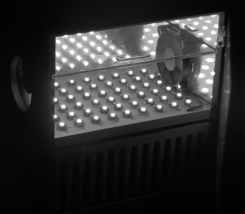
Light system with 96 LEDs delivering a uniform radiation (mirrored device) with coolers to avoid heating effect. LED, light-emitting diode.
Photodynamic treatment
The following groups were assigned: Group L−D−: no light, no drug (control group); Group L−D+: treated with curcumin (drug group); L+D+1 (PDT group 1, light intensity of 19 mW/cm2); and L+D+2 (PDT group 2, light intensity of 47.5 mW/cm2). The group L+D− (only light) was omitted according to previous experience of our group that observed unnoticeable variation with this level of light.
After the induction of dentine carious lesions, dentine slabs were relocated from the 24-well plates to eppendorfs containing 1 mL of the BHI culture medium and vortexed (60 Hz; Heidolph, Kelheim, Germany) for 10 sec to eliminate unadhered bacteria. The observable biofilms on the slab surfaces were removed to expose the carious dentine tissue. Then, the slabs were deposited to the 96-well plates for PDT. Performed in triplicate, curcumin was applied to group L−D+, L+D+1, and L+D+2 (0.75, 1.5, 3.0, 4.0, or 5.0 g/L) for 5 min. Groups L−D− and L−D+ were maintained at ambient temperature and covered with aluminum foil for a period equivalent to the irradiation time.
For PDT group 1, all wells were simultaneously irradiated with blue light from the diode laser for 5 min in the dark at room temperature. The light exposure was from below with an irradiance of 19 mW/cm2 in each well and a fluence of 5.7 J/cm2. For PDT group 2, the wells were irradiated for 2 min at 47.5 mW/cm2 with a fluency of 5.7 J/cm2. To avoid contamination, the plates were covered with polystyrene lids, and each 96-well plate contained only one group of three wells to avoid potential exposure from adjacent wells. In addition, the plates remained covered and undisturbed during illumination. After treatment, the carious bacterial colonies were dispersed into the medium through a forceful disturbance of the slabs in the wells. Then, the well contents were placed into an Eppendorf (the fragments were transferred with the aid of pliers and the remaining solution with Pasteur pipettes) and vortexed (60 Hz; Heidolph) for 60 sec. The supernatant was utilized to create serial dilutions with the BHI culture medium. Further, 100 μL aliquots of these dilutions were dispensed onto blood agar plates and incubated for 72 h under microaerophilic conditions.
The Research and Ethics Committee of the University of Pernambuco (Protocol No. 177/08) has approved these experimental procedures with human dental specimens.
Statistical analysis
The data were processed with version 13.0 of the Statistical Package for Social Sciences (SPSS, Chicago, IL, 2006). One-way ANOVA was used to evaluate the statistical significance of the differences between mean and Tukey test to all pairwise comparisons.
Results
The data are reported as the mean of a triplicate measurement. Survival fractions in each well were calculated by counting the colonies on the plates (L+D+) and dividing these by the number of colonies from the control group (L−D−).
The effectiveness of PDT to eradicate the S. mutans and L. acidophilus strains in carious dentine tissue is showed in Fig. 2. Through HSD (Honestly Significant Difference) it is demonstrated that PDT significantly reduced the number of bacteria (p < 0.05) in both the L+D+1 and L+D+2 groups more so than in any other group at 5.0 g/L curcumin concentrations.
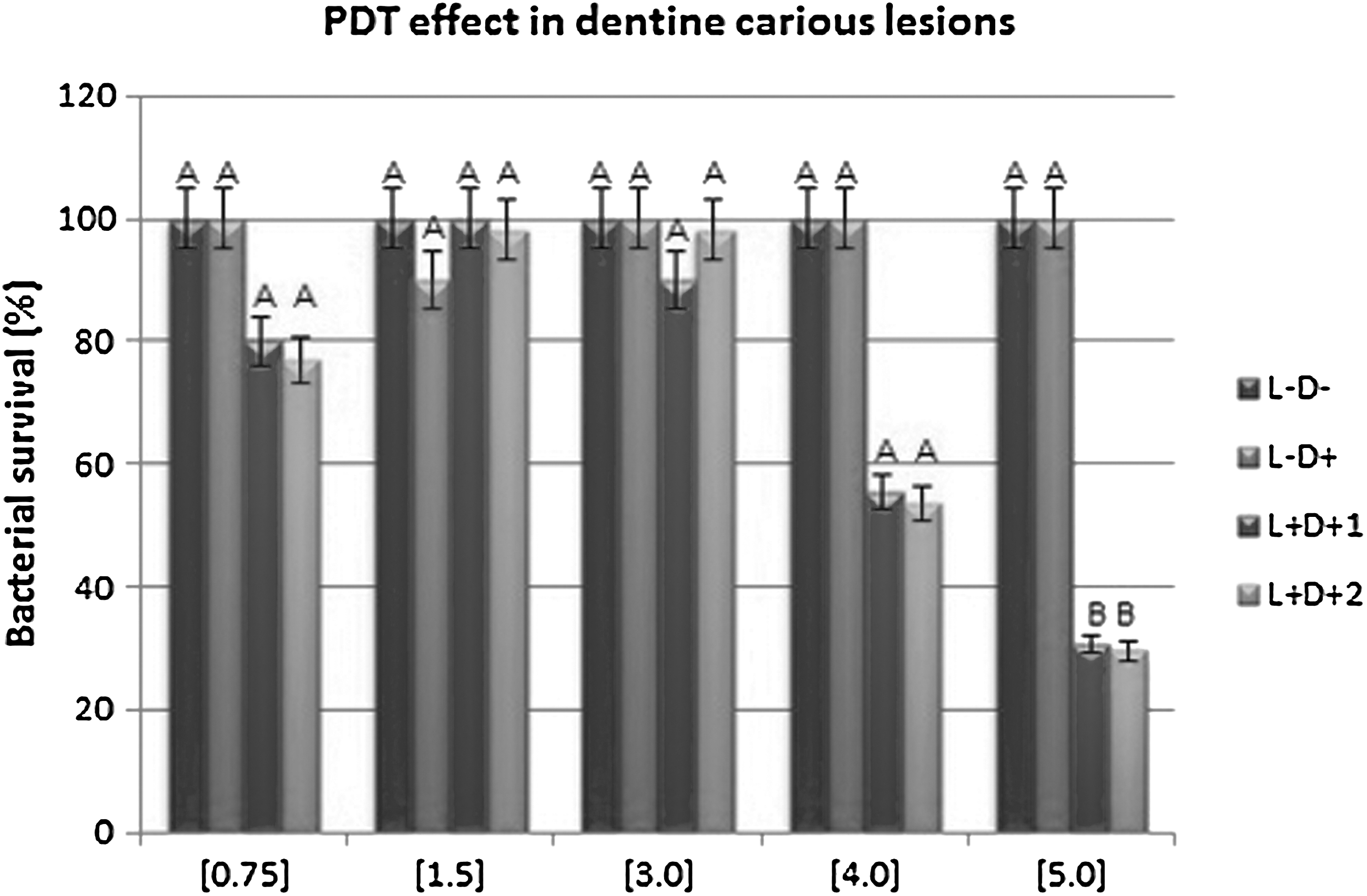
Percentage of survival fractions of Streptococcus mutans and Lactobacillus acidophilus grown as multi-species in dentine carious lesions after photosensitization with different curcumin concentrations followed by irradiation with blue light at a wavelength of 450 nm. Same letter (A) means no significant difference between the groups (p > 0.05).
For PDT group 1 and PDT group 2, a curcumin concentration exposure of 4.0 g/L induced a bacterial strain reduction of 44.4% and 46.4% during light exposure (p > 0.05). Moreover, at a concentration of 5.0 g/L, a significant reduction of 69.4% and 70.4% (p < 0.05) was evident. Curcumin concentrations of 0.75, 1.5, and 3.0 g/L were not sufficient enough to produce the desired toxic effect in either PDT groups. At all curcumin concentration levels, the dark assay group (L−D+) did not show toxicity, and the mean differences in the survival fractions of the drug-only group (L−D+) were low. The data are described as the mean of a triplicate measurement.
Discussion
This study demonstrated that PDT applied to artificially induced dental caries promoted a significant reduction in the viability of L. acidophilus and S. mutans. The use of curcumin followed by irradiation with blue LED light reduced the viability of microorganisms. This result is in agreement with previous studies that verified the photoinactivation of cariogenic bacteria with curcumin and blue light at 450 nm. 10 –14,21
Previously, our group utilized curcumin and an LED at 450 nm to eradicate oral bacteria with PDT, and a statistically significant reduction (p < 0.05) was noted. 21 The photodynamic effects of curcumin has also been investigated in planktonic cultures and biofilm conditions. 10,11 The present study indicated that PDT elicits a photoinactivation of cariogenic bacteria after the application of curcumin and an LED irradiation at 450 nm. Moreover, the phototoxic effect seems to depend on the curcumin concentration (Fig. 2).
Some photosensitizers have a low toxicity on biofilms 22 and collagen matrix, 23 due to their limited accessibility into the deeper layers of microorganisms. The study of Pereira et al. 22 demonstrated that lethal photosensitization mediated by methylene blue occurred predominantly in the superficial layers of the biofilms formed by Candida albicans, Staphylococcus aureus, and S. mutans. Although we have not used a methodology to identify the photosensitizer penetration depth, our results showed that under carious dentine conditions a high curcumin concentration (5.0 g/L) was required to obtain a significant reduction in bacteria cultures.
These results suggest that the concentration of the photosensitizer significantly affects the diffusivity, especially when microorganisms are in complex culture media. The organic structure of decayed dentine was probably able to reduce the photodynamic effects by both decreasing the photosensitizer penetrability, which diminishes the binding efficiency to bacterial colonies, and attenuating the potential irradiation penetration necessary to photoactivate the dye. In vivo evaluations are needed to confirm the clinical relevance of these results, since photodynamic toxic effects are potentially influenced by the microenvironment, the presence of other biomolecules, and the other probable cellular interactions. Further studies will be developed to determine the depth at which the photosensitizer and the blue light can penetrate into the dentine and to establish effective in vivo parameters for the treatment of dentine carious lesions.
This study also evaluated the effect of different curcumin illumination times and light intensities. The PDT group 1 utilized curcumin illumination time of 5 min and light intensity of 19 mW/cm2. For PDT group 2, curcumin illumination time of 2 min and light intensity of 47.5 mW/cm2 were applied. A similar reduction in the feasible cells was observed for both PDT groups. Different illumination time and light intensities did not affect curcumin photodynamic effects.
da Frota et al. 24 evaluated curcumin photodynamic effects on root canals contaminated with Enterococcus faecalis using two different illumination times (5 and 10 min). In the immediate posttreatment collection, only the group that irradiated over the course of 5 min showed greater bacterial reduction (p < 0.05). Curcumin was effective with 5 min LED irradiation, but not with 10 min irradiation PDT using LED light. This result can be related to curcumin photobleaching. Dovigo et al. 25 showed that irradiation fluencies larger than 5.28 J/cm2 had a negligible influence on the phototoxic efficiency of curcumin. Moreover, the light absorption and fluorescence of curcumin decreased as a function of illumination time, which may have been caused by photobleaching; this effectively reduces ROS production, as the authors noted after the long illumination period. 25,26 Accordingly, in the present investigation, the light intensity was adjusted to the illumination time to obtain the same fluency for both PDT groups (5.7 J/cm2).
The results showed that a curcumin illumination time of 2 or 5 min was equally effective in reducing bacteria. Evidently, only short durations of illumination are required to photoactivate the dye and induce the phototoxic effects. This represents an important parameter for clinical practice. A shorter time of clinic attendance means a lower cost treatment and higher patient satisfaction. Translational research involves a process known as translation of knowledge, in which the focus of scientific research in the area of health acquires a two-way flow of information between basic and clinical research. This results in a real applicability of knowledge and new technologies, improving clinical application of new therapeutic concepts, and, finally, providing direct benefits to the main interested in this process: patients. The aim of the translational research is to promote an interdisciplinary and two-way exchange of information from basic research findings in the laboratory to applied clinical settings involving patients and populations. In our study, the choice of curcumin as a photosensitizer allows our research to provide results that can be applied in the future of clinical practice. In dentistry, the choice of photosensitizer strongly depends upon the available light source. The PDT sources must produce adequate irradiation intensity in the activation spectrum of the photosensitizer. Commonly applied light sources in PDT are gallium–aluminum–arsenide diode lasers (630–690, 830, or 906 nm), helium–neon lasers (633 nm), and argon lasers (488–514 nm). However, the monochromatic and highly intense properties of these advantageous light sources may be outweighed by their cost. Thus, nonlaser sources, such as LED, have become an attractive alternative for PDT. Their inexpensive, flexible, and lightweight properties have made these instruments particularly conducive to the irradiation of superficial tissue surfaces. In addition, LED systems are present in common dental routine, and studies that use dental routine materials may facilitate the inclusion of PDT in dental practice without requiring the acquisition of new equipment. Therefore in this investigation, efforts were made to utilize an LED source (450 nm) to activate curcumin. Moreover, an effective study of an economically priced photosensitizer such as curcumin could promote the development of a PDT-type procedure into a clinical reality.
Further studies on the use of curcumin and blue light for eliminating S. mutans and L. acidophilus in dentine carious lesions should be encouraged. An efficient technique to sterilize the dentine tissue before a treatment is needed. PDT could deliver the solution to eliminate oral bacteria both deep in the root and away from the remaining dental tissues, and thus, contribute to the advancement of a conservative approach that effectively treats deep carious lesions.
Conclusions
These results demonstrated that PDT utilizing curcumin and blue LED light at 450 nm exhibited a sufficient toxicity against the bacterial strains of S. mutans and L. acidophilus grown as multi-species in dentine carious lesions. This study suggests that this technique may prove to proficiently treat caries-related diseases.
Footnotes
Acknowledgments
Funding for this research was provided by the Research Support Foundation of São Paulo State (FAPESP) and by the Science and Technology Support Foundation of Pernambuco State (FACEPE).
Author Disclosure Statement
No competing financial interests exist.