
Abstract
Introduction
I
Talaporfin sodium (TS) is a novel second-generation, water-soluble photosensitizer. In Japan, it is approved for the treatment of early lung cancer, brain malignancy, and recurrent esophageal cancer. 6 It has an absorbance peak of the Q band at 664 ± 10 nm. 2,7 TS binds strongly with albumin and is delivered through blood flow. 4,8 Photosensitivity (an important side effect of photodynamic therapy) is lower when using TS than a first-generation photosensitizer because the excretion rate is faster. 7 TS has deeper penetration and a higher molar absorption coefficient. 7 Various studies have reported VS by a photosensitization reaction against tumors. 9 –12 “Vascular patency” (i.e., the degree to which blood vessels are not blocked or obstructed) up to a diameter of 500 μm within healthy myocardial tissue 1 week after a photosensitization reaction that starts immediately after intravenous injection of TS in dogs has been reported. 13 We hypothesized that the difference in vascular patency already mentioned might arise from differences in injuries to VECs.
We studied VEC injury under a particular photosensitization reaction in vitro and in vivo. Dependence of cell lethality on drug contact time (DCT) and albumin concentrations with various radiation exposures was studied in vitro. The presence of normal VECs from the cervical veins of dogs ∼30 min after the photosensitization reaction was confirmed by observation in vivo.
Materials and Methods
Cell culture
Human umbilical vein endothelial cells (HUVECs; Promocell, Heidelberg, Germany) were cultured with a Cell Growth Medium-2 kit (Promocell) in a humidified atmosphere with 5% CO2 at 37°C for 5 days. HUVECs were seeded in collagen-coated cell culture flasks (Iwaki, Tokyo, Japan). All cells were studied at third passage.
Preparation of TS medium
TS (molecular weight, 799.69; Meiji Seika Pharma, Tokyo, Japan) was dissolved in two albumin concentrations to make TS medium of 20 μg/mL concentration. Dulbecco's modified Eagle's medium/nutrient mixture F-12 (DMEM/F-12; Invitrogen, Carlsbad, CA) with 10% fetal bovine serum (FBS; Invitrogen) was used to make 2.1 g/L albumin, which corresponds to the estimated albumin concentration in interstices. Human albumin was added to make 40 g/L albumin, which corresponds to the typical albumin concentration in plasma. 14 Thus, 40 g/L albumin comprised two types of albumin (mostly human).
Change of photosensitizer fluorescence in cell areas with DCT: in vitro
HUVECs were seeded in a collagen-coated cell culture dish (diameter, 35 mm; Iwaki) with 100% confluence. They were in contact with TS medium at a height of 2.8 mm with 2.1 g/L albumin for 0–120 min in a humidified atmosphere with 5% CO2 at 37°C. After contact with the drug, HUVECs were washed twice carefully with phenol-red-free cell culture medium. Images were obtained using a fluorescence microscope with a digital camera at an excitation wavelength of 400 ± 20 nm (corresponding to the Soret band of TS) and a fluorescence wavelength >600 nm (corresponding to the Q band of TS). Exposure time was set at 5 sec for all conditions. Also, microscopic bright field images of the same views as the fluorescence images were captured to confirm a cell area. We selected 10 cells in each field of view of the bright field image. Fluorescence intensity as a result of TS accumulated in cell areas was measured as bright-field images using ImageJ. 15
Changes in cell lethality because of DCT and irradiation conditions: in vitro
HUVECs were seeded in a collagen-coated black 96-microwell plate (diameter, 6.9 mm; Corning, Corning, NY). They were in contact with TS medium containing 2.1 or 40 g/L albumin at a height of 2.8 mm in a well in a humidified atmosphere of 5% CO2 at 37°C. In the study of dependence of cell lethality on DCT, cells were in contact with TS medium for 0–120 min and irradiated at 60 mW/cm2 for 10–40 J/cm2. In the study of dependence of cell lethality on irradiation conditions, cells were in contact with the TS medium for 60 min and irradiated at 30–120 mW/cm2 for 2.5–40 J/cm2. This was a unique situation in that cells were irradiated with TS inside and outside cells. Large quantities of TS were present outside the cells. A red diode laser (Rouge-LD; Cyber Laser, Tokyo, Japan) of wavelength 663 ± 2 nm was used. The laser beam was irradiated up to the bottom of the 96-well plate (Fig. 1). Cell lethality was detected using a water-soluble tetrazolium (WST) assay. After irradiation, cells were cultured with 100 μL/well DMEM/F-12 containing 10% FBS and 10 μL/well WST-8 solution (Doujin-kagaku, Kumamoto, Japan). Two hours after culture, absorbance at 450 nm was measured using a microplate absorbance reader (Sunrise™; Tecan, Männedorf, Switzerland). Cell lethality was defined using the absorbance of the well with no contact with TS and no irradiation of cells as 0%, and that with photosensitization-reaction-treated cells using 20 μg/mL TS and 72 J/cm2 radiant exposure as 100%. Then, 100% cell lethality was confirmed by staining of living and dead cells with Hoechst 33342 (Dojindo, Kumamoto, Japan) and dead cells using propidium iodide (Dojindo).
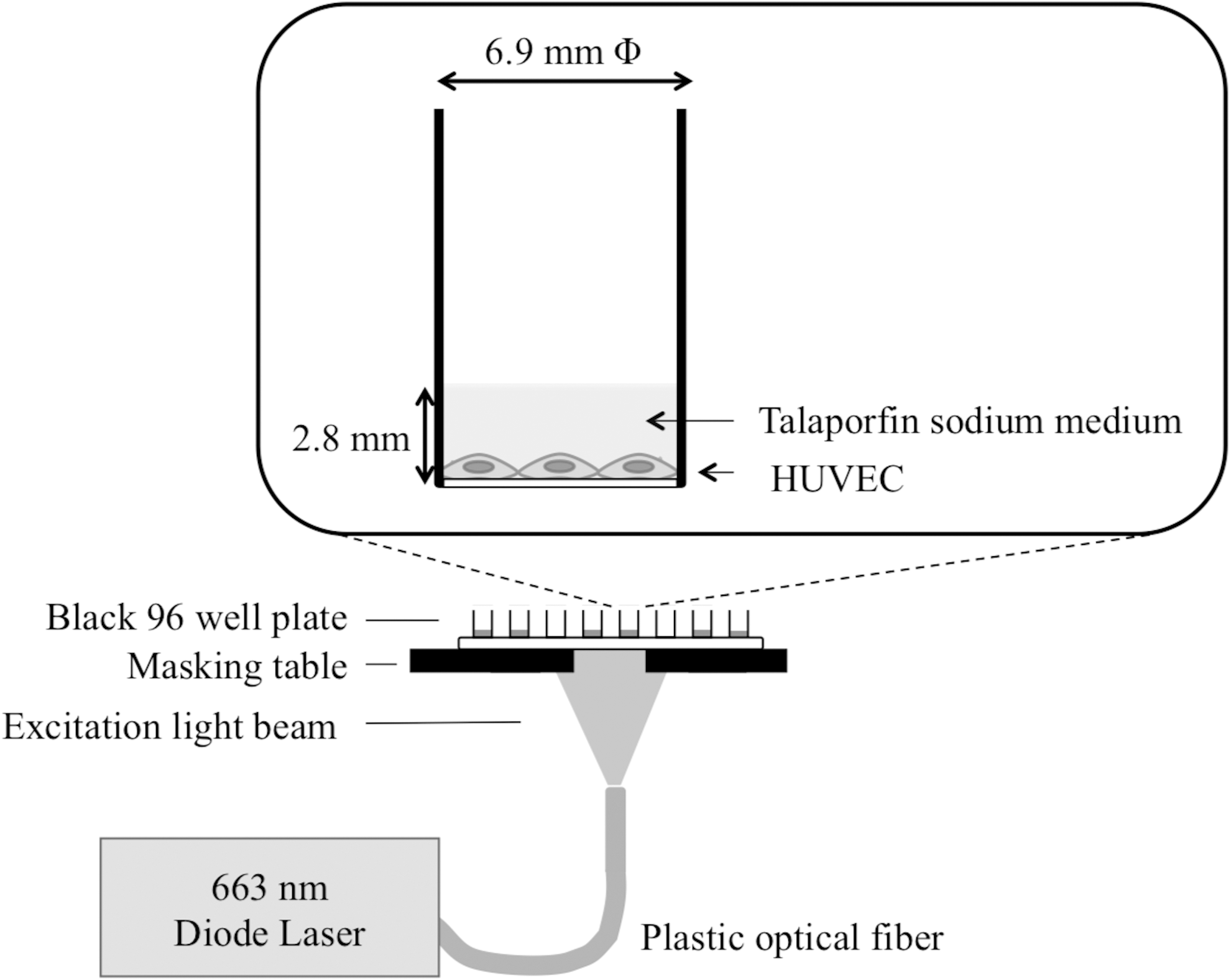
Experimental setup of laser irradiation for the photosensitization reaction.
Presence of canine VECs 30 min after irradiation in vivo
A male beagle dog (10 months; 9.2 kg) was used. After induction of local anesthesia, left and right cervical veins were exposed surgically. A diffuser probe comprising a thin transparent PEBAX® tube (diameter, 800 μm) and a thin laser diffuser comprising a plastic optical fiber (diameter, 250 μm) were obtained. Diffuser light was emitted uniformly in circumferential and longitudinal directions. The length of this diffuser was 70 mm. After mild vein ligation, diffuser probes were fixed on the outside of the venous adventitia along the direction of blood flow. TS (2.5 mg/kg) dissolved in 0.9% saline was injected into the radial vein. Thirty minutes after injection, the red diode laser was emitted from the diffuser probes at 21 mW/cm for 167 or 667 sec. Irradiance is shown in mW/cm because linear diffuser probes were used. Also, irradiance at 21 mW/cm corresponded to 84 mW/cm2 at the outer surface of the probe. Light fluence (mW/cm2)
16,17
in tissue at the positions of endothelial cells was calculated using the following equation:
18
where μeff is an effective attenuation coefficient considering light absorption and multiple scattering within tissue, Z is the axis of propagation of a light beam, and I (z) corresponded to laser intensity at depth z. Thickness of canine cervical veins measured in histology samples was 570 ± 40 μm. We could not obtain the attenuation coefficient of canine veins, so we used the μeff of the normal human aorta at a wavelength of 632.8 nm: 8 cm–1. 19 Estimated irradiance at VECs was 53 ± 2 mW/cm2. Blood samples were drawn from the left radial vein and centrifuged at 1917 g for 5 min to measure TS concentration before and after irradiation by absorbance at 670 nm with a microvolume spectrophotometer (Colibri Microvolume Spectrometer; Funakoshi, Tokyo, Japan). Albumin concentration in blood was measured by a commercial organization (SRL Medisearch, Tokyo, Japan). Approximately 30 min after irradiation, the dog was killed. Veins were extracted and fixed in 10% formalin for histology samples. Sample sections (thickness, 2 mm) were stained with von Willebrand factor to observe living VECs. Cross-sectional images of the vascular wall perpendicular to blood flow were taken under light microscopy (60 × magnification) and normal VECs were identified.
Results
Figure 2 shows the dependence of photosensitizer fluorescence in HUVEC areas on DCT using an albumin concentration of 2.1 g/L. Each plot in Fig. 2 was obtained from the average fluorescence of 10 cells selected in a fluorescent image. The intensity increased rapidly up to 30 min and gradually up to 120 min.
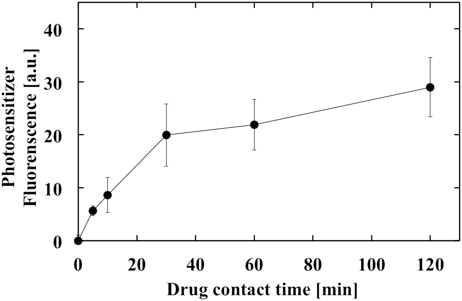
Dependence of photosensitizer fluorescence in cell areas of HUVECs on drug contact time with 2.1 g/L albumin (concentration of talaporfin sodium: 20 μg/mL; N = 10). HUVECs, human umbilical vein endothelial cells.
Figure 3 shows the dependence of cell lethality on DCT. There were no significant differences (p > 0.1) in cell lethality with DCT up to 120 min at 10, 20, and 40 J/cm2 using an albumin concentration of 2.1 g/L, except for cell lethality with DCT at 60 and 120 min at 40 J/cm2, which was 96.2% ± 0.5% and 94.3% ± 0.6%, respectively. There was no significant difference (p > 0.1) in cell lethality up to 120 min, except for DCT at 15 and 30 min, which was 2.1% ± 3.0% and 8.5% ± 4.4%, respectively, at 40 J/cm2 using an albumin concentration of 40 g/L.
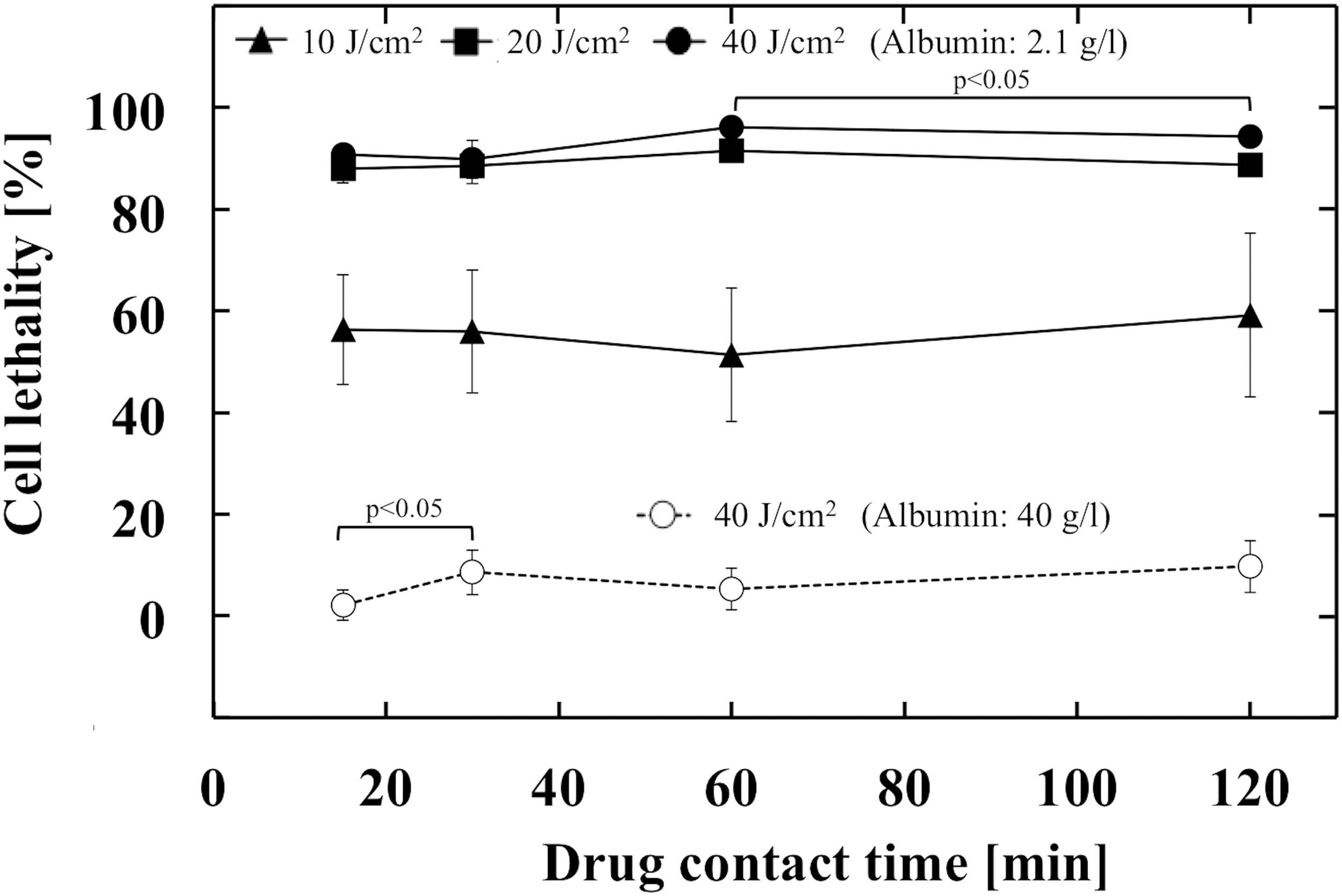
Dependence of cell lethality on drug contact time at various albumin concentrations and radiant exposure (concentration of talaporfin sodium: 20 μg/mL; irradiance: 60 mW/cm2; N = 4–8).
Figure 4 shows the dependence of cell lethality on radiant exposure at various albumin concentrations and irradiance values. Almost 50–60% of cells survived at 10 J/cm2 and virtually all cells were dead at 20 J/cm2 using an albumin concentration of 2.1 g/L. Almost all cells survived up to 40 J/cm2 using an albumin concentration of 40 g/L. Plots of 60 mW/cm2 for 10–40 J/cm2 for both albumin concentrations in Fig. 4 are shared with the plots of 60 min DCT and 10–40 J/cm2 at both albumin concentrations in Fig. 3.
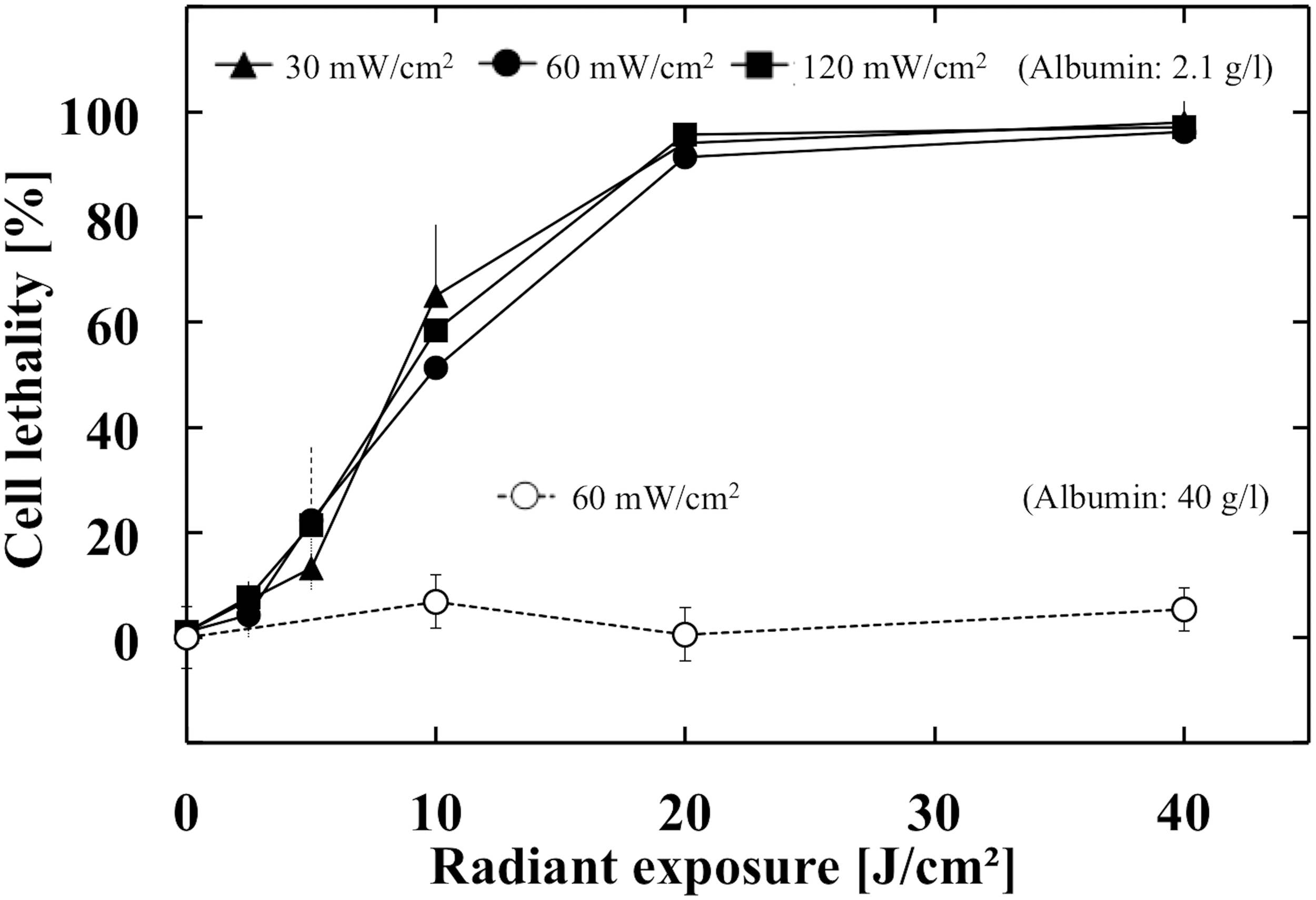
Dependence of cell lethality on radiant exposure at various albumin concentrations and irradiance values (concentration of talaporfin sodium: 20 μg/mL; drug contact time: 60 min; N = 8).
Figure 5 shows cross-sectional images of histology samples upon irradiation area stained with von Willebrand factor. TS concentration in plasma was 14 μg/mL, and albumin concentration was 30 g/L. We confirmed the presence of normal VECs after the photosensitization reaction at irradiation times of 167 and 667 sec. Comparison of images of zero irradiation with those for irradiation of 167 sec showed little morphological change in VECs. In the image for an irradiation time of 667 sec, few red blood cells adhered to VECs.
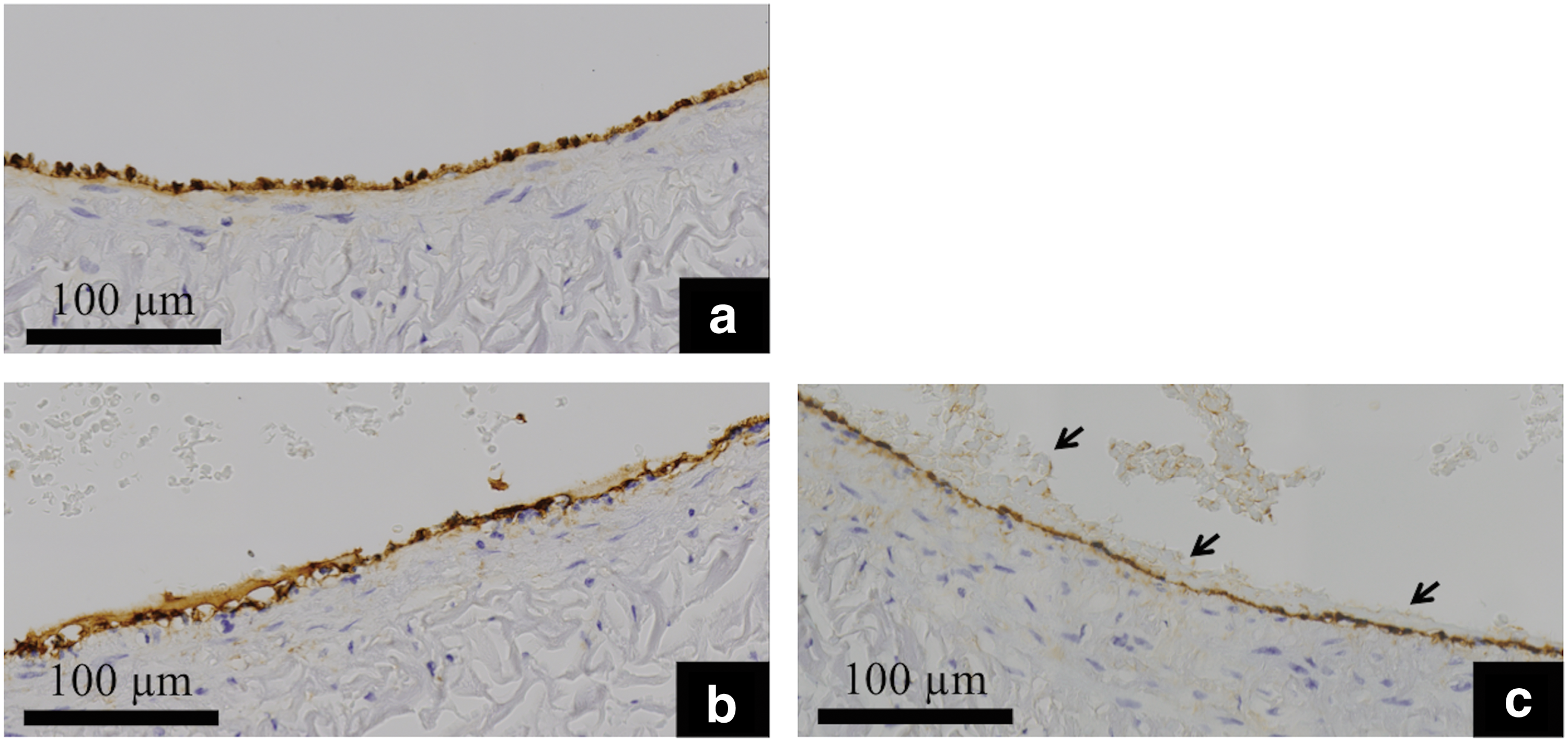
Histology of vascular endothelial cells ( × 60 magnification) in canine cervical veins.
Discussion
This basic science study focused on targeting healthy tissue and emitting irradiation 15–30 min after intravenous injection of a photosensitizer. Most studies focusing on vascular injury caused by a photosensitization reaction targeting tumor vessels start irradiation >4 h after intravenous injection of a photosensitizer. Kimbery et al. studied VEC injury in arterioles and venules in the normal cremaster tissue of rats, but they irradiated 4 or 24 h after intravenous injection of a photosensitizer. 20 Intracellular concentration of photosensitizer in VECs at a DCT of 4 or 24 h has been shown to be almost two- or threefold higher, respectively, than that for 1 h DCT. 9 Hence, the mechanism of VEC injury might differ between the study of Kimbery et al. and the present study.
Fluorescence as a result of TS in HUVECs increased, but not cell lethality, with DCT (Figs. 2 and 3). We used a unique method of irradiation, in which a considerable amount of TS was outside the cells. Hence, these results suggest that the TS inside HUVECs did not contribute to cell lethality. Therefore, major damage to HUVECs by the photosensitization reaction may have arisen from outside the cells for DCT up to 120 min under our experimental conditions in vitro. A Ca2+ dynamic study within myocardial cells using a photosensitization reaction with TS suggested that there was ion channel failure caused by membrane injury, which implies the same mechanism of damage as that already mentioned. 21
HUVEC damage by the photosensitization reaction was lower at 40 g/L albumin (corresponding to albumin concentration in plasma) than at 2.1 g/L albumin (corresponding to albumin concentration in interstices) (Fig. 4). This observation confirms the work of Sheyhedin et al., who concluded that albumin binds strongly with TS at a ratio of 1:1, and that this binding decreases the cytotoxicity of the photosensitization reaction. 8,22 VECs face blood and vessel walls. 23 Albumin concentration in the vessel wall is believed to be higher than in the interstices because of the diffusion process related to albumin. By considering the external milieu of VECs, we predicted that the highest severity of cell damage would be elicited with an albumin concentration between 2.1 and 40 g/L (Fig. 4). In a clinical setting, medium containing albumin is irradiated before the VECs, whereas, in our in vitro experiment, VECs were irradiated first. This difference suggests that the in vitro cell damage was more severe than that in vivo.
We confirmed that normal endothelial cells were present under an irradiation time of 667 sec, and that VECs survived at <34 J/cm2 in vivo (Fig. 5). Almost all cells were dead at 20 J/cm2 in 2.1 g/L albumin, but almost all cells survived at 40 J/cm2 in 40 g/L albumin in vitro. These in vitro and in vivo results suggest that VEC toxicity in vivo is more likely than that a result of high albumin concentration in plasma in vitro. This might also be because of the difference in irradiation milieu of VECs between the in vivo and in vitro conditions already mentioned. We predicted that acute injury to VECs was sufficiently low under our photosensitization reaction to explain the vascular patency within healthy myocardial tissue 1 week after photosensitization because of the low intracellular concentration of TS and reduced damage outside cells induced by a high concentration of albumin.
Our study had five major limitations. (1) HUVECs were used in vitro and canine cervical VECs were observed in vivo. Differences in endothelial function between these veins were not considered in our study. However, many studies of VEC function have used HUVECs, so we think the use of HUVECs may have been reasonable. 9,11 (2) The times after the photosensitization reaction differed between the in vitro and in vivo studies. Cell lethality 2 h after the photosensitization reaction was calculated in vitro, but canine veins were extracted ∼30 min after the reaction in vivo. We predict that the main mechanism of cell damage may be necrosis under our unique photosensitization reaction, and it is a short-term death reaction, lasting several tens of seconds to several tens of minutes. 24 The major damage to HUVECs caused by photosensitization may have arisen from outside the cells in our in vitro experiment. Therefore, we think that the cell lethality 2 h after photosensitization was mainly caused by necrotic cell death. So, we think there was only a small difference in timing of cell lethality after the photosensitization reaction between the in vitro and in vivo studies. (3) The presence of VECs was investigated in vivo, but not their function. (4) Interaction between albumin and the efficacy of the photosensitization reaction may be dependent on the animal species because binding sites change with this interaction. 25 Only bovine serum albumin was used to make 2.1 g/L albumin, but bovine serum albumin (2.1 g/L) and human albumin (37.9 g/L) were used to make 40 g/L albumin in the in vitro study. (5) Although the medium containing albumin is irradiated before the VECs under clinical conditions in general, VECs were irradiated first in our in vitro experimental scheme. Thus, we think that the cell lethality in vitro might have been higher than that in the clinical situation.
Conclusions
We conclude that VEC injury after a photosensitization reaction in healthy tissue shortly after TS injection might be low enough for the blood vessels to be patent.
Footnotes
Acknowledgments
This work was supported, in part, by the Adaptable and Seamless Technology Transfer Program (A-STEP; contract grant number: AS2415004P) of Japan Agency for Medical Research and Development and Japan Science and Technology Agency.
Author Disclosure Statement
No competing financial interests exist.