
Abstract
Introduction
P
Pulsed dye laser (PDL) currently represents the mainstream choice for PWS treatment in accordance with selective photothermolysis. 4,5 However, most PWS lesions cannot be removed. 6 Recent advances in laser technology have led to the development of PDL equipment with various settings for a more refined regulation of the laser beam during PWS treatment: wavelength, pulse duration, spot size, and cooling system. 7,8 PDL with 7 and 10 mm spot sizes is widely used on a regular basis to treat PWS lesions. Regardless of this, no studies have compared the safety and efficacy of PDL with different spot sizes in the treatment of PWS. 9,10 Accordingly, we conducted the current study to investigate the safety, efficacy, and outcomes of PWS treatments by PDL with 7 and 10 mm spot sizes.
This study was approved by the Investigational Review Board at Shanghai Ninth People's Hospital. All patients—minors and adults—provided verbal and informed consent or assent to participate in the study.
Materials and Methods
Subjects
A total of 35 untreated patients with PWS were included in the study. Each of two contiguous test sites of the PWS lesion in the same anatomical region and the same stain color was treated by PDL with 7 and 10 mm laser spot sizes for each patient. Facial anatomic areas were subdivided into several regions in accordance with the method used by Renfro and Geronemus.
11
All sites treated by PDL with 7 and 10 mm spot sizes were compared and subdivided into the same anatomical region for each patient. The sites for treatment using Vbeam with a 7 mm spot size and Vbeam with a 10 mm spot size were selected by randomization (Fig. 1): A random sequence software program was used (
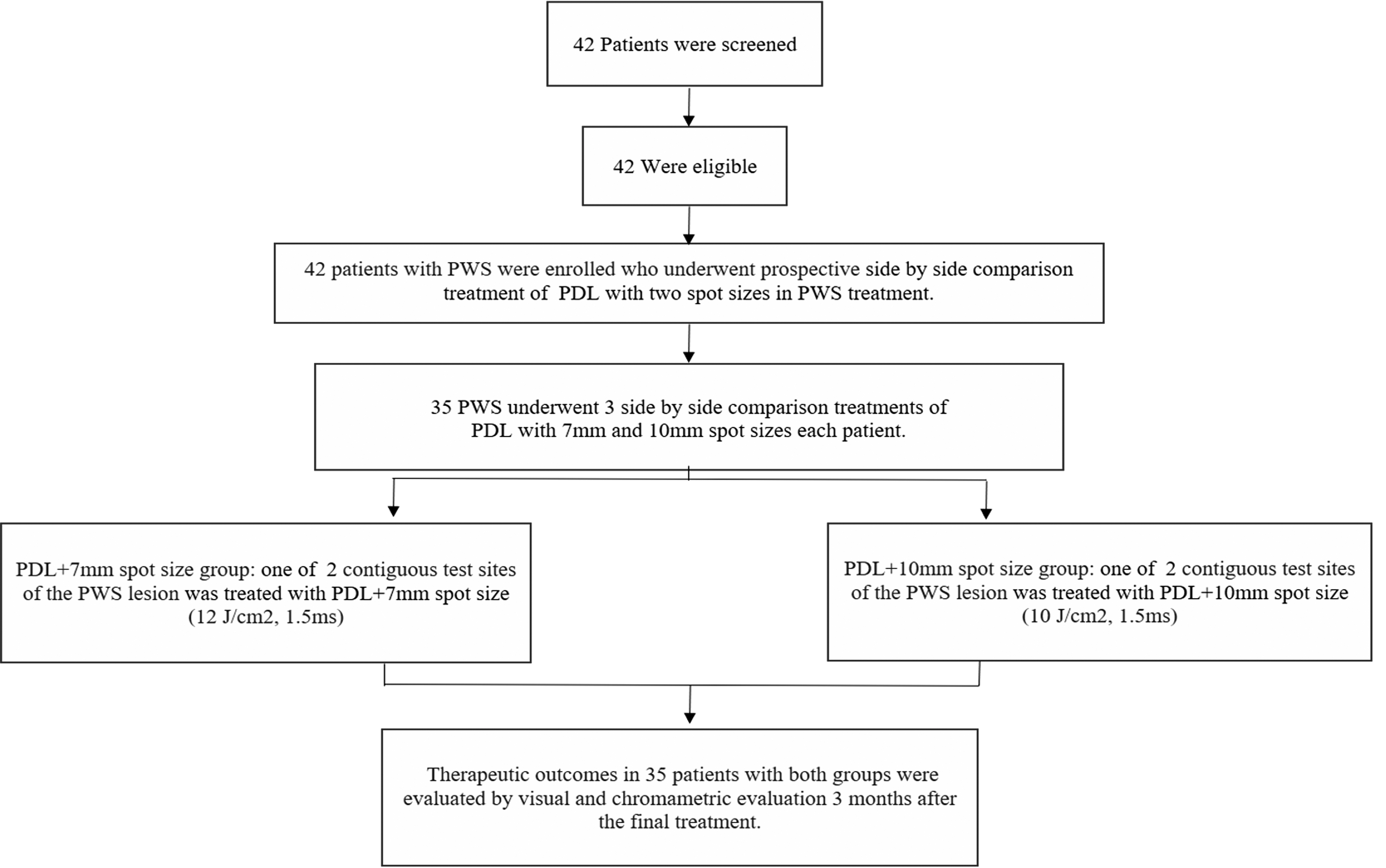
Participant enrollment, randomization, and analysis.
PWS, port-wine stain.
PDL treatment protocol: Vbeam with a 7 mm spot size
The Vbeam (Candela Corp., Irvine, CA) treatment protocol requires a 595 nm wavelength for PDL with 12 J/cm2 radiant exposure, 1.5 msec pulse duration, 7 mm spot size, and cryogen spray cooling (40 msec of cooling with a 20 msec delay).
PDL protocol: Vbeam with a 10 mm spot size
The Vbeam (Candela Corp., Irvine, CA) treatment protocol requires a 595 nm wavelength for PDL with 10 J/cm2 radiant exposure, 1.5 msec pulse duration, 10 mm spot size, and cryogen spray cooling (40 msec of cooling with a 20 msec delay).
Follow-up and assessment
All patients underwent three laser treatments of two test treatment sites by using Vbeam with 7 and 10 mm spot sizes at 6-week intervals. Follow-up was conducted 3 months after the final treatment to assess the efficacy of the treatment and color improvement, as well as to observe for side effects. Treatment efficacy was evaluated by chromametric and visual methods.
All test treatment areas were independently measured and assessed, despite their variation among patients. In addition, the test treatment sites for each patient were subdivided into similar anatomical locations to avoid bias attributed to site variation. 11
Chromametric assessment
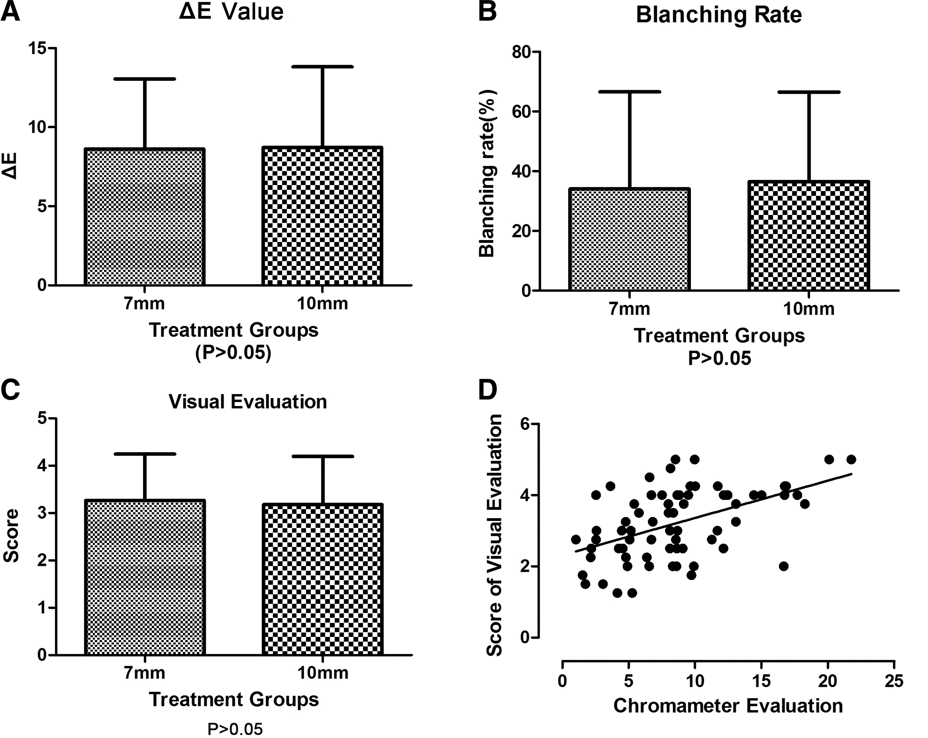
A chromameter (CR-400; Minolta, Japan) was used to assess the change in color at the two test treatment sites independently, before treatment and 3 months after the final treatment. The L*, a*, and b* color system of the International Commission on Illumination or Commission Internationale de l'Eclairage was used for objective assessment of PWS skin. 12,13 The change in color or improvement (ΔE) and the efficacy of color improvement (blanching rate) quantitatively measure efficacy in PWS treatment.
Visual assessment
Standard digital photographs (60D camera; Canon) of each patient were taken before treatment and 3 months after the final treatment. Four plastic surgeons evaluated the color improvement of the lesions in a double-blind manner. The color improvement of the PWS skin was graded as follows: score of 5 (75–100%, excellent); score of 4 (50–75%, good); score of 3 (25–50%, fair); score of 2 (1–25%, poor); and score of 1 (0%, no improvement). 14 The average score was used when the color improvement was scored differently.
Follow-ups on post-treatment responses and side effects were conducted.
Data analysis
SPSS ver. 19.0 (IBM Corporation) and GraphPad Prism 5 (GraphPad Software, Inc.) were used to analyze the therapeutic outcomes. For visual and chromametric assessment, the differences in efficacy outcomes of PDL with 7 and 10 mm spot sizes were compared and analyzed by using the Wilcoxon signed-rank test. Spearman's rank correlation coefficients were also used to compare the efficacy outcomes from the chromametric and visual evaluations for PDL with 7 and 10 mm spot sizes. p Value ≤0.05 was considered statistically significant.
Results
Patient information
A total of 35 patients with untreated PWS, consisting of 15 Asian men and 20 Asian women aged 1 month to 37 years (mean age = 8 years), participated in this study. The patient skin types according to the Fitzpatrick Skin Type were type III in 22 (63%) patients and type IV in 13 (37%) patients. No hypertrophic lesion was observed. PWS was observed in the facial region of 32 (91%) patients, neck region of 2 (5.7%) patients, and trunk of 1 (2.9%) patient. Patient information is summarized in Table 1.
Efficacy outcomes
Skin color assessment (ΔE, blanching rate) by chromametry
The ΔE and blanching rate were measured before treatment and 3 months after the third treatment and then compared. The average ΔEs for the sites treated by PDL with 7 and 10 mm spot sizes were 8.62 (SD = 4.43) and 8.73 (SD = 5.09), respectively (p = 0.635) (Fig. 2A). The average blanching rates for the sites treated by PDL with 7 and 10 mm spot sizes were 34.03% (SD = 32.65%) and 36.51% (SD = 30.05%), respectively (p = 0.719) (Fig. 2B). These results indicate no difference in efficacy outcome between PDL with a 7 mm spot size and PDL with a 10 mm spot size.
ΔE (color improvement) was measured pre-treatment and 3 months post-treatment. ΔE was calculated by:
Visual assessment
The mean scores for color improvement of PDL with 7 and 10 mm spot sizes were 3.26 (SD = 0.97) and 3.17 (SD = 1.01), respectively (p = 0.831). These results indicate that PWS treatment by PDL with both 7 and 10 mm spot sizes exhibits similar efficacy levels in improving the lesion color (Fig. 2C).
Relationship between visual and chromametric assessments
The Spearman's rank correlation coefficient was 0.488 (p ≤ 0.001) for the comparison of efficacy outcomes between the chromametric and visual evaluations for all treatment sites. This finding indicates no difference between the visual and chromametric assessments of the effectiveness of PDL with 7 and 10 mm spot sizes at the tested sites (Fig. 2D).
Photographs of representative patients with PWS from the group whose lesions were localized to the facial region and treated by PDL with 7 and 10 mm spot sizes are shown in Figs. 3 –5; the photographs were taken before treatment, immediately after treatment (to examine the immediate purpura responses), and 3 months after final treatments.
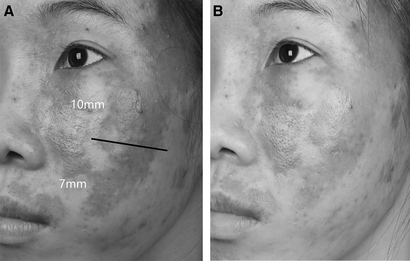
Patients with untreated facial PWS, before treatment
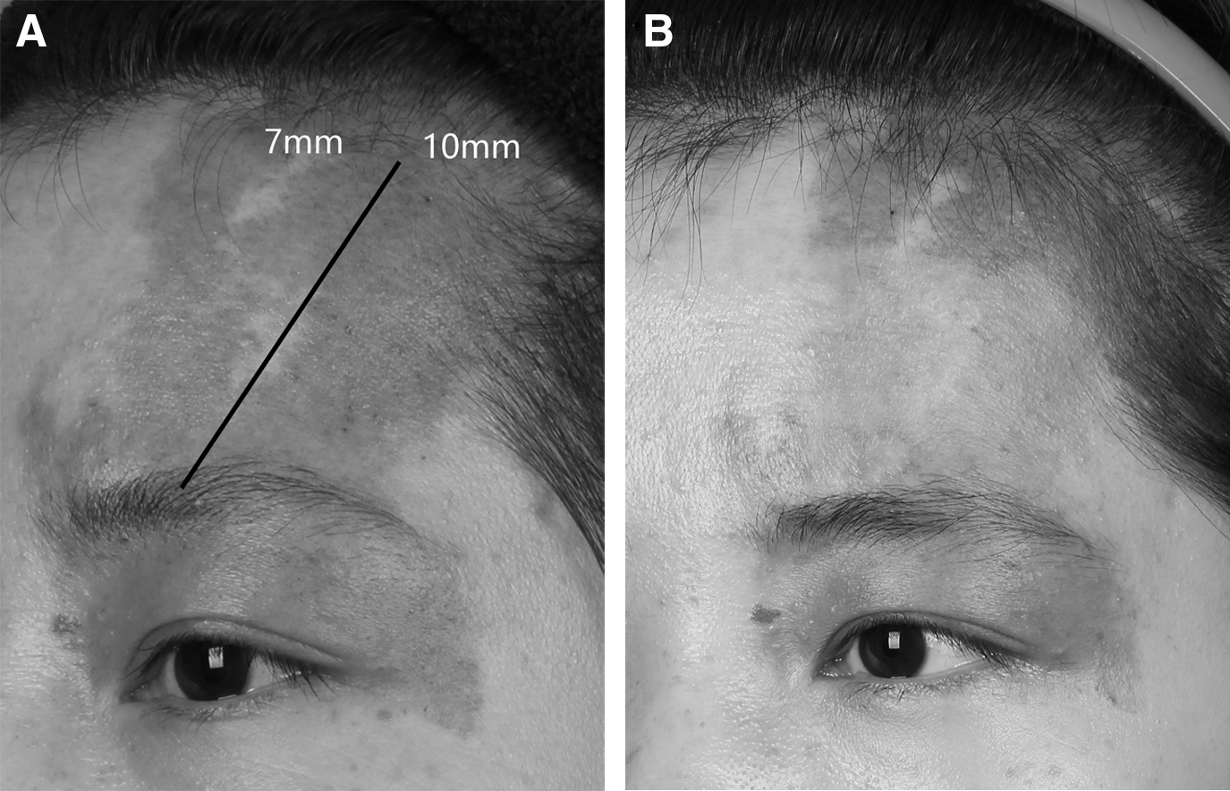
Patients with untreated facial PWS, before treatment
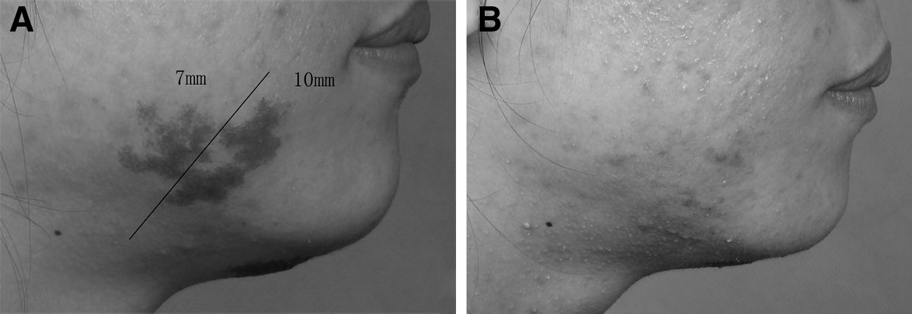
Patients with untreated facial PWS, before treatment
Side effects
Edema and purpura developed in all (100%) and 33 (94%) patients, respectively, at the sites treated with 7 and 10 mm laser spot sizes. Crusting and blistering occurred in 10 (29%) and 2 (5.7%) patients at the sites treated with 7 and 10 mm laser spot sizes, respectively. No patient developed pigmentation change, infection, scarring, or atrophy at either test site.
Discussion
At present, PDL systems are regarded as the gold standard for the treatment of PWS lesions in accordance with selective photothermolysis. Recent technological advances have led to an increase in the availability of choices in spot size, pulse duration, and epidermal cooling system. PDL systems with either 7 or 10 mm laser spot size are widely used for PWS treatment, and numerous studies on PWS treatment by PDL have been previously reported. 15 –17 In the current study, each of the 35 patients was treated by PDL with both 7 and 10 mm laser spot sizes. The outcomes of these treatments were compared.
Visual and chromametric assessments provided subjective and objective evaluations, suggesting a similarity in color improvement between sites treated by PDL with 7 and 10 mm spot sizes (which had higher and lower radiant exposure, respectively). The blanching rate of PWS with PDL treatment ranged from 1.38% to 66.68%. This variation in blanching rates may be attributed to the heterogeneity of blood vessel size in PWS in various patients. 18 –21
Reinisch proposed the use of variously sized beams to limit penetration into the dermis. By using the spot size arguments, the properties of spots can be used to change the behavior of particular wavelengths in the skin. For instance, a 1064 nm laser can be used to heat progressively larger depths of skin by increasing the spot size (Fig. 6). 22
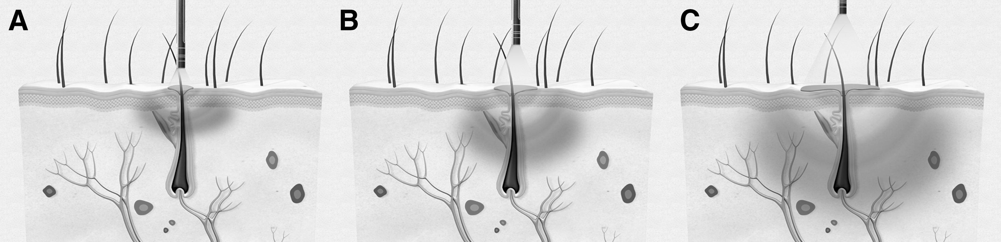
The size of laser spot size affected the depth of penetration. 0.5 mm laser spot size
Thus, in our study, PDL with the 10 mm spot size using low radiant exposure (10 J/cm2, 1.5 msec) acquired efficacy and safety outcomes similar to those of PDL with the 7 mm spot size using high radiant exposure (12 J/cm2, 1.5 msec). The larger spot size can provide uniform transmission of energy to treat lesions, increase therapeutic efficacy, and shorten treatment time for PWS.
The limitations of this study include the following: (1) The study included a limited number of subjects; (2) follow-up was conducted 3 months after the third treatment (longer follow-ups were lacking in this study); and (3) three sessions of laser treatment using two spot sizes were conducted (this number of treatments is too low to effectively compare the two spot sizes).
Conclusions and Summary
In summary, this study provides preliminary data of PWS treatment by PDL with 7 and 10 mm laser spot sizes that exhibited high and low radiant exposures, respectively. The outcomes of these treatment were compared. In addition, their safety and efficacy were evaluated. Thus, in our study, PDL with a 10 mm spot size, which has low radiant exposure (10 J/cm2, 1.5 msec), obtained blanching efficacy and safety similar to that of PDL with a 7 mm spot size, which has high radiant exposure (12 J/cm2, 1.5 msec: 120% of the radiant exposure of the 10 mm spot size). PDL using optimal laser parameters with the 10 mm spot size may provide efficacy and safety during treatment and shorten the process.
Footnotes
Acknowledgments
This study was supported by the Shanghai Health System Important Disease Joint Research Project (Grant No. 2013ZYJB0014).
Author Disclosure Statement
No competing financial interests exist.