
Abstract
Introduction
T
The mental nerve injury can be solved spontaneously, but when that does not occur, the available therapeutic modalities present restricted results to certain cases, confirming that the best is prevention, and the good anatomy knowledge of the area by the dentist is of fundamental importance. Among the therapeutic modalities, the photobiomodulation therapy (PBMT) has been studied, with results indicating improved function, acceleration of regenerative process, reduce the inflammatory response in the nerve, 7 metabolism increase in neurons, and better ability to produce myelin. 8
Low-level diode lasers most widely studied in Dentistry have as active component the Gallium-Aluminum-Arsenate (GaAlAs). 7,9 –12 Using specific laser and light-emitting diode irradiation parameters, cellular activities can be induced, such as cellular proliferation and viability while stimulating mitochondrial activity, thereby increasing adenosine triphosphate production, synthesis of DNA and RNA, and activating cell-signaling cascades, including the production of reactive oxygen species, nitric oxide release, activating cytochrome c oxidase, and modifying intracellular organelle membrane activity, calcium flux, and expression of stress proteins. 13 Due to its effects, PBMT is used in various procedures of oral surgery, 14 periodontics, 15 orthodontics, 16,17 pediatrics, 18 and endodontics, 19 among others.
In animal models, the use of PBMT resulted in stimulation of damaged fibers' axon growth. 19 However, several different parameters are observed like 45 J irradiation for 10 days 19 ; wavelength at 660 and 808 nm, with a 0.028-cm2 beam diameter, power output of 30 mW at 10 J/cm2 (0.27 J per point, irradiation for 9 sec), and 50 J/cm2 (1.41 J per point, irradiation for 47 sec) 10 ; energy density at the point: 10, 60, or 120 J/cm2 with wavelength at 660 or 780 nm, spot area: 4 mm2, power: 40 mW 9 ; and wavelength 901 nm (impulsive) and power of 10 mW, in square-shaped window type device (16 cm2). 8
Given the growing use of PBMT in dental offices and the search for a minimally invasive treatment for nerve injuries, this study aims to assess the regeneration of mental nerve after compression lesion and treatment with PBMT with different protocols.
Materials and Methods
Ethics considerations
This study was submitted to Ethics Committee for the use of animals of the Federal University of São Paulo, in which after analysis it was approved with the protocol number 213543.
Study groups
In this study, 48 Wistar rats with an average weight between 250 and 300 g were used. They were kept for 7 days housed in individual cages for acclimatization at 23°C room temperature with a 12-h light/12-h dark cycle.
The animals were randomly selected and divided into four study groups (n = 12). The groups NC (negative control—lesion by compression), GD (gradual dose—lesion by compression and PBMT with gradual), and CD (constant dose—lesion by compression and PBMT with CD) were submitted to lesion by compression of mental nerve; only animals of the PC (positive control) group were not submitted to injury. In the treatment phase, PC and NC groups composed the PC and NC groups, respectively, in which rats received no treatment with PBMT, only the laser pointer was positioned in the application points and not turned on; the animals of the treatment group CD received PBMT for 20 days, daily, starting 1 day after the surgery.
Surgery for compression of mental nerve
The surgical technique adopted in this study was based on Savignat et al. 20 and presented in Fig. 1. Under anesthesia with 2% xylazine (0.025 mL/100 g) and 10% ketamine hydrochloride (0.05 mL/100 g), submandibular incision was made to access the mental foramen. Then, the mental nerve was strongly crushed with an ultrafine forceps. The load for compression was standardized using the third ratchet of the ultrafine forceps for crushing the nerve for 15 sec. After surgery, the skin incision was sutured with a 3.0 Vicryl suture, and Betadine (Betadine, polyvidone iodee; VIATRIS, Merignac, France) was applied. The animals were maintained under analgesic (200 mg/L Paracetamol diluted in water) for 3 days and under antibiotic (Enrofloxacin—2.5 mg/kg) for 7 days.
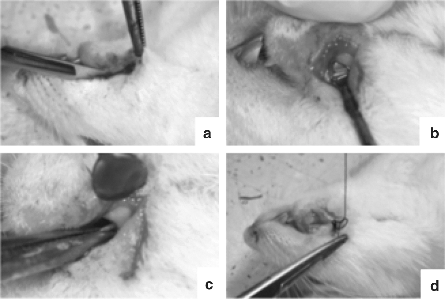
Surgical technique used in this study.
Treatment with PBMT
The PBMT device used was Whitening Lase II (DMC, São Carlos, Brazil), a low-power diode laser (GaAlAs). PBMT was performed in the animals of groups GD and CD in which were submitted daily during 20 days, starting 24 h after surgery. The application technique used was the punctual technique application for contact with tissue positioned perpendicularly to the skin at three equidistant points (with a distance of 1 cm between them) upon the suture. As showed on Table 1, for GD, parameters used were wavelength of 808 nm, 100 mW, and 22 sec (80 J/cm2 at each point; 2.3 J energy for point), 25 sec (90 J/cm2 at each point; 2.6 J energy for point), 28 sec (100 J/cm2 at each point; 3.0 J energy for point), 31 sec (110 J/cm2 at each point; 3.2 J energy for point), and 33 sec (120 J/cm2 at each point; 3.5 J energy for point), with a spot area of 0.028 cm2 and continuous mode of irradiation. For the CD group, parameters used were a wavelength of 808 nm, 100 mW, 33 sec (120 J/cm2 at each point; 3.5 J energy for point) and spot area of 0.028 cm2 with continuous irradiation.
CD, constant dose; GD, gradual dose; NC, negative control; PC, positive control.
Euthanasia
Three animals of all groups were euthanized at 3, 7, 14, and 21 days of study. The mental nerves were removed for analysis through transmission electron microscope (TEM).
TEM analysis
The collected mental nerves were prefixed in 4% PFA solution, postfixed in 1% osmium tetroxide, and included in spurr resin. Sections with 0.5 μm thickness were stained with toluidine blue, and images were acquired using AxioVision software (version 4.5). Ultrathin sections with 0.3 μm were collected on copper grids and contrasted in 5% uranyl acetate and 1% lead citrate and examined by TEM LEO 906E (Zeiss, Germany). Images of neural fibers were obtained with 20,000 × magnification (to count the number of myelinated fibers and thickness measurements of the myelin sheath, outer perimeter, and inner axonal diameter) and 100,000 × magnification (for evaluation of lamellar structure of myelin sheath). The measurements were performed using ImageJ software (version 1.47). The G-ratio was also evaluated, calculated by dividing the inner axonal diameter by the outer fiber diameter (that was composed by myelin sheath plus inner axonal diameter measurements), and the results were stratified in ranges of 0.4–0.5, 0.5–0.6, 0.6–0.7, 0.7–0.8, and 0.8–0.9. When ranges were used, the lowest portions were always included and the highest portions excluded (e.g., the 0–4 range includes 0 through 3.99, excluding 4). The results are expressed as means and considered significant values (p ≤ 0.05) after analysis of variance (ANOVA) and Tukey's test.
Results
The results were analyzed both qualitatively and quantitatively in a blind manner by two previously calibrated evaluators. The quantitative analysis was performed evaluating the number of myelinated fibers, thickness, outer perimeter, and inner area of the myelinated fibers in 20,000 × magnification photomicrographs. Qualitative analysis was performed with observations of the ultrastructure of rats' mental nerve with both 20,000 × and 100,000 × magnifications.
Qualitative analysis
For the ultrastructural analysis with 20,000 × magnification, it was observed that at 3 days the groups NC, CD, and GD presented large amount of myelinated fiber degeneration, which occurred at 7 days only in the group NC. At that same time, CD and GD already presented initial aspects of regeneration. At 14 days all groups showed increase of myelinated fibers regenerated. At 21 days, CD and GD reached a similar morphology to the PC group. Structures and organelles such as paranodes and macrophages were also observed (Fig. 2).
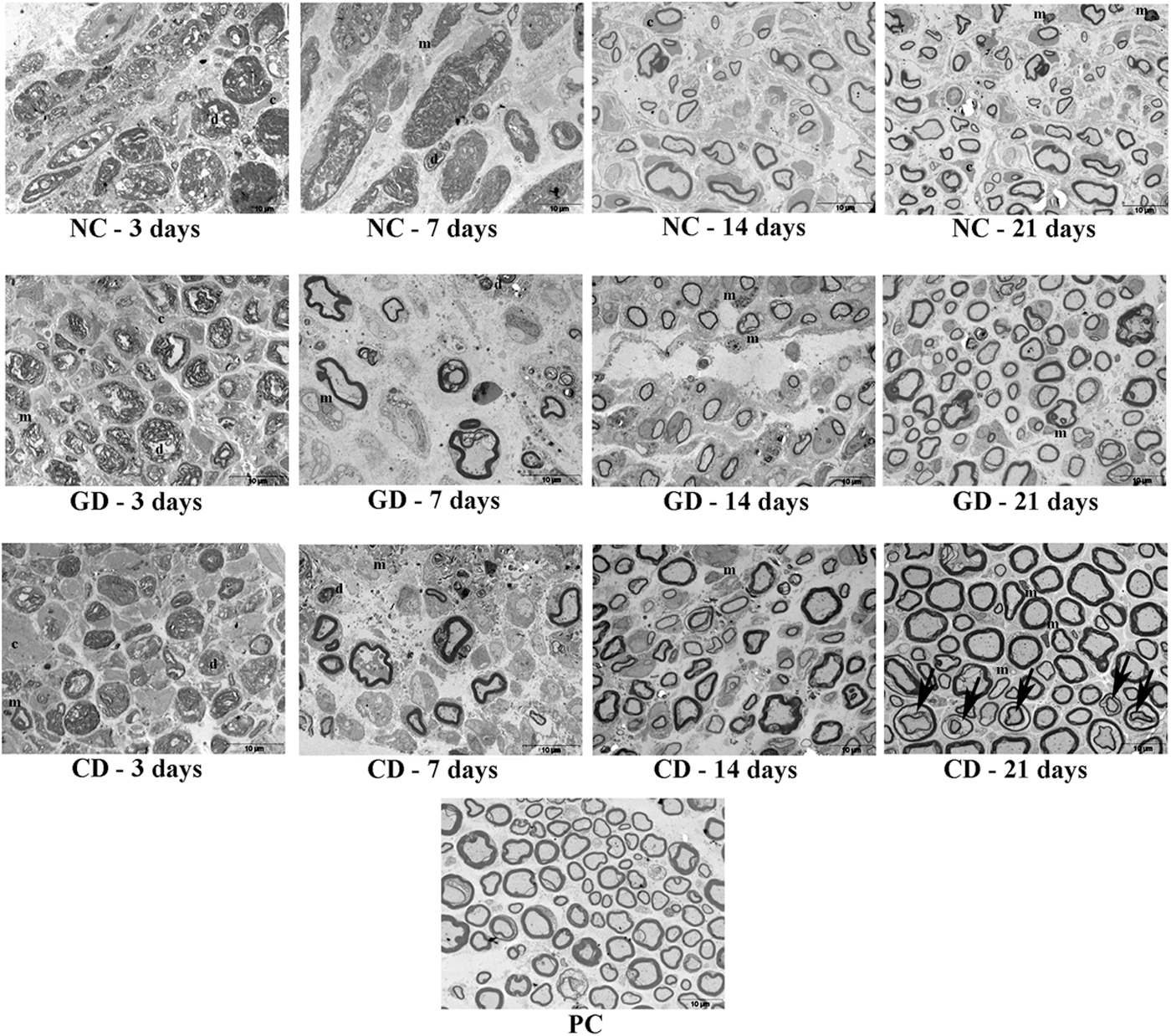
Cross-section TEM images of rats' mental nerve of groups PC, NC, GD, and CD on 3, 7, 14, and 21 days for ultrastructural analysis (20,000 × magnification). Black arrow, paranode; C, condensed connective tissue; CD, constant dose; d, axon degeneration; GD, gradual dose; m, macrophage; NC, negative control; PC, positive control; TEM, transmission electron microscope.
Ultrastructural morphologic analysis at 100,000 × magnification (Fig. 3) presented a better lamellar organization of myelin sheath at 7 days for GD and at 14 days for CD, similar to the patterns observed on PC. NC showed a large disorganization of lamellar structure in the first time span study, showing a similar pattern to PC only at 21 days. At the end of the treatment, there was no difference between CD or GD in this study.
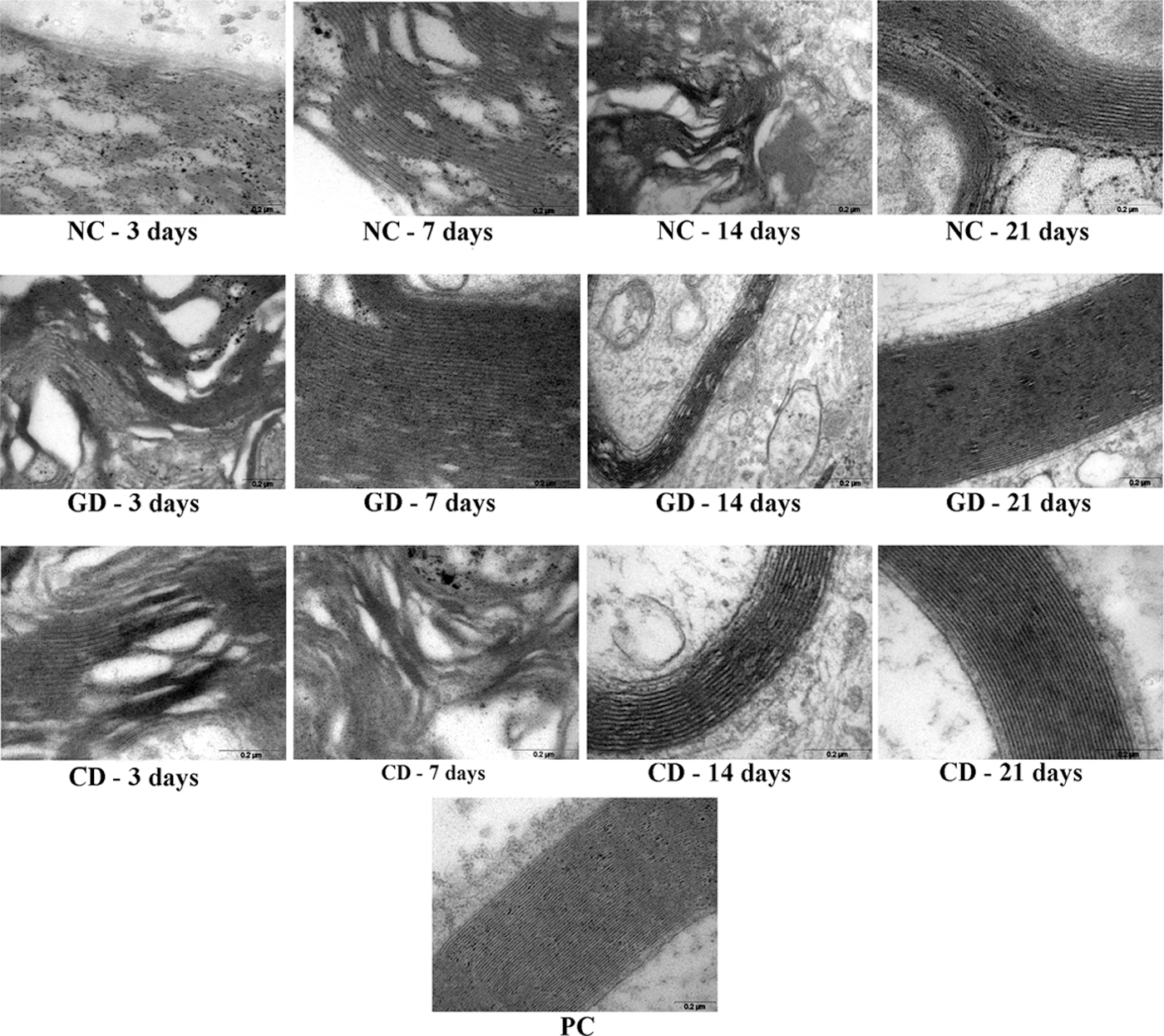
Cross-section TEM images of rats' mental nerve of groups PC, NC, GD, and CD on 3, 7, 14, and 21 days for ultrastructural analysis of lamellae of the myelin sheaths (100,000 × magnification).
Quantitative analysis
The myelin sheath thickness of the CD group at 7, 14, and 21 days was similar to PC and varied over time (Table 2). For the outer perimeter (Table 3), GD presented similar value at 3 days compared to PC. The number of myelinated fibers (Table 4) showed a significant increase starting at 7 days of treatment for group CD, which achieved statistically similar mean values at 14 days compared to PC. The means of internal area (Table 5) differed according to the time span studies, presenting the lowest average at 3 days for the CD, GD, and NC groups. After, at 7 days, groups GD and CD were similar to PC, which occurred again only for CD at 21 days. The G-ratio values were calculated and presented in Table 6. NC, GD, and CD presented an increase since day 7 and CD showed statistically similar values compared to PC at 7, 14, and 21 days. The mean in groups NC and CD was lower at 3 days compared to PC and GD.
Means followed by different letters (capital letters in horizontal and lower case letters in vertical) differ between them by ANOVA (p ≤ 0.05).
ANOVA, analysis of variance.
Means followed by different letters (capital letters in horizontal and lower case letters in vertical) differ between them by ANOVA (p ≤ 0.05).
Means followed by different letters (capital letters in horizontal and lower case letters in vertical) differ between them by ANOVA and Tukey test (p ≤ 0.05).
Means followed by different letters (capital letters in horizontal and lower case letters in vertical) differ between them by ANOVA (p ≤ 0.05).
Means followed by different letters (capital letters in horizontal and lower case letters in vertical) differ between them by ANOVA (p < 0.05).
Discussion
Lesions in peripheral nerves such as mental nerve may occur at the clinical practice in procedures such as infiltrative anesthesia and implant surgery. 4,5,21 Lesion by compression, classified as axonotmesis, is one of the most frequent in clinical practice and also on in vivo animal model studies, 7,9,22 justifying the injury by compression applied in this study.
Studies report the use of PBMT on neurological treatments like spinal cord or brain lesions and peripheral nerve regeneration in animal models and clinical trials on humans. 23 –25 This study used the GaAlAs laser, which is the most common laser device in dental offices and the most studied for nerve regeneration. 22
In the present study it was observed the presence of macrophages, which act by removing inhibitory substances associated with myelin and indirectly release cytokines and mitogen factors to Schwann and fibroblast cells, stimulating cellular repair. 26 Such structure may be responsible for the regeneration observed in group NC, showing that the nerve can regenerate by itself, but with a slower healing process than when treated with PBMT.
As in this study, Medalha et al. 10 also observed an increase in the density of myelinated fibers after laser irradiation of low level power with infrared spectrum (808 nm). Barbosa et al. 22 also reported after 14 days of treatment, a significant improvement in locomotion after functional evaluation in rats. However, even in the NC group, it was also observed an increase in the number of myelinated fibers over time, corroborating with the results presented by Gigo-Benato et al. 9,27 It can be explained by the endogenous immunoglobulin antibody that assist in the removal of axons in degeneration and contribute to axonal regeneration after injury by promoting entry of macrophages and their phagocytic activity. 28
Differences in internal area measurements can be explained by concomitant increase in the number of myelinated fibers, thereby reducing the interaxonal space and compacting the myelinated fibers in the perineurium. Moreover, the thickness measurements of the myelin sheath and outer perimeter differed between the groups, with the CD group showing similar values compared to PC starting at 7 days, corroborating with results obtained by Cantín et al. 29 The authors stated that a very low dose (5 J/cm2) led to the increasing density of the myelin sheath, the same related by Gigo-Benato et al. 9 that observed it with low and moderate doses.
For the outer perimeter, GD presented similar value at 3 days compared to PC, indicating that a low dose in the early stage of regeneration process may be indicated. According to Souza et al., 30 the mitochondrial activity of macrophages of the activated cells irradiated with PBMT decreased after 1 day of culture. After 3 days of culture, irradiation exerted a positive modulation of the mitochondrial activity of macrophages, which might denote an increase in cell activation. After 5 days of culture, laser irradiation no longer modulated the mitochondrial activity of the activated cells. In our study we observed that GD presented similar values of outer perimeter compared to PC, probably because the lowest dose used in the beginning of the experiment was more efficient to induce activity of macrophages than the dose used in CD, resulting in a quicker regeneration process. However, Medalha et al. 10 found that axon and fiber diameters were larger in animals irradiated with the highest dose compared to the control group. It can explain the differences between the groups CD and GD, in which the first one obtained higher values of myelin sheath thickness because it received the highest dose during all the experiment.
Our results indicated a positive effect of PBMT, both in CD and GD on mental nerve regeneration of rats after compression lesion, corroborating with several published studies. 7,9 –11,22,27,29 Comparisons among studies that evaluated the PBMT effects for peripheral nerve regeneration becomes complex due to the variety of treatment protocols used. The duration of treatment ranged from 10 days, 27 15 days, 10 and 21 days. 7,11,22 The wavelength ranged among 660 and 808 nm, 10 660 and 830 nm, 22 and 660 and 780 nm. 9 In addition, the type of laser used varied: Nd:YAG, 15 GaAlAs, 22 ArGaAl, 11 and AlGaInP 11 . Most of the studies assessed the sciatic nerve, 7,9 –11,22,27,31 followed by the inferior alveolar nerve 29 and the mental nerve. 20 As observed, there are several different parameters used in the literature, so in this study it was adopted as the parameters recommended by the manufacturer of the PBMT device for both CD or GD.
Despite the lack of optimal G-ratio for the mental nerve in the literature, the theoretical studies of Rushton 32 suggested that for a fixed outside diameter there should be an optimal myelin thickness for maximal conduction velocity. The arguments proposed that the G-ratio should have an optimal value at 0.6, maximizing conduction velocity, and that variation in G-ratio between 0.47 and 0.74 should result in a decrease of conduction velocity of not more than 5%. In addition, Chomiak and Hu 33 showed that an optimal G-ratio for central nervous system is around 0.77 and for peripheral nerves is around 0.6. However, Hodgkin 34 used similar analytic arguments to arrive at an optimal value of 0.7.
Most of literature studies adopted a CD use of PBMT, which is followed by most of the clinicians worldwide for the treatment of neural injuries. However, Karu 35 suggested that the exposure effects to PBMT are cumulative and that its clinical effectiveness has an intrinsic relationship with adequate energy doses, if applied correctly, gradually, and regularly. This relationship is due to the capacity of low doses or overdose does not produce effects or even generate damages. The choice for a GD group was due to the possibility of occurrence of a neuroplasticity of the affected tissue when it is applied with CD for a long period, in a way that a stimulus that initially generated positive effects does not generate the desired effects later. Thus, our intention was to compare morphological aspects between GD and CD, in a way to relate to clinicians if CD is the best protocol to be followed or if a GD could provide better stimulation of the nerve and should be considered.
Within the limitations of this animal model in vivo study, it was possible to observe that PBMT presented a positive effect in the regeneration of the mental nerve. Therefore, more experimental studies are needed, as well as more parameter standardization with the use of PBMT for the regeneration of peripheral nerves.
Conclusions and Summary
In this animal model in vivo study, based on quantitative and qualitative analyzes, it can be concluded that PBMT presented a positive effect on nerve regeneration of rats' mental nerve from 14 days after injury, and at 21 days there was no ultrastructural morphologic difference between treatments with GD or CD.
Footnotes
Acknowledgment
This study was supported by Coordenação de Aperfeiçoamento de Pessoal de Nível Superior (CAPES).
Author Disclosure Statement
No competing financial interests exist.