
Abstract
Introduction
W
At-home vital teeth bleaching is the most widely used technique; it stands out as a conservative and as relatively simple low-cost treatment. 1 Individual trays are used for the application of bleaching agents with 10–22% carbamide peroxide or 3–10% hydrogen peroxide on discolored teeth, promoting the chemical bleaching reaction by release of free radicals. 1 –5 Although this technique is a very common procedure in aesthetic dentistry, 1,2,6 the reported side effects from the chemical reaction are tooth sensitivity, 6 gingival pain, and surface morphological alterations. 2,7 Changes at the microscopic level were reported: damaging or denaturing protein components, the development of surface porosity, and erosion of tooth structure with increased surface roughness and decreased hardness. 2 –10
In addition, free radicals present in the enamel and dentin after bleaching treatment may be responsible for jeopardizing the adhesion to composite resins, by inhibiting the polymerization reaction due to the presence of free oxygen radicals in cases of replacement of esthetic restorations immediately after bleaching treatment. 11 –25
To obtain an adequate bond of esthetic restorations to bleached teeth, some studies recommend a waiting period of 1–3 weeks before final replacement. 12,13 During this period, the free radicals are naturally released from the dental structure. Several studies were conducted to test antioxidants agents such as catechins from green tea or sodium ascorbate to reduce the waiting period to bond procedures, 16 –20 but there is no standard treatment.
Lasers have been selected as an option to reverse the damage of dental bleaching in bonding procedures. 21 –25 Among dental lasers, the erbium and chromium-doped yttrium, scandium, gallium-garnet (Er,Cr:YSGG, λ = 2.78 μm) presents hydrokinetic characteristics that match the spectral range for absorption of water and hydroxyapatite, and when applied on dental hard tissues, it results in maximum absorption and complete transformation into thermal energy. 21,26 With minimal thermal damage to the surrounding tissues, minimal thermal-induced changes of dental hard tissue are produced after cavity preparation. 26
Since the thermal penetration is shallow, the Erbium lasers in non-ablative parameters result in the modification of dental tissues with reduced permeability, increased acid resistance and hardness without changing the subsurface. 23,26,27
In this context of laser irradiation, the energy absorbed by the dental tissues results in a temperature increase that may favor the rise of free radicals from the bleached tooth. 21,28,29 Er:YAG laser irradiation after bleaching has been described, 28,29 but limited studies have evaluated the effect of Er,Cr:YSGG on enamel. Thus, if free radicals are able to absorb the laser energy and therefore be completely eliminated from the enamel, it should be possible to get a bonding procedure in bleached teeth as effective as in non-bleached teeth. 22,24,25,29
Considering the increased use of dental bleaching and the need to replace restorations to obtain a good esthetic result, it is important to verify the conditioning with Er,Cr:YSGG laser and the effect of cavity preparation (abrasion) after bleaching treatment on tooth structure before composite resin bond.
As treatment with Er,Cr:YSGG laser after enamel bleaching and before adhesive restorative procedures simulating cavity preparation by abrasion have not been evaluated so far, the aim of this study was to investigate the effects of Er,Cr:YSGG laser irradiation on the microtensile bond strength (μTBS) on the interface restoration/enamel immediately after bleaching procedure and after a 14-day interval in non-abraded and abraded enamel.
Materials and Methods
Experimental design
The factors under study were “adhesion” after bleaching: immediate adhesion (B); after 14 days (14); and a control group (C) with adhesion on unbleached teeth; enamel “abrasion”: with (Ab) or without abrasion (NAb) simulating cavity preparation; “laser”: with (LA) or without (NLA) Er,Cr:YSGG laser irradiation (Fig. 1). The variable response was μTBS.
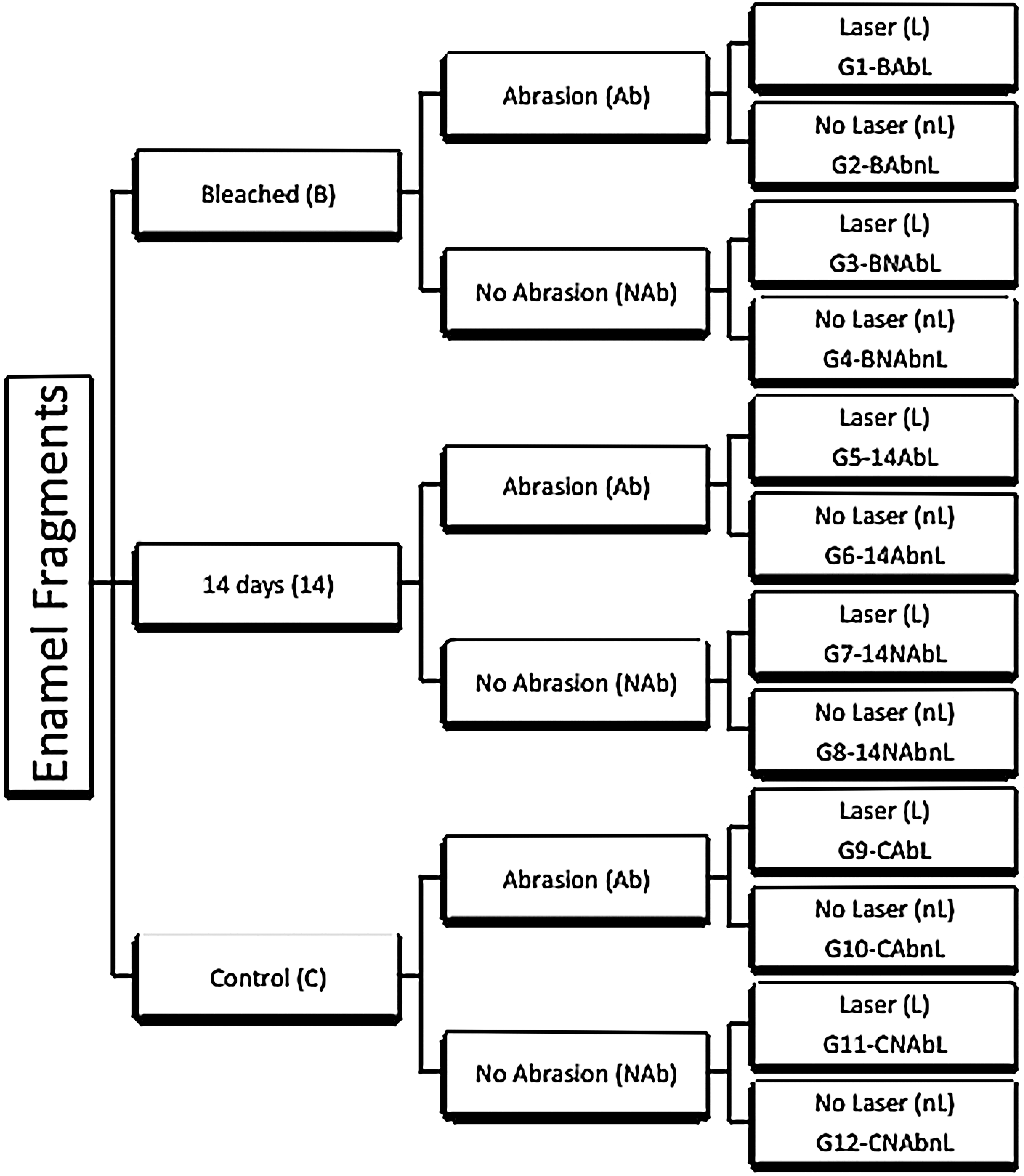
Experimental groups according to adhesion, enamel abrasion, and laser irradiation.
Specimen preparation
Two hundred twenty-eight bovine teeth were extracted and stored in 0.1% thymol solution at 4°C. Enamel from the buccal surface was cut with dimensions of 7 × 4 × 4 mm blocks with double-faced diamond disks (no. 7020; KG Sorensen, Barueri, Brazil) at low speed (Kavo, Joinville, Brazil) and under water cooling.
The buccal surface of enamel blocks was sequentially abraded with sandpaper (nos. 180, 320, and 600) in a polishing machine (LAB PL02 RB Com. Tecnica Ltda, São Paulo, Brazil) before the bond strength test was carried out.
Bleaching treatment
The enamel blocks of bleached groups (G1-BAbL, G2-BAbnL, G3-BNAbL, G4-BNAbnL, G5-14AbL, G6-14AbnL, G7-14NAbL, and G8-14NAbnL) were replicated with irreversible hydrocolloid (Jeltrate; Brazil Dentsply, Petrópolis, Brazil) and Type III Gipson (Asfer, São Caetano do Sul, Brazil) to obtain ethylene vinyl acetate with 0.4 mm-thick (EVA; Bio Art Dental Equipment Ltda, São Carlos, Brazil) individual trays for the bleaching treatment. After that, each enamel block was individually treated with 0.04 mL 11,20 of 20% carbamide peroxide (Opalescence 20%; Ultradent, Indaiatuba, Brazil) at 37°C for 8 h, followed by immersion in artificial saliva containing calcium and phosphate in a degree of saturation to mimic the remineralizing natural properties of human saliva (150 mmol/L potassium chloride; 1.5 mmol/L calcium; 0.9 mmol/L phosphate; 0.1 mmol/L trihydroxymethylaminomethane at pH 7.0), during 21 days, and renewed daily. 7,9,11,28,29
The unbleached groups and the groups restored 21 days after bleaching were stored during the period when the artificial saliva was renewed.
Enamel abrasion
After bleaching procedures, the abraded groups (G1-BAbL, G2-BAbnL, G5-14AblL, G6-14AbnL, G9-CAbL, and G10-CAbnL) had enamel surfaces abraded with silicon carbide sandpaper granulation 600 for 20 sec (Carborundum Abrasives Ltda., Valinhos, Brazil) by using a polishing machine (LAB PL02 RB Com. Tecnica Ltda) simulating the cavity preparation required for the removal of an unsatisfactory restoration. 28,29
Laser conditioning
The irradiation of the samples (G1-BAbL, G3-BNAbL, G5-14AbL, G7-14NAbL, G9-CAbL, and G11-CNAbL) was performed by using the Er,Cr:YSGG laser (Waterlase; Biolase Technology, San Clemente, CA) with a wavelength of 2.78 μm.
The handpiece with a sapphire tip was used for surface conditioning (type G4); a repetition rate fixed at 20 Hz, an average power of 0.5 W for 10 sec, and an air-water cooling proportion of 65%/55% were used for surface conditioning on the buccal surface of the enamel blocks. One single operator previously calibrated uniformly irradiated each enamel surface for 10 sec with manual grid movement. To standardize the working distance of 2 mm from the handpiece to the target surface, an endodontic K-file was fixed to the laser handpiece. The calculated energy density was 3.97 J/cm2.
μTBS test
After each treatment and respecting the adhesion period of time (“adhesion” factor), the enamel blocks were restored. The bleached groups G1-BAbL, G2-BAbnL, G3-BNAbL, and G4-BNAbnL were restored immediately after the bleaching procedure. The groups G5-14AbL, G6-14AbnL, G7-14NAbL, and G8-14NAbnL were restored 14 days after bleaching. The unbleached groups G9-CAbL, G10-CAbnL, G11-CNAbL, and G12-CAbnL were restored soon after immersion for 21 days in artificial saliva.
Restoration of the specimens was performed by using a two-step etch-and-rinse adhesive system (Single Bond II, 3M ESPE, St. Paul, MN) according to the manufacturer's instructions (Table 1). The enamel block surface was acid etched with 37% phosphoric acid for 30 sec, washed with water, and air dried. Then, the adhesive system was actively applied with a disposable brush (KG Brush; KG Sorensen, Cotia, Brazil). The excess of solvent was removed with a gentle stream of air; the adhesive was cured for 20 sec with a light-emitting diode (LED) curing unit average of 1411 mW/cm2 (Radii Plus-SDI Limited, Bayswater, Victoria, Australia).
Bis-EMA, bisphenyl glicol polyetilen diethil dimethacrylate; Bis-GMA, bisphenylglycidyl dimethacrylate; DMA, dimethacrylate; HEMA, 2-hydroxyethylmethacrylate; UDMA, uretane dimethacrylate.
After the hybridization process, the enamel was restored with 3 layers (±1.5 mm) of a microhybrid resin composite (Filtek Z 250, 3M ESPE, St. Paul, MN; shade A2) according to the manufacturer's instructions (Table 1), and it was light cured for 20 sec each. 28,29
The restored specimens were kept in 100% humidity at 37°C for 1 week. 28,29 After that, the enamel blocks were fixed onto an acrylic plate that was attached to a metallographic cutter (1000 Isomet; Buehler Ltd., Lake Bluff, IL) and sectioned under water cooling to obtain beans with ∼1 mm2 of interface bond area between composite resin and enamel. The beans were bonded to a microtensile testing jig by using a cyanoacrylate adhesive (Loctite Super Gel Bond, Henkel, Düsseldorf, Germany). 28,29
The jig was attached to the universal testing machine (EZ Test; Shimadzu Corp., Kyoto, Japan), and it was tested at a speed of 0.5 mm per minute until fracture. After fracture, the sticks had their cross-sectional area measured with a digital caliper (Mitutoyo Co., Tokyo, Japan) and the data were converted individually to megapascal (MPa) according to the cross-sectional area of each specimen. Five beans of each specimen were tested, and the average was used for statistical analysis. 28,29
The fracture mode was classified into three types: cohesive in enamel, cohesive in resin, and cohesive in adhesive (AD) at 100 × magnification by using a stereoscope (Pantec, Panambra Ind. Tecnica SA, Sao Paulo, Brazil). 28,29
Statistical analysis
The distribution of μTBS data was evaluated by Levene test, statistically analyzed by running a three-way analysis of variance at the 95% confidence level, and compared by Tukey post hoc test (α = 0.05). The data of each specimen obtained from each group were used as the value of bond strength of the experimental unit. The factors under study were “adhesion,” “abrasion,” and “laser”; and their interactions. The fracture mode was expressed in percentage by descriptive analysis.
Results
The raw data were characterized by a normal distribution using Levene test (α > 0.05). All specimens were used in statistical analysis, even the ones presenting cohesive failures. Table 2 presents the mean values of μTBS of each experimental group and the standard deviations. There was no statistically significant difference for the triple interaction (p = 0.30) and double interactions among factors Laser*Adhesion (p = 0.79), Laser*Abrasion (p = 0.60), and Abrasion*Adhesion (p = 0.66). There was no significant difference between the factors “adhesion” (p = 0.08) and “abrasion” (p = 0.44). A statistically significant difference was observed in factor “laser” (p = 0.045); laser irradiation resulted in significantly lower values of bond strength (Table 2). Regardless of adhesion interval time or abrasion, the Er,Cr:YSGG laser parameters evaluated in the present study impaired bond strength results.
Means followed by different capital letters in the column indicate statistical difference (p < 0.05).
Ab, abrasion; B, bleached; C, control; L, laser; MPa, megapascal; NAb, no abrasion; nL, no laser; SD, standard deviation.
It was observed that adhesive fracture was the predominant pattern (Fig. 2), but all groups presented cohesive failure in enamel and resin. The high percentage of cohesive failures was observed in G1-BAbL, G2-BAbnL, and G10-BNAbnL groups.
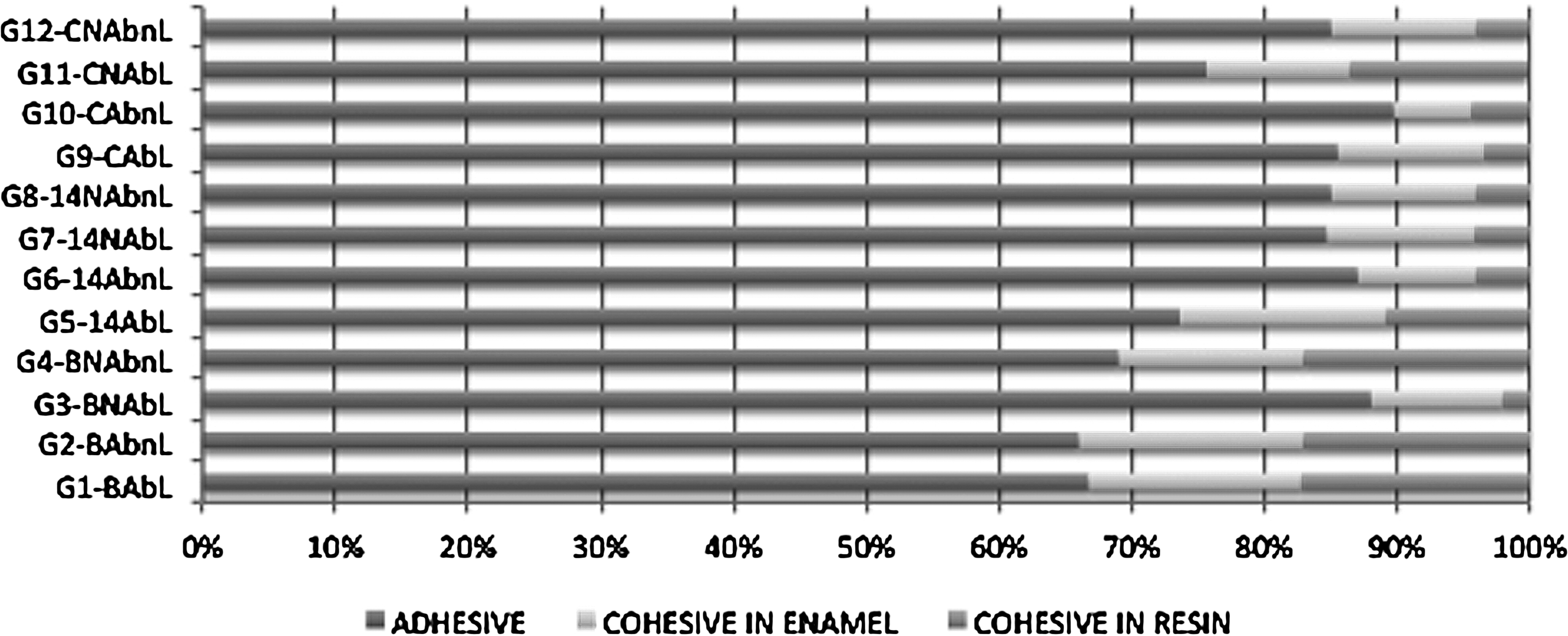
Distribution of failure modes within groups adhesion (B, bleached; 14–14 days; C, control), enamel abrasion (Ab, abrasion; NAb, no abrasion), and laser irradiation (L, laser; nL, no laser).
Discussion
Lower bond strength values of adhesive systems to bleached enamel have been reported due to oxygen-free radicals that interfere in the polymerization reaction, causing a reduction in the ability to form polymers and tags. 11 –25 Teeth are bleached by free radicals that are released from carbamide peroxide that breaks down into hydrogen peroxide. Instead of waiting for several weeks for the natural reversion of bleaching agents' detrimental effects, some strategies were proposed, such as the use of antioxidant agents sodium ascorbate, baking soda, and green tea; catalase; and the use of laser energy to remove free radicals of oxygen. 16 –25
The bleaching protocol used in this study did not jeopardize bond strength values. The bleaching agent 20% carbamide peroxide was applied for 8 h followed by immersion in artificial saliva for 21 days. Previous studies also found no influence of bleaching with 16% carbamide peroxide for 6 h during 21 days 28,29 or after 42 days of treatment with concentrations varying from 10% to 22% carbamide peroxide. 13 The 8 h home vital teeth bleaching with 20% carbamide peroxide protocol was used. 13 Although this concentration is indicated for 4–6 h of treatment, some patients prefer using the tray during sleep time and treatment could be extended to 8 h. The 8 h teeth bleaching did not produce decreased bond strength values in the present study.
Thus, detrimental effects reported by some studies may be the effect of continuous treatment with bleaching agents simulating vital and non-vital bleaching techniques. 11,30,31 At this point, it must be considered that an interval of time promoted by immersion in artificial saliva among bleaching exposures must be considered to be used in new studies, since patients are not allowed to apply bleaching gels all day long.
Perdigão et al. observed that 10% carbamide peroxide resulted in morphological alterations on the most superficial enamel crystallites. 32 Other studies also observed that after bleaching treatment enamel roughness 5,6 was increased and hardness was reduced, 2,7 –9 suggesting mineral loss. Thus, the abrasion of enamel directly exposed to bleaching agents could improve bonding procedures. However, the abrasion of enamel did not affect bond strength values and is in agreement with Leonetti et al. 28,29 Although this abrasion does not seem to be necessary, it is inherent to a composite restoration replacement. The artificial saliva is used as a remineralizing solution and could also favor the maintenance of a sound enamel surface to be acid conditioned.
The treatment with Er,Cr:YSGG laser aimed at conditioning the bleached enamel surface to become an enamel with minimal free radicals enhancing bond strength. Since free radicals released from the hydrogen peroxide molecules are very unstable, the Er:YSGG laser could heat enamel and accelerate their elimination. However, bleaching procedure did not affect the bond strength and the laser treatment jeopardized bond strength. Firat et al. showed that laser treatment with Er:YAG could both enhance and impair the μTBS to enamel and dentin depending on the pulse duration used and additional acid application. 21 The present study selected 3.97 J/cm2 Er,Cr:YSGG laser irradiation, and no visual enamel damage was observed. Er;YAG laser irradiation (4.42 J/cm2) before the adhesive procedure of teeth bleached with 16% carbamide peroxide showed no evidence of enamel ablation under scanning electron microscopy (SEM). 28
Lago et al. observed that Er:YAG irradiation of bleached surfaces may favor bonding procedures when performed 24 h after bleaching. 24 On dentin, Curylofo et al. observed that Er:YAG laser (20 sec, 400 mJ, 15 Hz) can restore the bond strength of the dentin treated with 38% hydrogen peroxide even if the restorative procedure is performed immediately after bleaching. 25 The same results were shown by Leonetti et al. 29 Er:YAG laser irradiation (25.56 J/cm2) before the adhesive procedure of teeth bleached with 16% carbamide peroxide did not affect μTBS on dentin and produced a dentin surface with no smear layer or open dentin tubules when observed under SEM. However, Er:YAG laser irradiation before the adhesive procedure of the non-bleached surface impaired μTBS compared with the control group. Mohammadi Bassir et al. 22 observed that treatment with other lasers can produce different effects on enamel. The surface treated by Nd:YAG laser was melted and recrystallized, whereas the surface treated by Er:YAG laser showed an irregular and a microporous surface with a flake pattern. Carbon dioxide laser resulted in a melting area and cracks. 32
Enamel surface abrasion tried to simulate cavity preparation with dental burs during composite restoration replacement. In the present study, the purpose of the enamel abrasion was to create a clinical condition similar to restoration replacement. As the objective was to treat enamel without its removal, the laser protocol was chosen to be non-ablative. However, non-ablative parameters are generally used in sound enamel to improve caries resistance and may jeopardize enamel acid conditioning. As this parameter was not evident to improve enamel bond strength, more studies are needed to test other parameters, including different powers, pulse repetition, and water irrigation.
To the best of our knowledge, there are no similar studies analyzing the influence of Er,Cr;YSGG laser irradiation in subablative energy density on the μTBS to enamel after dental bleaching.
Conclusions
Abrasion provided no benefit to improve the bond strength of composite resins to bleached enamel; treatment with the Er,Cr:YSGG (20 Hz, 0.5 W, 3.97 J/cm2) reduced the bond strength of composite resins to enamel.
Footnotes
Acknowledgments
This study was supported by Coordination for the Improvement of Higher Education Personnel (CAPES).
About the Authors
Dr. Oliveira has a PhD degree in Operative Dentistry, and Dr. Tenorio has a Master's degree in Operative Dentistry, Department of Restorative Dentistry, School of Dentistry, Guarulhos University, Brazil. Dr. Cassoni and Dr. Rodrigues are professors of the Dental Research and Graduate Studies Division, Department of Restorative Dentistry, School of Dentistry, Guarulhos University, Brazil. Dr. Brugnera Jr. is professor of the Biomedicine Engineering Division, Camilo Castelo Branco University, Brazil.
Author Disclosure Statement
No competing financial interests exist.