
Abstract
Introduction
T
The importance of root canal chemical irrigation has been firmly established since mechanical root canal instrumentation alone leaves 35% of the root canal walls intact. 3 Endodontic space disinfection depends not only on the type of root canal irrigant used but also on the way it is delivered and agitated in the root canal because irrigants have limited ability to reach all parts of root canal system. 4 The efficacy of irrigants in root canal disinfection has been improved with different agitation techniques using hand files, sonic, ultrasonic, and laser devices. 4
Bactericidal effect of laser irradiation occurs due to local heating inside the bacteria after absorption of laser light by bacterial pigments and laser light absorption by the substrate in which the bacteria are located, resulting in increased temperatures lethal for bacteria and photodamage (photochemical processes leading to the creation of reactive chemical species interfering with normal metabolic processes). 5 Besides their direct bactericidal effect, lasers could improve root canal disinfection and smear layer removal by producing intense streaming within an irrigant, which enhances penetration of irrigant into apical ramifications. 6 –9
The wavelength of the Nd:YAG laser (1064 nm) gets absorbed by bacterial pigments, while nonpigmented bacteria were shown to be transparent to near-infrared wavelength, suggesting that bactericidal effect of Nd:YAG laser on nonpigmented bacteria occurs through heating of the environment of bacteria. 5 The wavelengths of middle infrared lasers such as Er:YAG and Er,Cr:YSGG (2940 and 2790 nm, respectively) are very well absorbed by water and hydroxilapatite, resulting in heating in the bacterial surroundings within the infected root canal. 6 In the case of persistent periapical lesions due to microbial resistance to routine endodontics, such as the case with E. faecalis and C. albicans, laser energy increases efficacy in reduction of intraradicular microorganisms and success rate of root canal retreatments. 10
By using nonantimicrobial irrigant such as saline, it is possible to estimate purely physical effect of laser-activated irrigation (LAI). The purpose of this in vitro study was to evaluate the efficacy of Nd:YAG, Er:YAG, and Er,Cr:YSGG lasers in decontamination of root canal systems inoculated with E. faecalis and C. albicans and irrigated with saline.
Materials and Methods
This study was approved by the Ethics Committee of the School of Dental Medicine, University of Zagreb. Thirty extracted single-rooted human teeth were used (incisors and canines). Teeth with caries and fractures were excluded from the sample, and selected teeth were stored in 0.5% (v/v) chloramine solution until use. Before instrumentation, the teeth were curetted to remove periodontal ligament and calculus, and further stored in saline. The crowns were sectioned below cement–enamel junction using water-cooled diamond drill, and the length of roots was standardized to 16 mm. K-file of a size #15 (Dentsply Maillefer, Ballaigues, Switzerland) was put into each canal until it was just visible at the apical foramen, and working length was determined by withdrawing the instrument 1 mm. The root canals were instrumented with rotating instruments (ProTaper, Dentsply Maillefer, Ballaigues, Switzerland) to size F3. Rotation speed was 300 rpm and torque was adjusted according to the manufacturer's instructions. Glyde root canal lubricant (Dentsply Maillefer, Ballaigues, Switzerland) was used during instrumentation. After the use of each file, each canal was irrigated with 2 mL of 2.5% sodium hypochlorite using a disposable syringe and a needle, No. 27-gauge needle (BD Microlane 3; BD, Drogheda, Ireland). To remove smear layer, root canals were rinsed with 2 mL of 17% ethylenediaminetetraacetic acid (EDTA) (pH 7.7), each for 1 min. Final irrigation of all samples was carried out with saline, after which the canals were dried using ProTaper paper points, size #30 (Dentsply Maillefer, Ballaigues, Switzerland).
Apical foramen of each specimen was sealed using composite resin material (G-aenial, GC, Tokyo, Japan) and the root surface was sealed using adhesive resin (G-aenial Bond, GC; Tokyo, Japan). The specimens were placed into 1.5-mL Eppendorf tubes (Eppendorf, Hamburg, Germany). All prepared specimens were sterilized in autoclave at 121°C for 20 min.
Suspension for inoculation was prepared by mixing pure cultures of E. faecalis and C. albicans, which were grown on blood agar for 24 h, with 2 mL of saline [0.9% (w/v) NaCl]. The turbidity of suspension was 0.5 McFarland units (Densimat, BioMerieux, l Etoile, France). Root canals were filled with 10 μL of suspension with sterile insulin syringe and a needle. The teeth were incubated at 37°C at 100% humidity for 7 days. During the inoculation period, the root canals were reinoculated with fresh bacterial and C. albicans suspension every 2 days (Fig. 1). After incubation, the sample was taken with a sterile paper point from each root canal and planted on the blood agar plate to determine the number of E. faecalis and C. albicans colony-forming units (CFUs) before the irrigation and disinfection protocols. The colonies of C. albicans and E. faecalis were differentiated based on their morphology (Fig. 2).
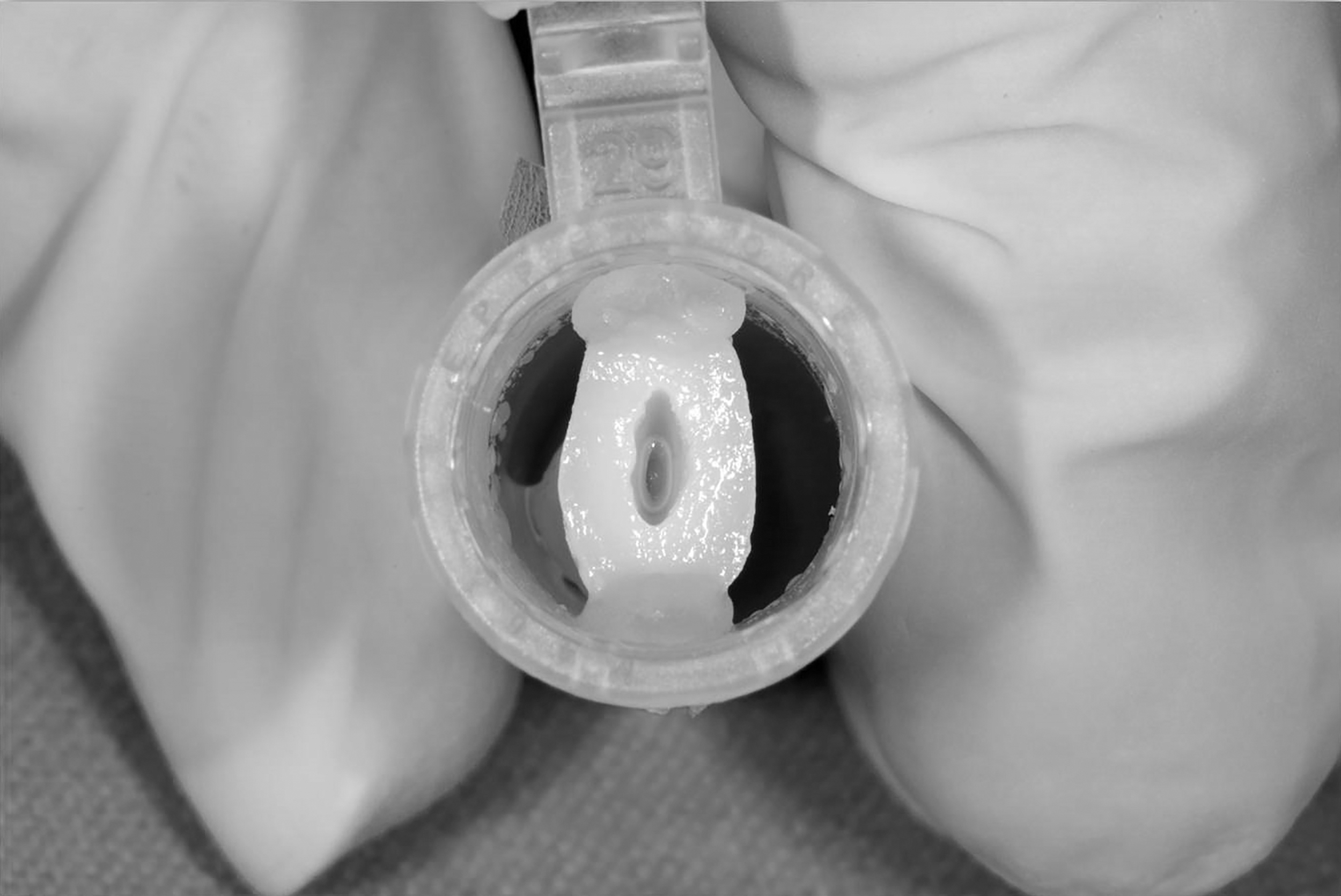
Sterile specimens fixed in Eppendorf tubes were inoculated with fresh suspension of Enterococcus faecalis and Candida albicans every 2 days.
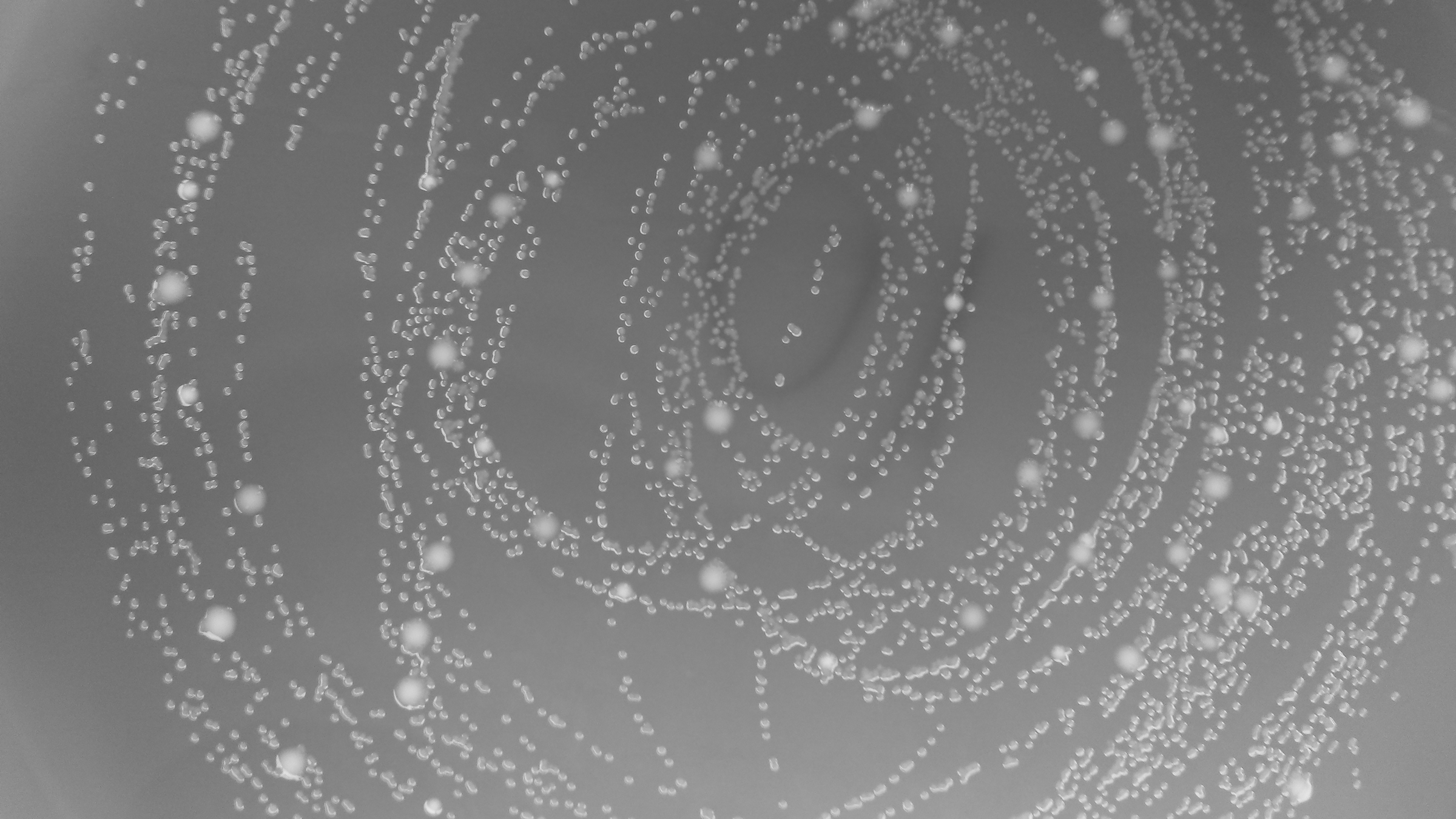
Blood agar plate with colony-forming units of E. faecalis and C. albicans colonies. The colonies of C. albicans are bigger than colonies of E. faecalis.
The teeth were randomly divided into three experimental groups (n = 10). In the first experimental group, so-called photon-induced photoacoustic streaming (PIPS) protocol was applied (Fig. 3). Er:YAG laser (LightWalker, Fotona, Ljubljana, Slovenia) was used with 14-mm-long 400 μm diameter tapered tip and with 4 mm of the polyamide sheet stripped back from the end. Pulse energy of the laser was 20 mJ, power 0.3 W, pulse duration 50 μsec, and frequency 15 Hz. The laser was used for 40 sec with 5 mL saline injected into the root canal using disposable syringe and a needle, No. 27-gauge needle (BD Microlane 3; BD, Drogheda, Ireland). The laser file tip was placed into the coronal access opening. In the second group, Nd:YAG laser (LightWalker, Fotona, Ljubljana, Slovenia) was used with R23 (200 μm in diameter), medium short pulse (MSP) mode (100 μsec), power 1.5 W, and frequency 15 Hz. The fiber-optic beam delivery tip was moved slowly (2 mm/sec) in circular spiral-forming movements from the apical to the coronal part. This was done four times for each root canal. In the third experimental group, Er,Cr:YSGG laser (Waterlase, Biolase, Irvine, CA) with radial firing tips RTF 2 (200 μm) was used in combination with 5 mL of saline, and pulse duration was 150 μsec. The power was set to 1.25 W, frequency 15 Hz, with 34% of air and 24% water levels. Laser tip was positioned 5 mm apically from the coronal access. After different disinfection protocols, the number of E. faecalis and C. albicans CFUs was determined for each root canal in the same manner as previous disinfection protocols: the samples from root canals were taken with sterile paper points and planted on the blood agar plate. They were incubated at 37°C for 24 h.
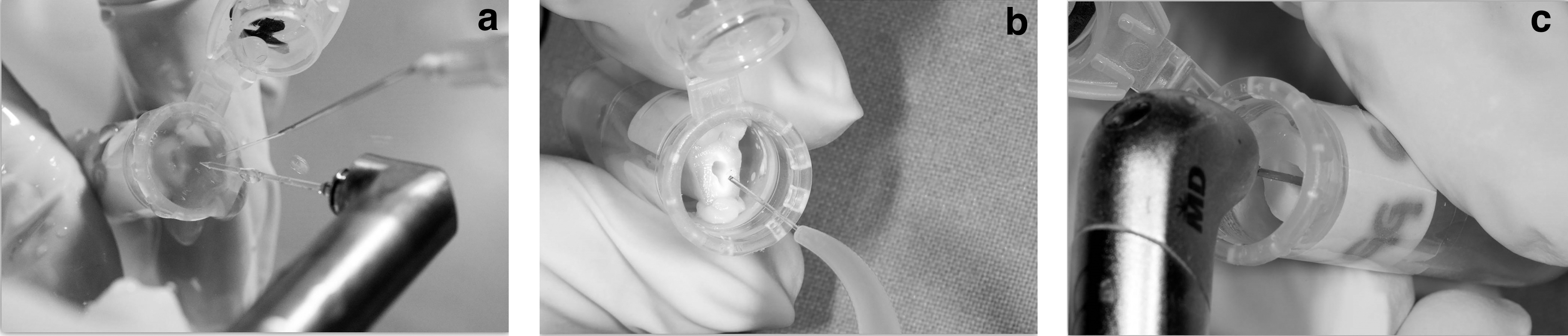
Different root canal disinfection protocols using saline and irradiation with
Results were statistically processed in SPSS version 16.0 (IBM, Chicago, IL) using t-test (paired samples and independent samples tests) with 5% level of significance (p < 0.05).
Results
Of the three lasers tested, Er,Cr:YSGG showed best results in terms of reduction of the number of bacterial colonies of E. faecalis and C. albicans in root canals. The disinfecting efficacy of Er,Cr:YSGG laser was significantly better in comparison with Er:YAG and Nd:YAG lasers (p < 0.05) (Table 1). Er:YAG laser showed higher efficacy in eradication of microorganisms from root canals in comparison with Nd:YAG laser (p < 0.05) (Table 1).
Different superscript letters refer to statistically significant difference between experimental groups.
CFU, colony-forming unit, SD, standard deviation.
Discussion
The aim of our study was to test the effect of three different lasers on reduction of bacterial load organized in biofilms. The biofilm model was considered better than liquid with suspended planctonic microorganisms, since bacteria in biofilms are more resistant, and is more similar to the actual situation in vivo. 11 Further, during conventional chemomechanical debridement, a significant proportion of the root canal wall remains untouched, and biofilm on the root canal wall presents a reservoir of microorganisms potentially compromising endodontic treatment. 3,12 Intraradicular infection has been recognized as the most prevalent endodontic treatment failure, and microbial species reported to be found in such cases are resistant and fewer than microorganisms in primary infections. 1,2
The biofilm in our study was formed during a period of 7 days. There are relevant recent studies where the incubation period lasted only 48 h. 13 It has been reported that microorganisms in mature biofilms that are 3 weeks old were more resistant to antimicrobial therapy than the bacteria in biofilms 2 weeks old or less. 14 This categorizes the biofilm in our study as young biofilm, but since the thickness of the biofilm continually grows, the biofilms of 2 and 7 days are expected to behave differently and cannot be compared. According to the available literature, there is no unequivocally accepted protocol concerning the development of biofilm model in in vitro or ex vivo studies, and the simultaneous inoculation of a single root canal with two microorganisms has not been used previously. 15
Saline is not considered an appropriate endodontic irrigant because it is not effective in dissolving either organic or inorganic material. 16 Saline syringe irrigation protocol was shown to be inferior to NaOCl irrigation, either syringe or laser activated. 7 Nevertheless, it was reported that the removal of smear layer and antibacterial effect of saline in root canal significantly improved upon laser activation when compared with syringe irrigation alone. 15,17 Protocols using saline, even with laser activation, were reported to be clinically insufficient. On the other hand, NaOCl is a disinfecting agent, and when used as an irrigant in endodontics, it reduces intracanal infection through chemical action, but if additionally activated using laser energy, the turbulent motion of irrigant contributes much to the antimicrobial effect. 7,15 Besides, it was reported that laser activation may increase chemical reaction kinetics of NaOCl. 18 The use of saline in our study enabled us to test only the physical effect of LAI on the reduction of microorganism load in the root canal.
The results of our study indicate that middle infrared lasers Er:YAG and Er,CR: YSGG are more effective in root canal disinfection than Nd:YAG laser. It has been reported that both Er:YAG and Er,Cr:YSGG lasers create effects within the irrigant, enhancing apical debris and smear layer removal. 8,9,17,19,20 Further, Er,Cr:YSGG performed better than Er:YAG, which was used in a PIPS protocol. 17 We can assume that better performance of Er,Cr:YSGG laser compared with Er:YAG was due to the laser tip positioned more apically. We can make this assumption based on previous findings that the apical fluid pressure was significantly higher when the laser tip was positioned 5 mm apically than when it was just at the coronal access orifice. 20 The most intense fluid motions in laser activation are located near the fiber tip, 8 and this could explain why more apical placement of the fiber tip results in better decontamination. It was also reported that the apical pressure increased as the laser power increased. 20 The power of Er.Cr:YSGG was set at 1.25 and 0.3 W for Er:YAG laser.
With Nd:YAG laser, the reduction of bacterial load in root canals was insignificant. This poor performance of Nd:YAG in root canal disinfection can be attributed to the fact that the wavelength of 1064 nm does not get absorbed by unpigmented bacteria such as E. faecalis. 5,21 Although the effect on C. albicans colony reduction was somewhat better, it was also low, and this is in accordance with the study by Meire et al. 22 Because the Nd:YAG wavelength does not get absorbed well, neither by the microorganism used in the study nor by the surroundings of the biofilms (dentine and water), we can speculate that the only way for the Nd:YAG to exhibit antimicrobial effect would be through the increase of temperature beyond lethal temperature for E. faecalis and C. albicans. This would surely imply higher laser energy output. However, this can be harmful for the tooth structures in in vivo conditions.
On the other hand, both erbium lasers used in our study have the highest absorption in water and hydroxyapatite. 23 Since it is known that biofilms have high water content, 24 it is expected that the wavelength of these lasers gets well absorbed in biofilms and exhibits antimicrobial effect at relatively low energy density, as already reported, especially with gram-negative bacterial species. 25 This explains why both lasers had significantly better antimicrobial effect than Nd:YAG laser in our study as well as in previous studies. 22,26 –29 Nevertheless, almost every study had a unique irrigation protocol, and it is hard to compare the results.
The wavelengths of Er:YAG and Er,Cr:YSGG lasers are readily absorbed not only by water in biofilms and hydroxilapatite but also by commonly used solutions in root canal irrigation. Commonly used irrigants such as NaOCl, chlorhexidine, EDTA, and citric acid have absorption peaks around 1450 nm and above 2500 nm. 30 This makes Er:YAG emitting at 2940 nm and the Er,Cr:YSGG laser emitting at 2780 nm suitable for LAI due to cavitation phenomenon. 19
In our study, we could not expect an antimicrobial effect due to the chemical action of irrigant, therefore the reduction in microbial count can be attributed purely to the mechanical effect of rapid fluid movement. The De Meyer et al. 13 study resulted in an interesting finding revealing that better biofilm removal was achieved when saline was activated than when NaOCl was activated, although overall microbial reduction was better with activated NaOCl. The results of some recent studies suggest a positive bactericidal effect of PIPS of irrigants, generated by a pulsed Er:YAG laser. 15,31 This agrees with our findings. Pedulla et al. 15 reported on significant difference between root canal samples syringe irrigated with bidistilled water compared with the samples where bidistilled water was laser activated (PIPS). The results of study by De Meyer et al. 13 are in accordance with the results of our study. In the part of their experiment where they did not use antimicrobial agent for irrigation, but irrigated canals with saline, reduction of microbial counts was the highest when saline was activated with pulsed Er:YAG laser compared with syringe irrigation and ultrasound-activated irrigation. In fact, syringe irrigation resulted in insignificant reduction of bacterial counts. This better efficacy of LAI compared with ultrasound-activated irrigation and syringe irrigation was explained by turbulent action of irrigant within the canal after activation. 13
The results of our study clearly indicate that erbium laser-activated irrigation significantly improves antimicrobial action against E. faecalis and C. albicans biofilms in vitro, probably through physical effect of rapid irrigant movement and direct biofilm irradiation. Low power outputs and optimal absorption of these lasers' wavelengths make them more suitable for root canal disinfection in endodontics than Nd:YAG laser. When comparing all lasers used in the present study, Er,Cr:YSGG laser was the most efficient in eradication of E. faecalis and C. albicans biofilms.
Summary
The objective of this study was to compare the efficacy of three different lasers in disinfection of root canals inoculated with E. faecalis and C. albicans biofilms. Thirty single-rooted human teeth were inoculated with E. faecalis and C. albicans. After 7 days of incubation period, the number of E. faecalis and C. albicans CFUs was determined for each root canal. Laser-assisted root canal disinfection (Nd:YAG, Er:YAG, Er,Cr:YSGG) was performed. After different root canal disinfection protocols, the number of E. faecalis and C. albicans CFUs was determined again for each root canal. Er:YAG and Er,Cr:YSGG laser eradication was significant (p < 0.05), while Nd:YAG laser irradiation did not result in statistically significant reduction (p > 0.05). Er,Cr:YSGG laser was the most efficient tool in eradication of E. faecalis and C. albicans biofilms.
Footnotes
Author Disclosure Statement
No competing financial interests exist.