
Abstract
Introduction
B
Laser systems, especially those using erbium lasers, are able to create surface irregularities similar to those of acid-etched surfaces and have been widely studied since 1964. 3,4 Laser irradiation causes thermal changes of enamel surfaces to a depth of 10–20 μm, depending on the type of laser and the energy delivered. Irradiation of enamel surfaces occurs through a process of continuous microexplosions due to the vaporization of water trapped within the hydroxyapatite matrix. 5
The literature reveals abundant information about shear bond strength (SBS) and adhesive remnant index (ARI) scores for etching enamel surfaces with Er:YAG or Er,Cr:YSGG laser systems. 6 –9 In general, laser energy is delivered to the area to be etched manually using a handpiece in a sweeping motion. A novel digitally controlled Er:YAG laser handpiece (X-Runner, Fotona, Ljubljana, Slovenia) that is similar to a standard noncontact Er:YAG laser handpiece has been found to be unique in its ability to digitally control the size, shape, and depth of irradiated enamel areas.
To date, the laser etching research includes studies on power output, 5,10 pulse settings, 11 and various surface applications. 12 However, there are no data about the effects of Er:YAG laser etching of enamel surfaces using an X-Runner handpiece. Therefore, the aim of this study was to compare the SBS values, enamel surface characteristics, and ARI scores of acid etching and manual or digitally controlled laser irradiation of enamel surfaces. For the purposes of the study, the null hypothesis assumed that different methods of enamel etching would not result in any statistically significant differences in the parameters evaluated.
Materials and Methods
Ethical approval was obtained from the Clinical Research Ethics Committee of Bezmialem University in Istanbul, Turkey (No. 71306642/050-01-04/208). Ninety-eight human maxillary premolar teeth without caries, restorations, surface malformations, or cracks were used in this study. Samples were stored in distilled water at room temperature and changed weekly to prevent bacterial growth. Cold-curing acrylic was used for embedding the teeth vertically in plastic ring molds. The buccal enamel surfaces of the teeth were cleaned and polished for 20 sec using a rubber cup and a slurry of pumice and water. Eighty of the 98 teeth were randomly divided into 4 experimental groups of 20 teeth each for SBS testing. To observe the changes in etched and debonded, restored enamel surfaces, 16 teeth were divided into the same four experimental groups and scanning electron microscopy (SEM) and atomic force microscopy (AFM) scans were taken after etching and following debonding/restoration. The remaining two samples were not etched and served as controls (Table 1).
AFM, atomic force microscopy; ARI, adhesive remnant index; SBS, shear bond strength; SEM, scanning electron microscopy.
Group 1: enamel surfaces were conditioned with liquid 37% phosphoric acid etchant (Liquid Etchant, Reliance Orthodontic Products, Inc., Itasca, IL) for 30 sec, rinsed with water for 20 sec, and dried for 15 sec.
Group 2: enamel surfaces were manually irradiated using an Er:YAG laser handpiece (2940 nm wavelength; LightWalker, Fotona) for 15 sec (120 mJ, 10 Hz, 1.2 W, 40% water, and 50% air) in medium short pulse (MSP) mode. A contact-type handpiece was held perpendicular to the enamel surface at a distance of 1 mm. The beam spot size was 0.0132665 cm2, the power density was 90.4 W/cm2, and the energy density was 9.04 J/cm2.
Group 3: enamel surfaces were irradiated using an Er,Cr:YSGG laser handpiece manually (2780 nm wavelength; Waterlase MD, Biolase Technology, Inc., Irvine, CA) for 15 sec (MGG 6 laser tip, 45 mJ, 50 Hz, 2.25 W, 30% water, and 60% air). Laser settings were 45 mJ, 50 Hz, 2.25 W, 30% water, and 60% air according to the manufacturer's recommendation for laser etching on enamel surface (MGG 6 laser tip). The beam spot size was 0.002826 cm2, the energy density was 15.92 J/cm2, and the power density was 795.77 W/cm2. A contact-type handpiece was held perpendicular to the enamel surface at a distance of 1 mm.
Group 4: A new Er:YAG laser with an X-Runner handpiece (2940 nm wavelength; LightWalker, Fotona) that is based on digitally controlled scanner technology and able to irradiate a precise area in advanced program was used. The following settings can be changed on the touch screen control panel of the laser device: • the shape of the area to be irradiated (circular, rectangular, or hexagonal); • the size of the area to be irradiated (the width and length of the rectangle shape and the diameter of the circle and hexagon shapes); • the number of scans (a function for the requested ablation depth); and • the pause duration between sequential scans.
The enamel surfaces were irradiated using an X-Runner handpiece (2940 nm wavelength, 100 mJ, 10 Hz, 1 W, 40% water, and 50% air) in MSP mode. The size and shape of the ablation area were determined to be 5 × 5 mm and rectangular. Two vertical scans and two horizontal scans were performed to obtain a homogenously ablated enamel surface. The distance to the enamel was 10 mm, which was accomplished using a designed system.
Stainless steel upper premolar brackets (Mini-master series, American Orthodontics, Sheboygan, WI) were bonded to etched enamel surfaces using Transbond XT primer and resin (3 M Unitek, Monrovia, CA). The samples were light cured with a light-emitting diode (Valo, Xtra power mode; Ultradent, South Jordan, UT) for 3 sec from the occlusal direction. 13 They were stored in distilled water at room temperature for 24 h and then thermocycled (SD Mechatronics Thermocycler, Westerham, Germany) for 5000 cycles between 5°C and 55°C, with a dwell time of 30 sec in each bath and a transfer time of 15 sec.
After thermocycling, SBS testing was performed using a universal testing machine (Shimadzu AG-X, Tokyo, Japan) operating at a crosshead speed of 0.5 mm/min. The chisel edge was aimed in the occlusogingival direction parallel to the long axis of the tooth. The maximum shear force required to debond the bracket was recorded in newtons and then converted into megapascals using the upper premolar bracket base area (10.8 mm2, calculated using a digital caliper).
The debonded enamel surfaces were examined under a stereomicroscope (SMZ 1000 Nikon; Tokyo, Japan) at × 10 magnification. The amount of resin remaining on the teeth was evaluated using Artun and Bergland's 14 ARI classification.
The adhesive remaining after debonding was removed with an eight-fluted tungsten carbide bur (Komet Gebr Brasseler Gmbh & Co. KG, Lemgo, Germany) operated at low speed by air cooling. Great care was taken not to damage the surrounding enamel surface. The adhesive cleaning procedure was continued until no adhesive remained upon visual inspection. After adhesive removal, Sof-Lex polishing discs (3 M Unitek, St. Paul, MN) were used in the polishing procedure. Enamel surfaces were finished using rubber prophy cups and 1.23% fluoride containing polishing paste (Mydent International, Suffolk, NY) at low speed.
SEM images of two representative teeth from each experimental group were taken at × 3000 magnification to observe the changes to the enamel surface after the etching and debonding/restoring procedures (Evo LS10, Carl Zeiss, Cambridge, England). One sample that did not receive any surface treatment was used as a control. Etching patterns were evaluated according to Galil and Wright 15 classification.
To observe the surface roughness, AFM images (NTEGRA Solaris, NTMDT, Russia) of two teeth from each experimental group were taken after the etching and debonding/restoring procedures. The AFM images of etched and restored enamel surfaces were compared with those of untreated enamel surfaces serving as controls.
Statistical analysis
Descriptive statistics, including mean, standard deviation (SD), and minimum and maximum values, were calculated using the SPSS software program (ver. 22.0; SPSS, Inc., Chicago). The normality of the bracket bond strength results were evaluated using a Kolmogorov–Smirnov test. One-way analysis of variance and Kruskal–Wallis tests were used to compare the means of SBS and ARI scores. To determine significant differences in the ARI scores between the groups, Dunn's post hoc multiple comparison tests were used. p Values of less than 0.05 were considered statistically significant.
Results
Descriptive statistics and coefficient of variation values of SBS are shown in Table 2 and Fig. 1. The differences among the groups were not statistically significant (p = 0.148). The mean SBS results for all groups remained at or above the 6–8 MPa range. The number of individual samples below or above 6 MPa, which was discussed by Reynolds, 1 is summarized in Table 2. The results demonstrated that 15% of the Er:YAG laser group samples, 25% of the phosphoric acid group and X-Runner group samples, and half of the Er,Cr:YSGG laser group samples yielded clinically inadequate bond strength results.
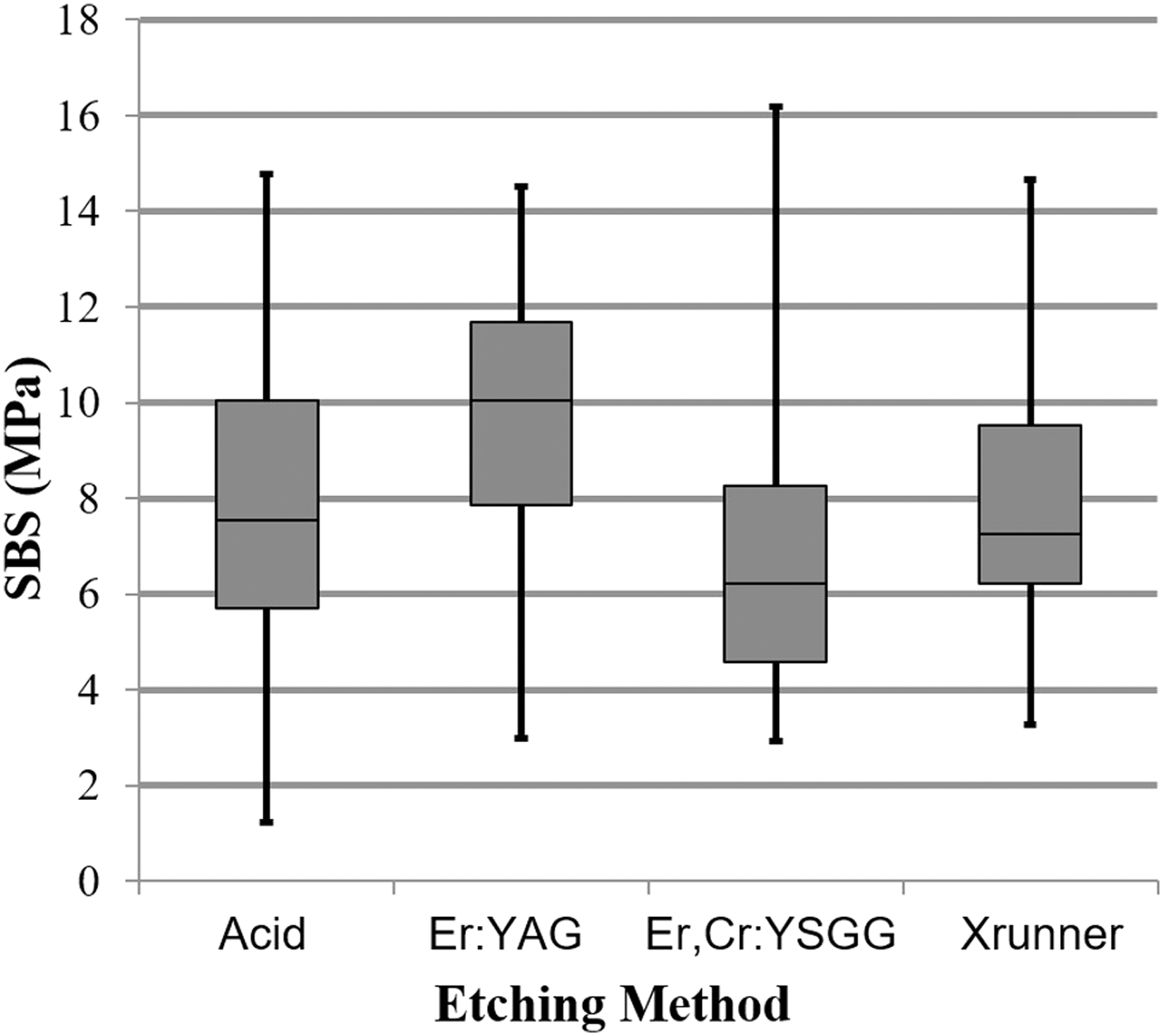
Mean SBS values and SDs for different etching methods. SBS, shear bond strength; SD, standard deviation.
One-way analysis of variance.
SBS, shear bond strength; SD, standard deviation.
There were no statistically significant differences among the ARI scores of the laser groups, while the acid etching group demonstrated significantly higher ARI scores compared to the other experimental groups (p < 0.001). Regarding fracture patterns, in the acid etching group, most failures (11% of 20% or 55%) were in the bracket–resin interface. For the remaining experimental groups, all bracket failures were in the resin–enamel surface (Table 3).
Dunn's post hoc multiple comparison.
Kruskal–Wallis test.
ARI, adhesive remnant index.
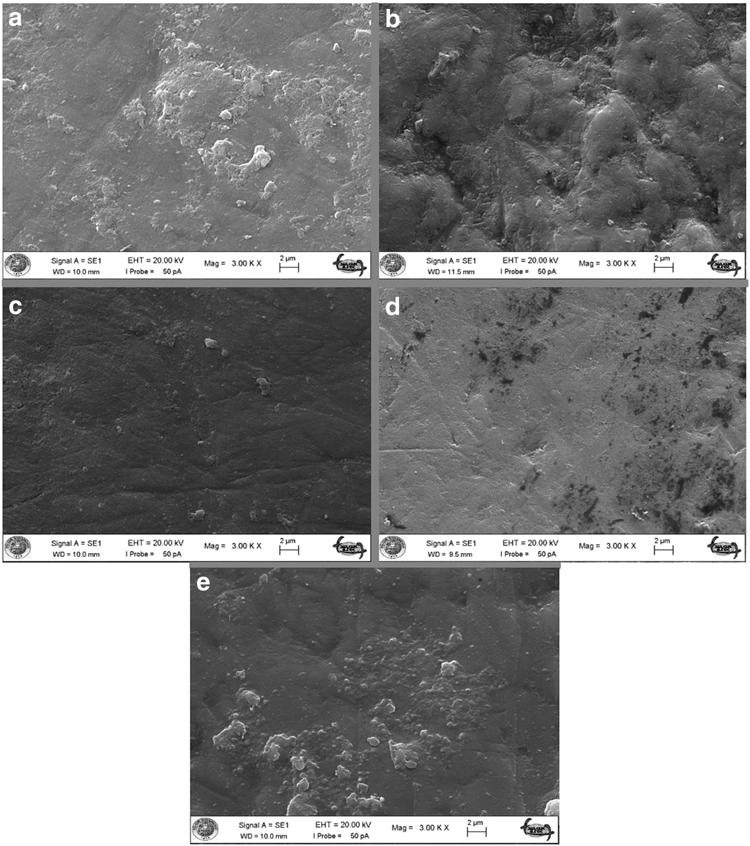
AFM scans showed that all etching procedures caused enamel surface irregularity (Fig. 2a–d). Relatively smooth surfaces were observed in the Er,Cr:YSGG and X-Runner groups, while a rougher surface pattern was observed in the Er:YAG laser etching group (Table 4). The AFM evaluation of the experimental groups after debonding and the control group, which was not subjected to any surface treatment, are shown in Figures 3a–e. All experimental groups except the Er:YAG laser group demonstrated surface irregularity similar to that of the control group. The Er:YAG laser-etched sample demonstrated a rougher surface compared to other groups, while AFM evaluation demonstrated that the maximum surface roughness value of restored enamel was lower compared with the controls for all test groups (Table 4).
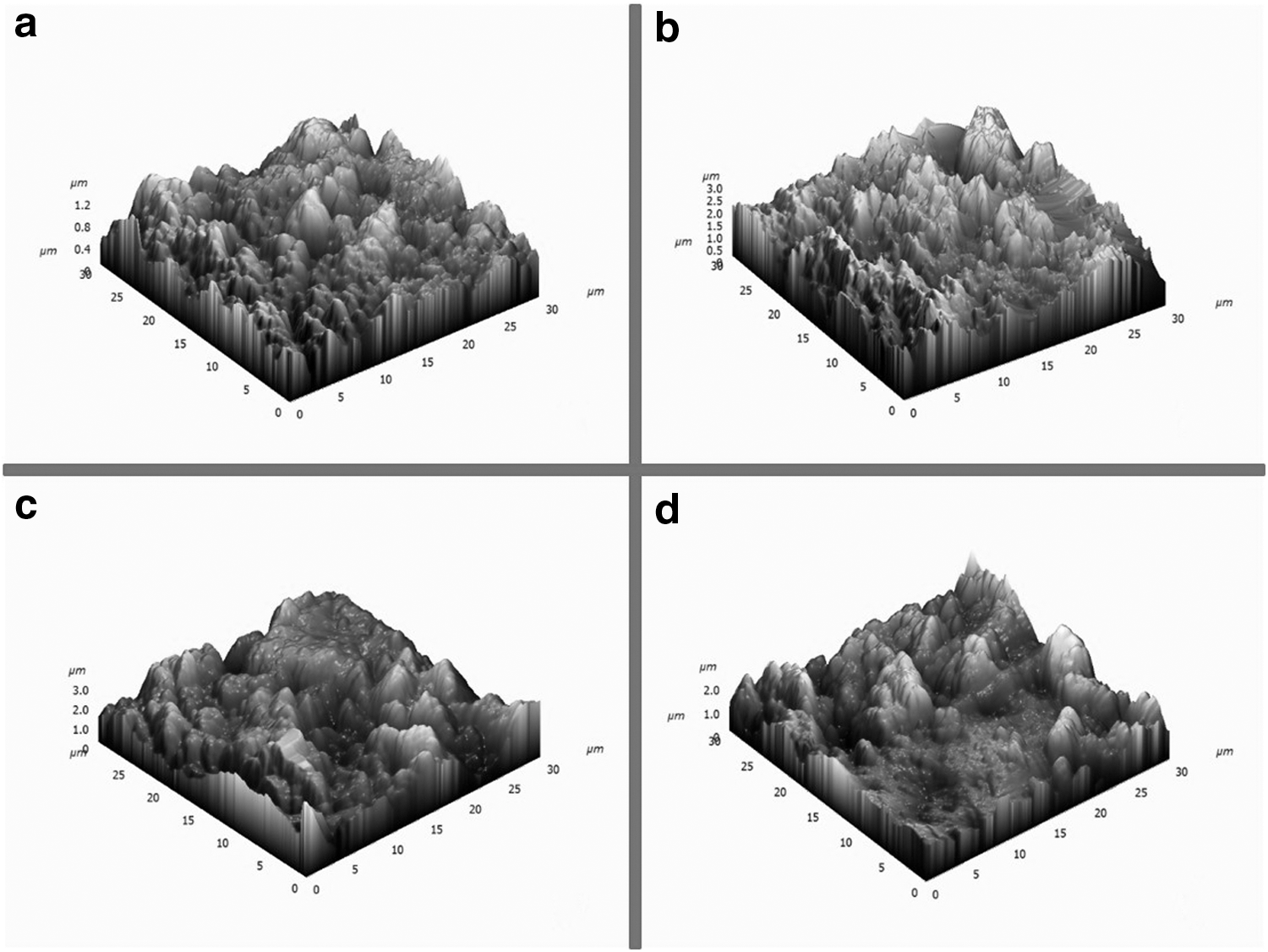
AFM scans of etched
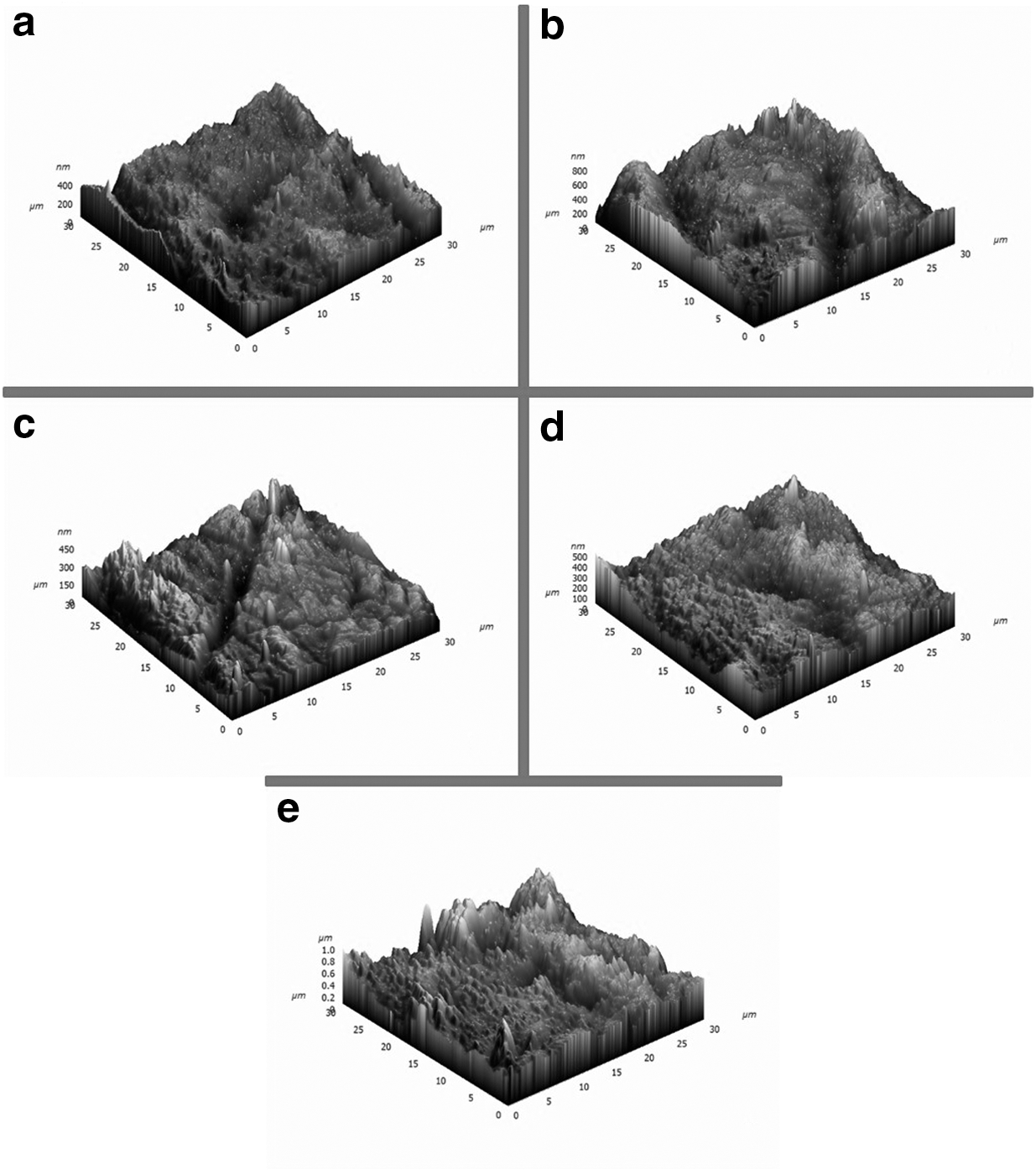
AFM scans of restored
AFM, atomic force microscopy.
According to SEM examination, the surface characteristics of the acid-etched enamel are similar to the type IV etching pattern, whereas the enamel surface irradiated using the Er:YAG laser had a pattern resembling the type II etching pattern described by Galil and Wright. 15 In the Er,Cr:YSGG laser etching group, the enamel surface was completely affected, but the etching pattern could not be determined. The enamel surface etched with the X-Runner demonstrated a more geometric and homogenous surface pattern compared to the enamel surfaces in the other experimental groups (Fig. 4a–d). The SEM evaluations of the experimental groups after debonding and the control group, which was not subjected to any surface treatment, are shown in Figs. 5a–e. The restored/debonded experimental samples, except the Er:YAG laser group sample, demonstrated surface characteristics similar to those of the control group. However, the restored Er:YAG laser-etched sample demonstrated a more irregular surface than the control group samples, which is in accordance with the AFM evaluation.
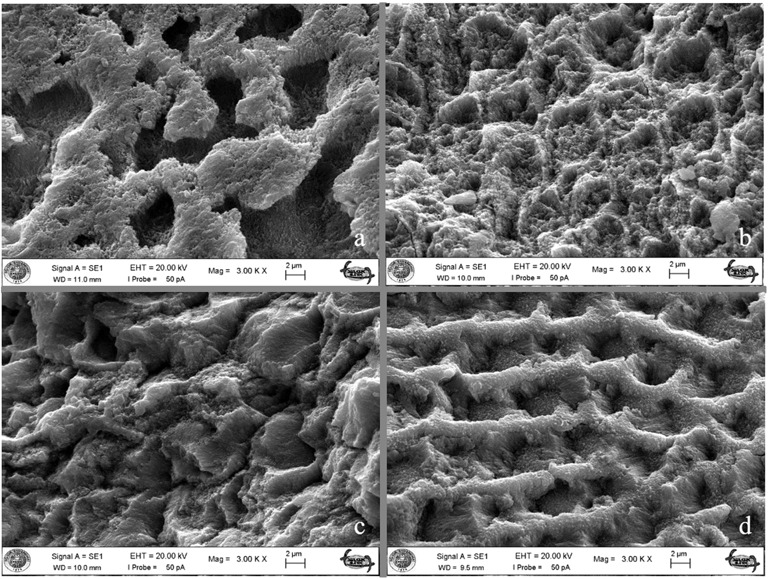
SEM examination of etched
SEM examination of restored
Discussion
There are only a few case reports in the literature about using X-Runner handpieces for etching procedures. 16,17 Therefore, before conducting the experiments, a pilot study was performed to determine the ideal setting parameters for the X-Runner handpiece by taking SEM and AFM scans and comparing the SBS values achieved with different power outputs, irradiation shapes, and number of scans. The settings that provided the most acceptable results were used in the main study (100 mJ, 10 Hz, 1 W, 40% water, and 50% air in MSP mode with two vertical scans and two horizontal scans). In literature, numerous studies reported the bond strengths of enamel surfaces irradiated with Er:YAG laser. Before orthodontic bonding, enamel irradiation with 1.2, 1.5, or 2 W Er:YAG laser resulted acceptable SBS in different studies. 10,11 According to that, in our study manual, Er:YAG laser handpiece's settings were 120 mJ, 10 Hz, and 1.2 W.
The results of this study indicate that no significant differences were found among the groups based on SBS values (p = 0.148). In addition, all experimental groups demonstrated mean SBS values within the range of 6–8 MPa mentioned by Reynolds 1 for adequate bond forces in orthodontics. The null hypothesis was therefore accepted. This result is in accordance with the studies by Basaran et al. 18 and Sagir et al., 11 but is in contrast with those of Usumez et al., 8 von Fraunhofer et al., 5 Hosseini et al., 19 and Martinez-Insua et al., 12 most probably due to the different power and irradiation settings employed.
Usumez et al. 8 reported that unevenly distributed high and low values were seen in laser-etched groups, whereas the values were in a narrower range in the acid-etched group. They commented that this is the result of nonuniform etching pattern on enamel surface because of the hand-controlled sweeping motion of the laser handpiece during the irradiation. 8 However, in this study, the SD and coefficient of variation calculations indicate that SBS values remained in a narrow range in the X-Runner group where there is no need for a hand-controlled sweeping motion. The difference is probably the result of the X-Runner handpiece, which provided a more standard and reproducible etching pattern not only on the irradiated area but also between the different samples tested, as confirmed by the SEM images (Fig. 4).
In our study, the ARI scores showed significant differences among different etching procedures (p < 0.001). Generally, more adhesive remained on the enamel surface with acid etching, which indicates that the bond failure was within the bracket–resin interface. However, in the laser-irradiated groups, less adhesive remained on the enamel surface. Despite the fact that less adhesive left on the enamel surface may save some chair time for cleaning teeth after debonding, 10 some authors are in favor of bond failure at the bracket–resin interface to avoid enamel fracture and crazing, which is occasionally reported to occur with bracket debonding. 20
AFM and SEM scans were acquired to allow a visual observation of surface roughness. The SEM scans demonstrated that all etching procedures caused surface irregularities. Following debonding, adhesive removal, and polishing, it was possible to restore the enamel surface to its original gloss or even better. However, not all the samples have a similar smooth pattern, and although the roughness of restored enamel was less in all test groups compared to the control group, the Er:YAG laser-etched images showed a relatively rougher surface compared to the other test groups. It should be noted that the AFM and SEM evaluations of both the etched and polished samples should be evaluated with caution due to the limited sample size and the fact that the scans were only of representative parts of a larger enamel surface. Also, the duration and pressure of polishing procedure or the amount of enamel removal were not standardized among the groups and this difference can cause different surface patterns. AFM and SEM scans of polished samples should be evaluated by considering these limitations.
Ultimately, laser etching seems to be a viable alternative to acid etching. However, this is an in vitro evaluation of a novel method under controlled conditions, and despite the fact that thermocycling was employed, it may not reflect the actual oral environment and real-life loading patterns. Therefore, the results may not be directly extrapolated to in vivo conditions. Clinical success is the final test, and prospective clinical trials should be conducted to confirm the in vitro results.
Conclusion and Summary
Taking the limitations of this study into consideration, the following can be concluded: 1. Etching of enamel surfaces with Er:YAG or Er,Cr:YSGG lasers at the given settings using either manual or digitally controlled scanning can yield SBSs comparable to those of acid etching 2. Metallic orthodontic brackets bonded to laser-etched surfaces always fail at the resin–enamel interface, while those bonded to acid-etched surfaces tend to fail at the bracket–resin interface, 3. It seems possible to restore enamel to its original gloss after debonding and polishing using all the etching methods tested.
Footnotes
Acknowledgment
This research was supported by Scientific Research Projects Unit of Bezmialem University, Istanbul, Turkey (Project No. 12.2013/10).
Author Disclosure Statement
No competing financial interests exist.