
Abstract
Introduction
S
Collagen fibers are structural proteins that confer strength in muscle and tendon tissue to the mechanical deformations imposed on the muscle. 2 According to Kannus, 3 the organization of these fibers differs throughout their trajectory, which initiates in the muscle tissue and extends to the tendon-to-bone insertion. The fibers may be arranged either in parallel or in a cross pattern. Changes in the direction, organization, and deposition of collagen fibers seem to be related to the load imposed by the muscle and remodeling after an injury. Moreover, the mechanical factors of these fibers are important to the prevention of injuries, such as a ruptured tendon and tendinopathy. 4,5 These conditions result from excessive functional overload and training, which generate considerable elastic tension. The mechanical behavior corresponds to a tension-deformation curve, in which resting collagen fibers in an undulating configuration reach their greatest degree of stretching at 4% of their length. When a tendon is submitted to forces that exceed this 4% stretch, such as in the case of muscle hypertrophy, the collagen fibers change their arrangement, forming intermolecular cross-linking in an attempt to diminish the tension. At forces exceeding 8% of fiber stretching, interfibril shearing of the collagen fibers may occur, leading to local inflammatory processes and even the rupture of the tendon. 6
Muscular hypertrophy stemming from athletic training or daily repetitions of movements imposes an acute overload on the muscle and tendon, resulting in an increase in the cross-sectional area of the muscle fibers, which is accompanied by remodeling of the extracellular matrix and a rearrangement of collagen fibers (degradation and synthesis). 7 According to Abate et al., 6 factors other than muscular hypertrophy may also be involved in tendinopathies. Epidemiological data demonstrate that 30–50% of elderly individuals exhibit shear stress in the tendons of the lower limbs at postmortem exams. Such alterations are often associated with sports involving lower limb movements, such as the tendons employed when playing tennis, the patellar tendon in repetitive activities of jumping (basketball and volleyball) and kicking (football) and the tendon of the tibialis posterior muscle in runners.
In many fields, low-level laser therapy (LLLT) is used as a photobiomodulation agent due to its effects on different clinical and experimental conditions. Laser treatment is capable of modulating the inflammatory response and myonecrosis, 8 –10 stimulating the production of growth factors, 11 –14 increasing the proliferation of satellite cells, 15,16 increasing the proliferation of fibroblasts, 17,18 modulating collagen synthesis and distribution, 7,14,19,20 stimulating the remodeling of the extracellular matrix 7,21 and modulating angiogenesis 7,20,22 and pain. 23
Studies involving human subjects have demonstrated that LLLT at the infrared (780 nm) wavelength is capable of reducing muscle fatigue, improving one's training performance, increasing peak muscle torque, diminishing oxidative stress, and enhancing the recovery process following a muscle injury. 22,24 –26 In a previous study by our research group, LLLT during the process of compensatory hypertrophy induced an increase in the weight of the plantar muscle in rats after 14 days in comparison to both the control group and group submitted to hypertrophy without laser radiance. Moreover, an increase in the cross-sectional area of the muscle fibers was found 14 days following the ablation of synergist muscles in comparison to the hypertrophy group without laser radiance. 27
The effect of LLLT on the alignment of muscle collagen is not yet fully understood. According to some studies, 24 –26 the absorption of light by mitochondrial chromophores induces the activation of numerous intracellular signaling pathways, thereby increasing the proliferation of fibroblasts and muscle collagen fibers. However, the effect on the alignment of these fibers appears also to be related to regulatory factors in the extracellular matrix. Enzymes such as metaloproteinases, blood vessels, and muscle hypertrophy can cause the disorganization of muscle collagen fibers. Regarding the collagen fibers in tendons, the mechanism of action of LLLT on the deposition and alignment of these fibers remains unknown. Studies suggest that the most important factor to the alignment and arrangement of fibers is the transmission of muscle force to the tendon (mechanical stress).
Few studies in the literature have addressed the effect of low-level laser during the hypertrophy process with regard to the remodeling of the connective tissue between muscles and tendons. Such knowledge is of fundamental importance to the establishment of more effective strategies during the process of compensatory hypertrophy, which is clinically seen in athletes who perform high-intensity eccentric exercises and in muscles that need to achieve a rapid functional return by increasing their demand without the assistance of synergist muscles, as in cases of trauma, prolonged immobilization, peripheral nerve injuries, and others.
The aim of this study was to evaluate the effect of infrared laser (780 nm) on the deposition and organization of collagen fibers in muscle and tendon tissue during the process of compensatory hypertrophy in the plantar muscle of rats.
The hypothesis tested here is that LLLT can improve the organization of collagen fibers in both tendons and muscles undergoing a process of adaptation in cases of compensatory overload.
Materials and Methods
This study was conducted at the research laboratory of the Master's and Doctoral Program in Biophotonics Applied to Health Sciences in compliance with international ethical norms on animal experimentation and received approval from the Animal Research Ethics Committee of Universidade Nove de Julho, Brazil (AN007/2013). All experiments were performed in accordance with the guidelines of the Brazilian National Concil for the Control of Animal Experimentation.
Animals
Wistar rats weighing 242.5 ± 13.59 g were divided into three groups. Since bilateral surgery was performed, each group corresponded to one of the hind paws of the animals. The right paw was submitted to hypertrophy (H) without LLLT (n = 10) and the left paw was submitted to hypertrophy with LLLT (H+LLLT) (n = 10). The two experimental groups (H and H+LLLT) were analyzed 7 and 14 days after surgery. A control group (n = 2) was not submitted to either hypertrophy or laser radiance.
Surgery
The animals were anesthetized with an intraperitoneal injection of 1 mL/kg of xilasine (2%) (Anasedan; Vetbrands, São Paulo, Brasil) and ketamine HCL (1%) (Dopalen; Vetbrands). Ablation was performed as described by Terada et al. 28 and consisted of the partial removal of the synergists (gastrocnemius and soleus) of the plantar muscle, thereby overloading the plantar muscle.
Laser irradiation parameters
The LLLT was performed immediately following surgery using the protocol described by Alves et al. 7 with an active medium arsenide-gallium-aluminum (AlGaAs) laser device (Twin laser®; MM optics, São Carlos, São Paulo, Brazil). Table 1 and Fig. 1 display the radiance parameters and application points, respectively, according to the World Association of Laser Therapy (WALT). 29
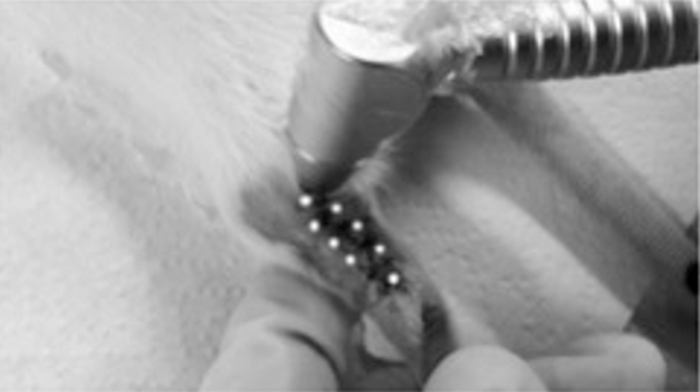
Laser application points around postsurgical suture.
At the end of the experimental protocol, the animals were euthanized with an overdose of anesthesia and the plantar muscles and tendons were removed for analysis. The groups were euthanized on days 7 and 14 days after surgery.
The muscle samples were fixed in 10% formalin. The histological cuts (7 μm) embedded in paraffin were strained with Picro Sirius Red (Sigma, St Louis, MO) and hematoxylin-eosin. A polarized light microscope (Zeiss Axioplan, Germany) and photographs were used for the morphologic analysis. The captured images were analyzed with the aid of the ImageJ program (National Institute of Health—NIH—EUA) for determination of the area related to the distance between muscle fascicles, percentage of collagen deposition, and quantification of the fiber types. The first analysis involved use of the Binary/Threshold/Black and White tool. The second analysis was performed using the Binary/Make Binary process. In the third analysis, quantification was performed using the windows imaging component (WIC) plug-in of the ImageJ program in Color Function/Color Deconvolution, with the selection of red and green. 30
The tendon samples were frozen. Cuts (5 μm) were made with a cryostat, placed on silanized slides, and deparaffinized to avoid affecting the interpretation of data for birefringence analysis (optical retardation). The slides were immersed in distilled water for 30 min, as described by Vidal. 31 Measures were obtained with the axis of the tendon at a 45° slant, which permitted greater brightness and analysis of the organization of fibers throughout the tendon. The angles (°) obtained in the reading with the polarized light microscope were converted into wavelength (nm) and multiplied by 0.758 for reading at λ = 560 nm (green light). Larger angles denote a greater degree of collagen fiber organization and parallelism. 31 The data were evaluated by two experienced blinded examiners, since the samples were identified by random numbers, which enhances reliability of the findings.
Statistical analysis
The data were analyzed with the aid of Minitab 14 (Minitab, Inc.). The Shapiro–Wilk test was used to determine the normality of the data distribution. Data with parametric distribution were submitted to two-way analysis of variance followed by Bonferroni test for the comparison of the groups. The significance level α = 5% was adopted for all comparisons (p < 0.05).
Results
Collagen deposition in plantar muscle
The quantitative analysis of the cuts stained with hematoxylin-eosin (Fig. 2) revealed an increase in the space between muscle fascicles in the irradiated group (H+LLLT) when compared to the control group at 7 days and a reduction in H+LLLT when compared to the control group at 14 days (p < 0.05). Moreover, an increase in the percentage of the area of space between fascicles was found in H+LLLT when compared to the nonirradiated group (H) at 7 days and a reduction in this area was found in H+LLLT when compared to the nonirradiated group (H) at 14 days (p < 0.01).

Photomicrograph of cross-sectional cuts stained with hematoxylin-eosin (magnification: 40 × ) and with inversion of colors using ImageJ software of space between muscle fascicles. Figure shows quantification of area of spaces in percentage values. Data expressed as mean and standard deviation (two-way ANOVA followed by Bonferroni test for comparison of groups) (p < 0.05). The different letter denotes a significant difference. ANOVA, analysis of variance.
The quantitative analysis of the cuts stained with Picrosirius Red and photographed under polarized light (Fig. 3) revealed an increase in the total area of collagen in the H+LLLT group when compared to the H group (p < 0.05) at 7 days and a reduction in the collagen area in the H+LLLT group when compared to the H group (p < 0.01) at 14 days.
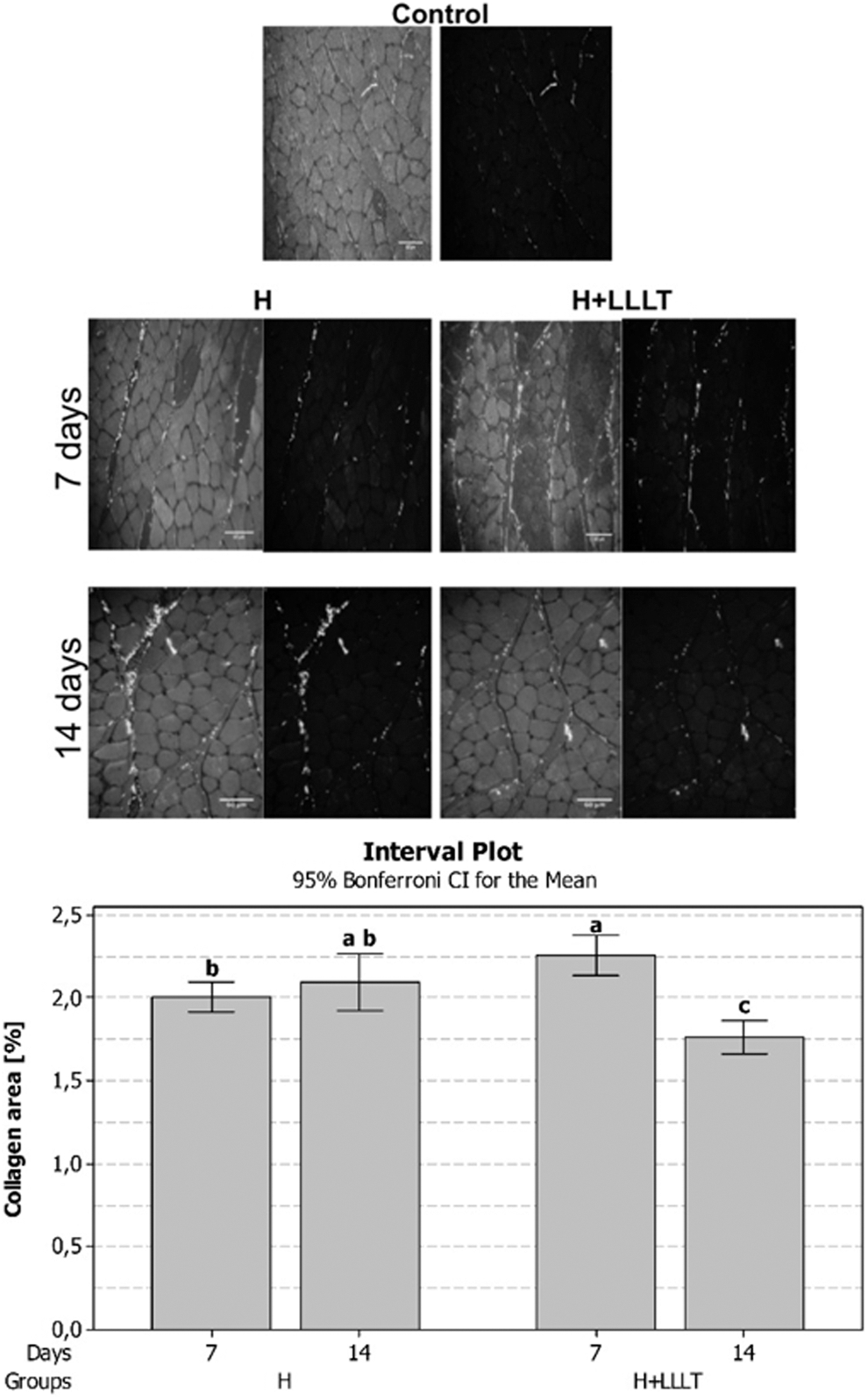
Photomicrograph of histological cuts of muscle tissue stained with Picrosirius Red (magnification: 40 × ). Figure shows quantification of total collagen area (μm2). Data expressed as mean and standard deviation (two-way ANOVA/Bonferroni test). The different letter denotes a significant difference.
Following the separation of colors using the Image J program (Fig. 4), a significant increase in the relative area of mature collagen fibers (type 1–stronger) and in the relative area of fibrils (type III–thinner, more elastic fibers) were found in both H+LLLT groups when compared to the H groups and control group at 7 days (p < 0.05). Moreover, significant reductions in the relative area of both thin and thick fibers were found in the H+LLLT group when compared to the H group at 14 days (p < 0.01).
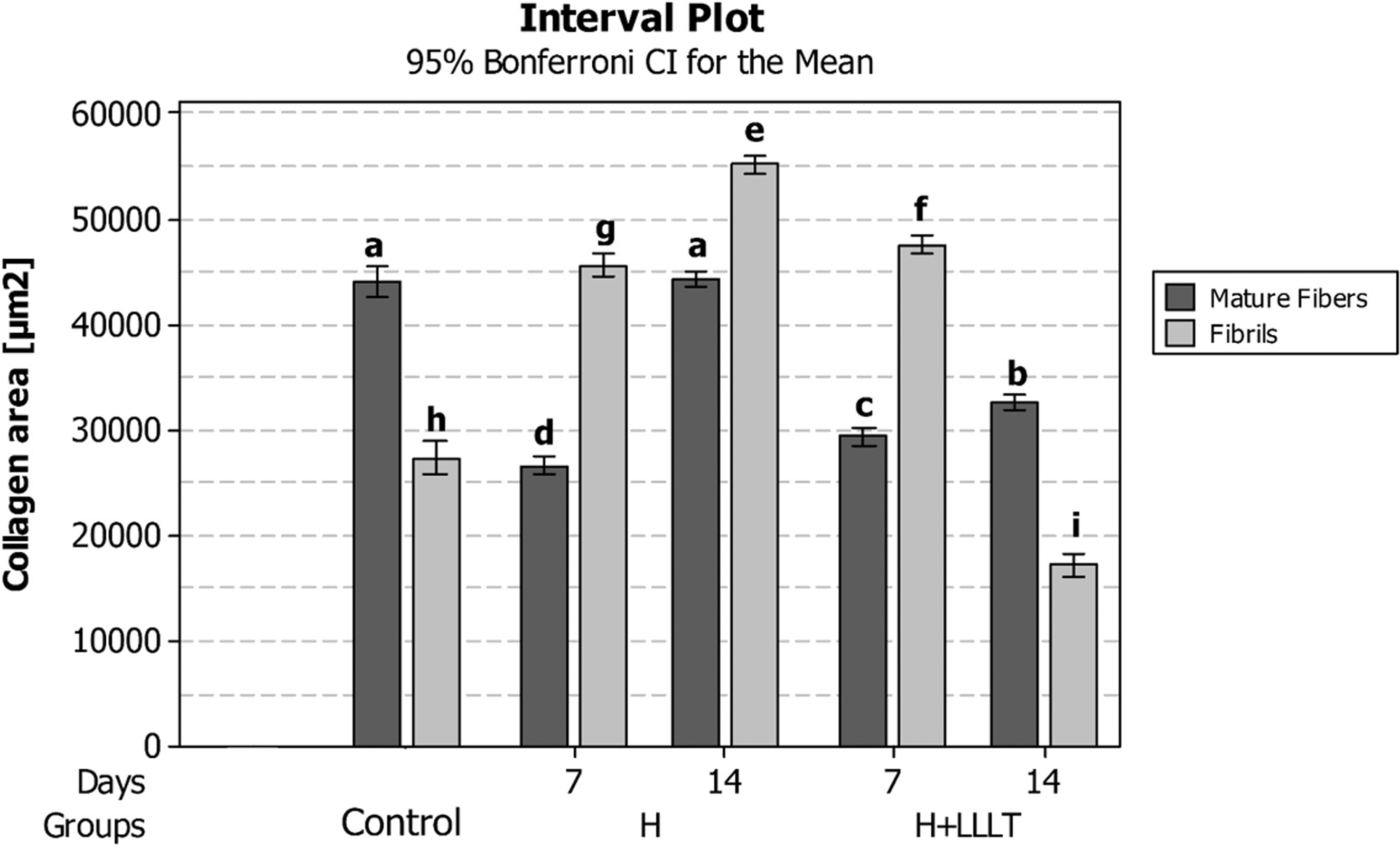
Quantification of collagen types and distribution in experimental groups. Significant differences in both mature fibers and fibrils in both groups when compared to control group at both evaluation times (p < 0.05). Significant differences between H and H+LLLT groups without laser radiance (H) and with radiance (H+LLLT) at 7 days. Significant differences with regard to both types of fibers in the H+LLLT in comparison to H group at 14 days. The different letter denotes a significant difference. H, hypertrophy; LLLT, low-level laser therapy.
Organization of collagen in tendon of plantar muscle
The qualitative birefringence analysis of the collagen revealed an improvement in the organization of the fibers in the H+LLLT group when compared to the H group (arrows and circles in Fig. 5). The quantitative birefringence findings also revealed an increase in the organization of collagen fibers in the tendon in the H+LLLT group when compared to the H group at both 7 and 14 days.
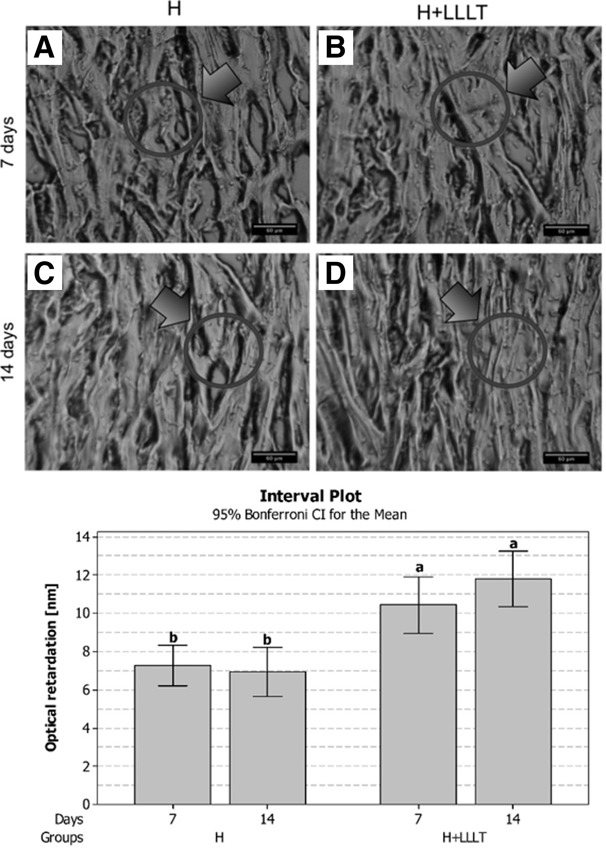
Qualitative analysis of images based on birefringence of collagen in tendon of plantar muscle (conventional polarized light microscope; magnification: 40 × ). At 7 days, greater organization of fibers in irradiated group
Discussion
Functional overload imposed on a muscle induces hypertrophy and remodeling of the extracellular matrix closely involved in this tissue. Such events contribute to the integrity of the basement membrane, improving the contractility of muscle fiber, and the transmission of force from muscle to tendon. 32,33 Collagen (the main protein of the extracellular matrix) is organized in fibers and the increase in functional demand induces adaptation in the deposition and organization of these fibers in the muscle-tendon unit to permit an increase in the cross-sectional area of the muscle fiber, which is the main result of muscular hypertrophy. 2,5
Studies have demonstrated that the lack of adequate remodeling of the extracellular matrix, which involves the degeneration, synthesis, and organization of collagen fibers during muscular hypertrophy, appears to be related to a greater occurrence of injuries among athletes and tendinopathy and ruptured tendons. 3,4,6 In muscle biopsies of 163 runners with signs of tendinopathy, Astrom and Rausing 34 used birefringence to demonstrate the derangement of collagen fibers, with the loss of parallel bands and an increase in cross-linking among the fibers.
This study is important for the development of light-based therapies and for understanding mechanisms involved in photobiomodulation, especially in tendon and muscle during the hypertrophy process. 35 LLLT led to an increase in the total area of collagen in comparison to the nonirradiated group at 7 days and a reduction in the total area of collagen in comparison to the nonirradiated group at 14 days. Using a cryoinjury model in the tibialis anterior muscle, Alves et al. 7 found a reduction in the total area of collagen in the group submitted to LLLT in comparison to the nonirradiated group after 7 days. Despite the different models used in the two studies, the laser parameters were the same.
The reduction in the distance between fascicles and the total area of collagen is believed to be favorable, as its permits an increase in the cross-sectional area of the fiber that occurs during the process of muscular hypertrophy. In a previous study using the same parameters, Terena et al. 27 found that the ablation model employed in the present investigation was effective at increasing the cross-sectional area of the muscle fibers and that the area was larger in the group that received LLLT.
With regard to the type of collagen fibers that constitute muscle tissue, significant increase differences in the amount of type I (thick, mature, strong fibers) or type II (thinner, more elastic fibrils) were in the H+LLLT when compared to the H group at 7 days after ablation surgery. However, a reduction in both types of fibers was found in the H+LLLT group in comparison to the H group at 14 days. Moreover, an inversion was found with regard to fiber types, with a predominance of fibrils in the nonirradiated hypertrophy group and a predominance of thick fibers in the LLLT hypertrophy group at 14 days. This finding may reflect a change in functional property of the muscle, as the type of collagen fiber is directly related to elasticity and strength of muscle tissue. The increase in the percentage of thick collagen fibers in studies evaluating tendon remodeling during the process of hypertrophy has been considered a protective factor against the recurrence of injuries by conferring greater strength to this tissue. 2,3
In the tendon, LLLT led to an improvement in the parallel organization of the fibers, as evidenced by the increase in the angle of birefringence (or optical retardation) analysis in the irradiated group at 7 and 14 days. According to Vidal 31 and Aro et al. 36 the angle of birefringence is directly proportional to the state of organization of collagen fibers.
It is important to note that no studies were found in the literature evaluating the effects of LLLT on this experimental model (compensatory hypertrophy following ablation of synergists). Previous studies mainly address the effects of photobiomodulation on remodeling of tendons and muscles following injury.
In accordance with present results, Arruda et al. 37 evaluated the influence of LLLT using different parameters (904 nm, pulsed emission, pulse frequency 2000 Hz, pulse duration 180 sec, peak power 15 W, 3 J/cm2 and 670 nm, continuous emission, 30 mW, 3 J/cm2, energy of 3 J), on the organization of collagen fibers (evaluated using birefringence) during the repair process of the Achilles tendon following complete tenotomy in rats. The results showed that all irradiated groups demonstrated an increase in the angle, regardless of the laser parameters employed. Moreover, the group irradiated with infrared laser exhibited better organization of the collagen fibers in comparison to the group irradiated with red laser.
Furthermore, Marcos et al. 38 and Joensen et al. 39 reported the positive effects of LLLT on tendon remodeling following an injury in accordance with our findings. Marcos et al. 38 induced tendinitis with an injection of collagenase in the muscle-tendon junction of the Achilles tendon and found that LLLT (810 nm, spot size = 0.028 cm2, power density of 3.57 W/cm2 and energy of 1 and 3 J) was capable of diminishing the inflammatory process and increasing the strength of the tendon after 7 days. However, collagen fiber organization was not evaluated using the angle of birefringence. Joensen et al. 39 induced an injury by Achilles tendon trauma with a mini guillotine in groups of wistar rats and irradiation (904 nm, energy of 3 J) initiated 30 min after injury and demonstrated that LLLT increased tendon thickness in the irradiated group after 15 h. In addition, Jesus et al. 40 also report the effects of LLLT were positive to alignment of collagen fibers as exposed in present results. The authors performed an injury to the Achilles tendon of rats and used the infrared laser (780 nm) during 10 sec and energy of 0.7 J and obtained in 3 and 7 days increase in the production of fibers of type I collagen and greater alignment of collagen fibers. Although it is another experimental model, using only one application with energy lower than the total energy used in our treatment, the findings are in agreement with the present study.
Based on the present findings, LLLT contributed to better collagen fiber deposition and organization in both muscle and tendon tissue during compensatory hypertrophy of the plantar muscle following the ablation of synergist muscles. This better organization reflects the better quality in the muscle hypertrophy process that contributes significantly to this tissue in terms of functional performance and protection against recurrence of injury.
Conclusions
In conclusion, infrared laser irradiation (780 nm) induces an improvement in collagen organization in tendons and a reduction in the total area of collagen in muscles during compensatory hypertrophy following the ablation of synergist muscles, both of which are beneficial to tissue adaptation.
Footnotes
Acknowledgments
This work was supported by UNINOVE and the Brazilian fostering agencies: National Council for Scientific and Technological Development (
Author Disclosure Statement
No competing financial interests exist.