
Abstract
Introduction
T
Approaches combining mechanical and chemical steps are typically used in dentistry to promote the control of oral biofilms. 7,8 These approaches aim at removing adherent biomasses from hard surfaces of teeth and dental materials, to effectively disinfect biofilm-free surfaces and to remineralize enamel's outermost layers. Recently, promising results were reported regarding the use of antimicrobial photodynamic therapy (aPDT) to control cariogenic biofilms. 9 This therapy has its biocidal mechanism based on a photophysical process that generates cytotoxic reactive oxygen species (ROS) 10,11 that are capable of killing viruses, bacteria (both gram-positive and -negative), and fungi. 12 Detailed information regarding aPDT's biocidal mechanism can be found in the extensive review published by Ormond and Freeman. 13
In brief, when photosensitizers (PSs) are exposed to adequate wavelengths, their molecules are promoted from the ground state (S0) into an excited state (S1). Excited molecules (S1) are very reactive and unstable, and they may spontaneously decay to the ground state (S0) either by releasing the excess energy in the format of heat or by emitting light (fluorescence). If the PS is sufficiently stable, then excited molecules can undergo intersystem crossing to the triplet state [T1, (where S0 < T1 < S1)]. Molecules in this longer-lived energy state may react with surrounding biomolecules (e.g., tissue oxygen) through two types of reactions (types I and II). 14 Type I reactions are based on electron transfer between PS-excited molecules, resulting in the generation of free-radical species (O2 −•, H2O2, and OH•). 15,16 Type II catalytic reactions are believed to be the dominant process in aPDT treatments 13 whereby the PS's excess energy is transferred to surrounding triplet oxygen molecules ( 3 O2), which results in a spin orientation change to further excite 3 O2 into singlet oxygen ( 1 O2). 16,17 These highly reactive and oxidizing-capable 1 O2 can participate in numerous reactions to cause critical damages to DNA, organelles, and cellular membranes, ultimately leading to cell death. 18
First-generation PSs are considered the most commonly used drugs in photodynamic therapy. 13 However, despite their proven success, 13 these drugs have been demonstrated to have limited applicability due to their low intensity of light absorption (ɛmax at 630 nm ∼3000 M−1 cm−1) and the presence of high levels of aggregates and impurities. 13 These limitations have precipitated the development of second-generation PSs. Several in vitro studies have investigated the utility of second-generation drugs such as curcumin, 19 –21 hypericin, 20 and methylene blue (MB) 22 as antibacterial agents against planktonic cultures of oral pathogens. However, limited information is available regarding the antibacterial efficacy of MB against cariogenic oral biofilms when irradiated with a visible and low-intensity level laser. In this study, Streptococcus mutans was chosen to be the model microorganism because of its ability to form and grow biofilms and its uncontested ability to produce organic acids that are capable of demineralizing the tooth structure.
The objective of this study was to assess the efficacy of antibacterial photodynamic therapy mediated by MB PS and low-intensity laser irradiation (LI) against intact S. mutans biofilms. The null hypothesis tested was that experimental aPDT treatments would have similar antibacterial efficacies when compared with the positive control group (100% viability, no treatment).
Materials and Methods
Specimen fabrication
Specimens (diameter 6.0 mm, height 1.9 mm) of Point 4™ microhybrid resin composite (shade A2; Kerr, Corp., Orange, CA) were fabricated in a custom-made metallic mold. Specimens were polymerized against glass slides (40 sec) by using a light-curing unit (Ultra-Lume LED 5; Ultradent Products, Inc., South Jordan, UT). Specimens were sequentially wet-polished (180–1200 SiC disks; final polish 0.5 μm diamond suspension) by using a MultiPrep system (Allied High Tech Products, Inc., Rancho Dominguez, CA). Specimens were ultraviolet-sterilized (254 nm, 1.6 mJ/cm2; model CL-1000 UVP Crosslinker; UVP, LLC, Upland, CA) and were stored in sterile ultra-pure water (37°C, 72 h) for monomer extraction. Specimens (n = 15/group) were then randomly assigned to experimental groups (Table 1).
Specimens in this group were kept hydrated by using 3 μL of sterile ultra-pure water during LI procedures.
aPDT, antimicrobial photodynamic therapy; LI, laser irradiation; MB, methylene blue; PS, photosensitizer.
Bacterial strain and growth conditions
A recombinant and bioluminescent S. mutans strain (JM10) was used in this study. The strain is a derivative of the S. mutans wild type (UA159) obtained by insertion of a constitutively firefly luc gene reporter placed under the control of the lactate dehydrogenase enzyme (Φ::(ldh-luc), SpcR) by using plasmid transformation. 23 The luc-reporter presence was confirmed by the selection of spectinomycin-resistant colonies on Todd-Hewitt (TH; BD Difco, Franklin Lakes, NJ) agar plates supplemented with 0.3% yeast extract (Y; EMD Millipore, Billerica, MA), referred to as THY medium, that was supplemented with 800 μg/mL of spectinomycin (MP Biomedicals, Santa Ana, CA). S. mutans colonies were passaged under anaerobic conditions (37°C, 48 h).
Biofilm growth conditions
Planktonic cultures of S. mutans were grown in THY medium supplemented with spectinomycin (32 μL) for 16 h (anaerobic, static cultures, 37°C). Planktonic cultures with desired optical densities (OD600 ≥0.900) were used as inocula for biofilm growth. Biofilms were grown by using a 1:500 inoculum dilution added to 0.65 × THY medium supplemented with 0.1% (w/v) sucrose (growth medium). Aliquots of inoculated growth medium (2.5 mL) were dispensed into the wells of sterile 12-well microtiter plates (Falcon; Corning, Corning, NY) containing sterile specimens. Specimens incubated in sterile growth medium served as the sterility control. All biofilms were grown under static and anaerobic conditions at 37°C for 24 h. The spent growth medium was then aspirated, and biofilms were replenished with 2.5 mL of sterile THY medium supplemented with 1% (w/v) glucose (recharge medium). Biofilms were then incubated for 1 h at 37°C in preparation for bioluminescence (BL) testing.
BL assay
A novel firefly luciferase assay, recently developed and validated by our laboratory, was used in this study to determine the aPDT's antibacterial efficacy against intact S. mutans biofilms. Detailed information regarding the assay's mechanism of action can be found in our previous publication.
24
Bioluminescent measurements of cellular metabolic activity before experimental treatments (baseline) were performed for all specimens by adding 40 μL of
Antibacterial treatments
Treatments were performed according to conditions described in Table 1. Two consecutive LI (3 J/cm2, 60 sec/irradiation, ≅5 cm high, total energy dose/specimen = 42.4 J/cm2 for an irradiation spot = 0.2827 cm2) was individually provided to each specimen by a portable hand-held device (100 mW ±20%, diode laser, 660 ± 10 nm, 0.094 cm optical fiber; Therapy XT; DMC, São Carlos, Brazil). The photosensitizing agent used was a plaque disclosing solution containing MB in concentrations of 0.005% 25 and 0.01% 26 (Chimiolux; DMC). Irradiation parameters may be also found in Table 2. After treatments, biofilms were incubated in fresh recharge medium for 1 h at 37°C in preparation for post-treatment measurements, which were obtained identically to baseline measurements.
Viable colony counts
After the post-treatment measurements, biofilms were removed from the surfaces of specimens by using a sonicator (4 min, 15 sec interval between cycles; power 310 ± 10 W; Q700; QSonica, LLC, Newtown, CT) connected to a water bath (4°C). Aliquots (10 μL) from each microcentrifuge tube were diluted in 90 μL of recharge medium (10−1). Five 10-fold serial dilutions (10−6) were then carried out in recharge medium for all specimens. Each dilution was plated in triplicate (total: 30 μL/specimen/dilution) by using spectinomycin-containing THY plates (800 μg/mL).
Confocal laser scanning microscopy
Additional specimens were subjected to the fabrication, polishing, monomer extraction, biofilm growth, and treatment procedures described in sections Specimen Fabrication, Bacterial Strain and Growth Conditions, Biofilm Growth Conditions, BL Assay, and Antibacterial Treatments in preparation for confocal laser scanning microscopy (CLSM). All biofilms were then stained by using LIVE/DEAD® BacLight™ Bacterial Viability Kit (Molecular Probes, Eugene, OR; 1.67 μM each of Syto® 9 and Propidium Iodide). A TCS SP2 microscope (Leica Microsystems, Inc., Buffalo Grove, IL) with Ar (488 nm) and He/Ne (543 nm) excitation lasers was used to acquire full-thickness biofilm images at three random locations per specimen. Volocity software (PerkinElmer, Boston, MA) was used to render representative 3D images of the distribution of live and dead/damaged cells within the biofilms of all groups. Detailed information regarding the CLSM and rendering protocols can be found in our previous publication. 27
Statistical analysis
BL and viable colony counts (VCC) data were tested for normality, sphericity, and homogeneity of variances by using Shapiro-Wilk, Mauchly, and Levene tests (α = 0.05), respectively, which indicated that both data sets were not normally distributed (p < 0.05). BL data were then corrected by using the Greenhouse-Geisser method and were analyzed by using repeated-measures analysis of variance and post hoc Bonferroni test (α = 0.05). VCC data were analyzed by using Kruskal–Wallis for independent samples and Dunn's multiple-comparisons post hoc tests (α = 0.05). Statistical analyses were performed by using SPSS software (version 19.0; IBM, Corp., Armonk, NY).
Results
Figure 1 shows the metabolic activity of intact S. mutans biofilms in terms of luciferase activity. Time points BL1–BL18 (baseline) and TRT1–TRT18 (post-treatment) denote the evolution of time in 9-min increments after the addition of
where Madiff stands for mean metabolic difference at each time point; X stands for the specific time point considered (e.g., either 1, 9, or 18 min); Maf stands for final metabolic activity measured (post-treatment, TRT1–TRT18); and Mai stands for initial metabolic activity measured (baseline, BL1–BL18).
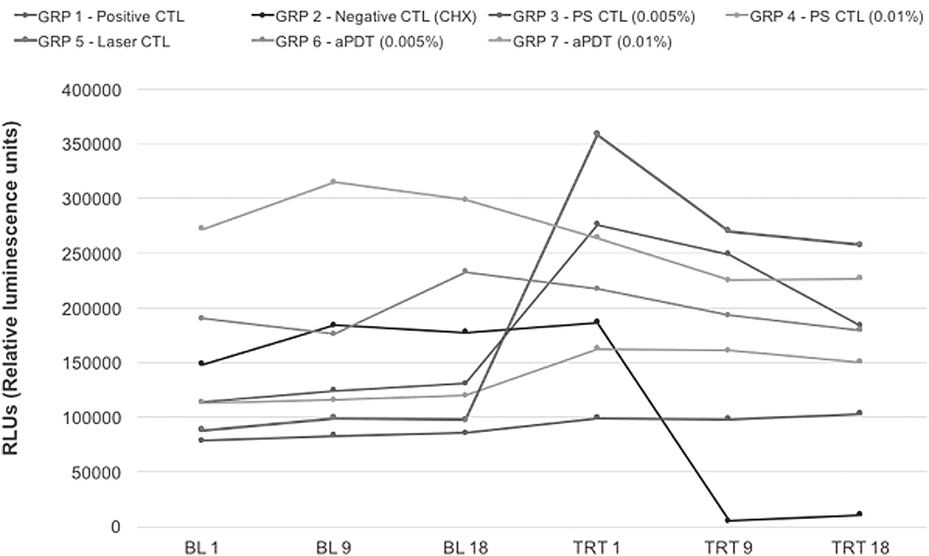
Time-dependent expression of luciferase activity (n = 15/group) was quantified in terms of bioluminescence. Initial metabolic activity values (BL1–BL18) demonstrated discrete metabolic differences for biofilms of different groups. All biofilms were grown under the same conditions by using the same culture medium. Post-treatment metabolic activities (TRT1–TRT18) demonstrate that chlorhexidine was the most efficient antibacterial treatment investigated. aPDT, antimicrobial photodynamic therapy; CTL, control; GRP, group; PS, photosensitizer.

Antibacterial efficacy of experimental treatments against Streptococcus mutans biofilms in terms of metabolic activity differences calculated for each specific time point combination by using the following formula
Figure 2 demonstrates the efficacy of each antibacterial treatment in terms of calculated relative metabolic differences (MAdiff1, MAdiff9, and MAdiff18) in each specific time point combination. Data analysis demonstrated the presence of differences (p < 0.0001) for the parameters “time points” and “groups,” and for the interaction between the parameters considered (“time points*groups”). Significant differences (p < 0.0001) were also found between MAdiff1 and Madiff9 in all groups; however, no differences (p = 0.57) were found between MAdiff9 and MAdiff18. These findings suggest that after the cellular uptake, the
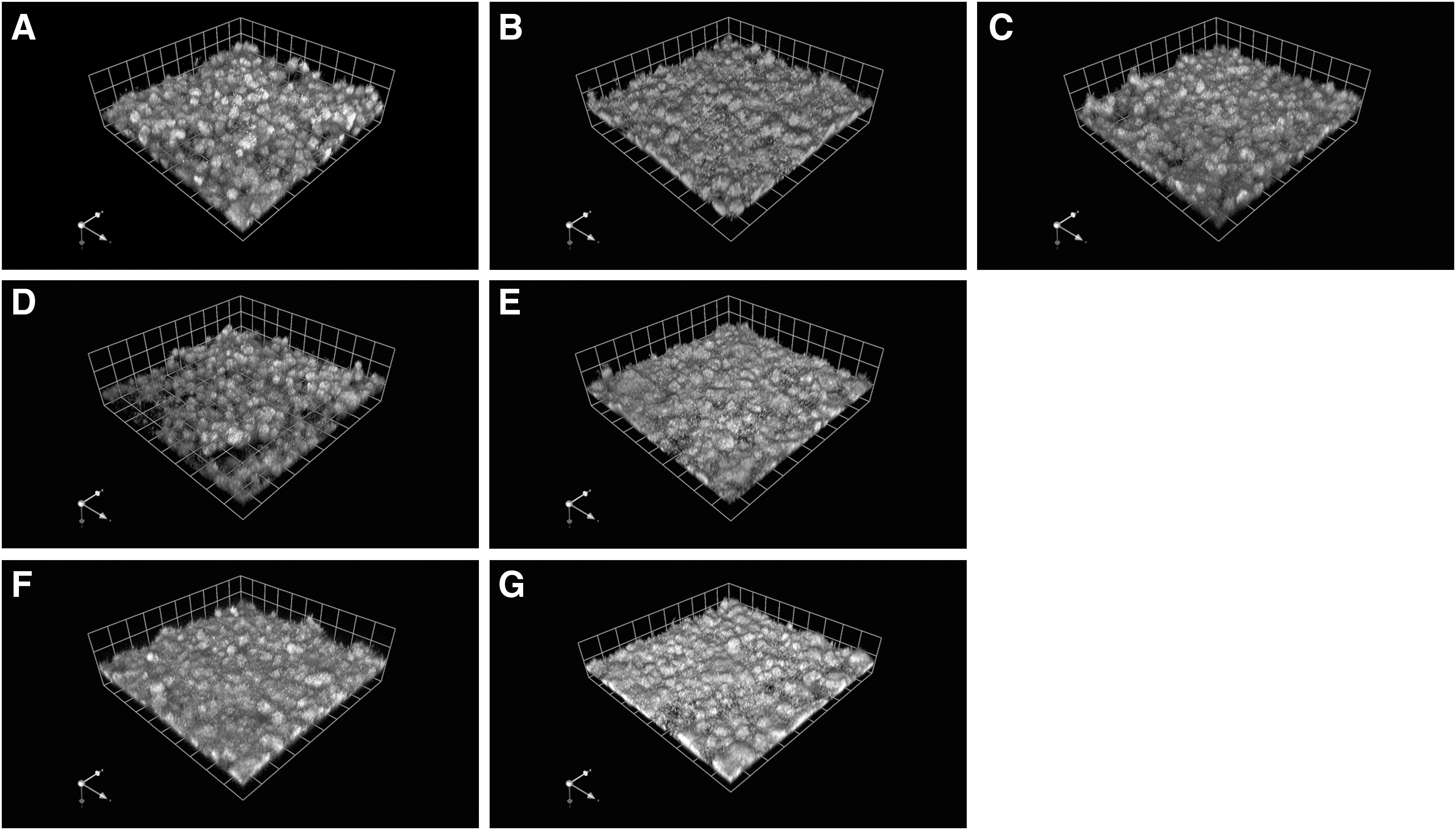
Figure 3 displays the mean and standard deviation values of VCC results (CFU/mL) for sonicated bacteria. Kruskal–Wallis non-parametric test detected differences (p < 0.0001) among groups tested. As expected, VCC of groups 1 and 2 displayed the highest and lowest viability levels among the groups tested. Biofilms exposed to experimental treatments (groups 3 through 7) had significantly lower mean VCC values (p < 0.05) when compared with the mean values of group 1. In addition, no differences (p > 0.05) were found among the colony counts of groups 3 through 7. Figure 4 shows 3D rendering of representative confocal images of living and dead/damaged cells within the biofilms of each group. The largest distribution of live cells was found on biofilms pertaining to group 1, whereas the largest volume of dead/damaged cells was observed in biofilms of group 2. Confocal microscopy confirmed the findings of the BL and VCC assays.
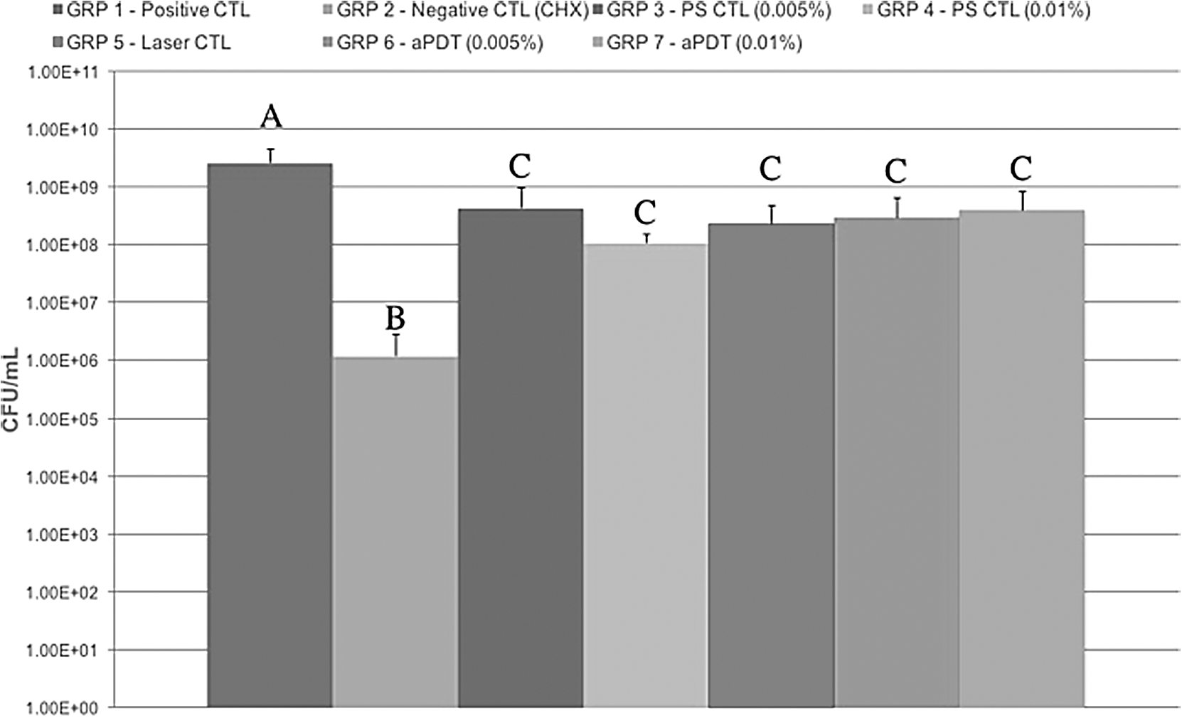
Mean viable colony counts of sonicated bacteria. Capital letters (A–C) denote mean values of colony counts that were statistically different based on Kruskal–Wallis for independent samples (α = 0.05) and Dunn's multiple-comparisons post hoc tests. The results shown demonstrate the behavior of the experimental antibacterial treatments. It becomes evident that group 1 (positive control) and group 2 (negative control) had the highest and lowest colony counts, as expected. Experimental treatments groups 3 to 7 had displayed significant viability reductions. However, the observed reductions were not comparable with the traditional 2% chlorhexidine gluconate (group 2, negative control) aqueous solution.
Discussion
The ability of microorganisms to adhere to surfaces (biotic or abiotic) in the oral cavity was previously demonstrated. 10 Other studies have shown that surface properties such as wettability, electrical charges, and surface roughness 28 can upregulate bacterial attachment 29 and modulate biofilm composition 10 to favor the selection of pathogenic bacteria. 5,30 These observations promoted the development of alternative approaches to prevent oral diseases via improvements in the control of oral biofilms. 31
aPDT has drawn significant attention from the scientific community 10 due to its minimally invasive 9 and ultraconservative features and effectiveness against resistant bacteria, 32 –34 viruses, and fungi. 35,36 Other aPDT's advantages over traditional disinfection techniques include the non-specific biocidal mechanism, ability to kill cells in biofilms, 37 and a localized treatment delivery. This atraumatic therapy was shown to not disturb the patient's sense of taste and was found to be painless, 38 which are clinically relevant attributes for the treatment of children and patients with special needs.
The efficacy of aPDT against S. mutans was determined for intact biofilms in terms of BL levels measured with a novel non-destructive and real-time luciferase assay recently developed and validated in our laboratory. 24 Figure 2 demonstrates the presence of significant reductions (p < 0.05) in biofilm metabolic activity after experimental aPDT treatments. Mang et al. 39 investigated the efficacy of porfimer sodium (25–125 μg/mL) against S. mutans biofilms by using the Alamar Blue metabolic assay.
In their study, biofilms grown in fluid shear conditions were incubated in the PS for 5 min before LI (630 nm, 30 J/cm2). 39 Their findings demonstrated that aPDT significantly reduced the metabolic activity of S. mutans biofilms. This study reported similar levels of metabolic activity reduction even though biofilms were incubated in a more diluted PS (0.005% or 0.01%; 5 min) and were irradiated with energy doses that were fivefold weaker (6 J/cm2). The similar reductions in metabolic activity observed in both studies could be explained by the differences in biofilm growth conditions (shear vs. static), which may have influenced biofilms' density and cells' physiology, thereby affecting the overall antibacterial efficacy of aPDT treatments. Their results could also have been adversely influenced by Alamar Blue's intrinsic and critical limitations, such as its requirement for the use of large number of cells (>5- or 6-log CFU) and the reduction of resorufin (fluorescent) into hydroresorufin (non-fluorescent). 40 –43
Viability of sonicated bacteria was determined in terms of VCC (CFU/mL; Fig. 3). Results presented suggest that all antibacterial treatments investigated significantly reduced S. mutans viability (p < 0.05) when compared with the viability levels of the positive control. As expected, biofilms from groups 1 and 2 have displayed the highest and lowest VCC levels among the groups investigated. However, no significant differences (p > 0.05) were observed among experimental antibacterial treatments. Zoccolillo et al., 10 while investigating aPDT's efficacy against S. mutans biofilms (24 and 48 h) grown on relevant dental substrates, have reported the effective killing (>5-log) of cells in biofilms by using a purpurin-based PS [(25 μg/mL) in dimethyl sulfoxide] and LI (664 nm, 15–45 J/cm2), thereby corroborating the results of this study.
Panhoca et al. 21 investigated the antibacterial efficacy of aPDT by using curcumin and blue-light irradiation in orthodontic patients. Their results demonstrated that surfactants may be used synergistically to improve the reduction of viability of S. mutans in saliva. This study's short irradiation protocol (2 min) is corroborated by Dovigo et al., 44 who demonstrated that PSs undergo photobleaching that results in decreased ROS generation by impacting PS's triplet and singlet quantum yields. In this direction, Chignell et al. 45 suggested that photobleached PS may have fewer optically active molecules, thereby further corroborating the irradiation protocols herein recommended.
The present findings are corroborated by several studies that demonstrated the effectiveness of various aPDT treatments in decreasing the viability of cells in oral biofilms. 34,39,46 Figure 3 displays the CLSM results, where it can be observed that biofilms in groups 1 and 2 displayed the highest biovolume of live and dead/damage cells, respectively. It is also noticeable that low-intensity LI (energy dose = 6 J/cm2) was not able to significantly damage cells within biofilms. Conversely, biofilms exposed to PSs (0.005% or 0.01%) without LI were shown to be more porous and less viable when compared with biofilms from group 1. Contrary to previous studies reporting that PSs are not cytotoxic without light irradiation, 20 this study has demonstrated that MB exhibited some levels of dark cytotoxicity (groups 4 and 5). CLSM imaging of aPDT-treated intact biofilms have demonstrated intermediate biovolumes of live and dead cells that are consistent with the results of both the BL and VCC assays performed.
Major limitations of this study include the use of single-species biofilms in in vitro growth conditions and the absence of saliva pre-conditioning. This study's findings have rejected the null hypothesis that experimental photodynamic therapy with MB and LI would have similar antibacterial efficacies when compared with the positive control group.
Conclusions
This study has demonstrated for the first time the utility of a non-destructive and real-time BL assay to screen the efficacy of different aPDT treatments mediated by MB and low-intensity LI against intact S. mutans biofilms. aPDT treatments for 2 min were shown to be effective <2% chlorhexidine gluconate treatments used for 7 min. More studies are necessary to improve aPDT's efficacy against oral cariogenic biofilms.
Footnotes
Acknowledgments
The authors thank the DMC company for graciously providing the photosensitizers and light sources used in this study.
Author Disclosure Statement
No competing financial interests exist.