
Abstract
Introduction
D
Minimum intervention dentistry is a key term in contemporary dentistry and regards choosing the least invasive treatment option to minimize tissue loss and patient discomfort. 4 The development of more conservative techniques for the removal of carious tissue from teeth enables the greater preservation of sound dental tissue during the treatment of decayed teeth. 5
As an alternative for the conventional caries removal method, we can apply a proteolytic substance that softens dentinal tissue that is called chemical–mechanical method of caries removal. With this method, the dentist preserves the affected dentin layer, which is capable of remineralization, and removes the infected dentin layer, which is incapable of remineralization. 6
The combination of papain and chloramine originated a papain-based gel denominated Papacarie™, which has the cleaning properties of papain combined with the disinfecting characteristics of chloramine. Collagen fibrils exposed by the dissolution of the surrounding minerals in the dentin due to the action of bacteria are susceptible to interaction with the papain-based gel, which softens the disorganized infected layer, enabling its removal with a blunt manual instrument. This gel has been used successfully in clinical trials for removal of carious tissue for the minimally invasive method. 7 –9 According to Silva Júnior, 10 Papacarie can be safely used in minimal intervention dentistry, because it does not cause the degradation of collagen, which is essential to the tooth restoration process.
Antimicrobial photodynamic therapy (PDT) is a variant of conventional PDT that emerged in minimal intervention dentistry due to the need to reduce the microbiota in the oral cavity and avoid the progression of caries. 11 –14 Antimicrobial PDT combines a photosensitizing agent and a suitable light wavelength at the maximum absorption peak of the photosensitizer to produce reactive oxygen species, which are toxic to bacterial cells. 15,16
To combine the advantages of the papain gel and antimicrobial PDT, a change in the composition of Papacarie was made so that this product could be used for the removal of infected dentin tissue and simultaneously as an antimicrobial agent. The change involved the addition of methylene blue, which is a well-known photosensitizer activated by red light (660 ± 10 nm), giving rise to PapaMBlue™. This low-cost innovation has been used in an experimental study with positive results. 17 By preventing and treating caries in the best possible way, dentists can minimize tooth loss and improve the quality of life of patients.
The aim of the present study was to evaluate the degradation of type I collagen fibers following treatment with a papain-based gel containing a blue dye (PapaMBlue) combined with antimicrobial PDT for minimal intervention dentistry.
Materials and Methods
In studies involving spectroscopy analysis, sponges of type I collagen from bovine Achilles tendon can be used as an efficient substitute for demineralized human dentin. 18 Sixty bioabsorbable membrane sponge discs of type I collagen (Technodry Liofilizados Médicos, Brazil) were used for the analysis of the possible degradation of collagen fibers by the papain gel. The membranes were cut into discs with a biopsy punch with an inner diameter of 5 mm. The discs (5 × 2 mm, in diameter and thickness, respectively) were randomly distributed among six experimental groups (n = 10), as described in Table 1. The laser irradiation parameters are listed in Table 2.
CW, continuous wave; FWHM, full width at half maximum.
The sample spectra can be altered by dehydration, then to avoid this, all membrane treatments were performed immediately prior to the infrared spectroscopy compositional analysis of each sample by ATR-FTIR (attenuated total reflectance-Fourier transformed infrared spectroscopy—Varian 610; Agilent Technologies, CA). The compositional analysis was made using a diamond crystal coupled to the spectrometer and the spectra, of each sample, were obtained: 4.0 cm−1 resolution (80 scans, range of 4000–500 cm−1). After the subtraction of the background, one spectrum was collected for each sample. After the subtraction of a baseline, the spectra were submitted to vector normalization and the peaks of each infrared band were analyzed. The Varian Resolutions Pro software program (Agilent Technologies) was used for record and convert the absorption spectra. Baseline subtraction, spectrum normalization, and the analysis of the peaks were performed using the Origin Pro 8 (OriginLabCorp., USA) software program.
Absorbance ratio of the 1235/1450 cm−1 peaks was considered for the analysis of the integrity of collagen triple helix. If the result ratio is closer to 1, it denotes the maintenance of the integrity of amide III and CAH bond present in the pyrrolidine ring of type I collagen triple helix. 19 The data were statistically analyzed by ANOVA and the pairwise comparisons among groups were analyzed by Tukey's test (Minitab 14; Minitab Inc.) with 5% significance.
Results
Neither classic Papacarie nor the product modified with a photosensitizing agent (PapaMBlue) caused collagen degradation, as demonstrated by FTIR analysis (absorbance ratio values ≥0.8 for all treatments) (Fig. 1).

Mean absorbance ratio of infrared spectra of peaks 1235 cm−1 (amide III) and 1450 cm−1 (pyrrolidine ring) and integrity of collagen triple helix of pure collagen membrane in treated groups. Different letters indicate a significant difference. Black line represents limit value of 0.8 of denaturation of three-dimensional structure of the type I collagen triple helix.
Figure 2 shows the mean infrared spectra of the untreated collagen membranes as well as collagen treated with Papacarie and PapaMBlue. After treatment, we did not identificate the disappearance of bands no new bands. However, an increase in absorbance of the 3000–3750 cm−1 band was found after the application of Papacarie and PapaMBlue. A decrease in the absorbance of peaks of amides I, II, and III (1630, 1550, and 1235 cm−1, respectively) was observed. A decrease in the absorbance of the 1450, 1400, and 1340 cm−1 bands was also observed after treatment with Papacarie, but these effects were not evident with the application of PapaMBlue.
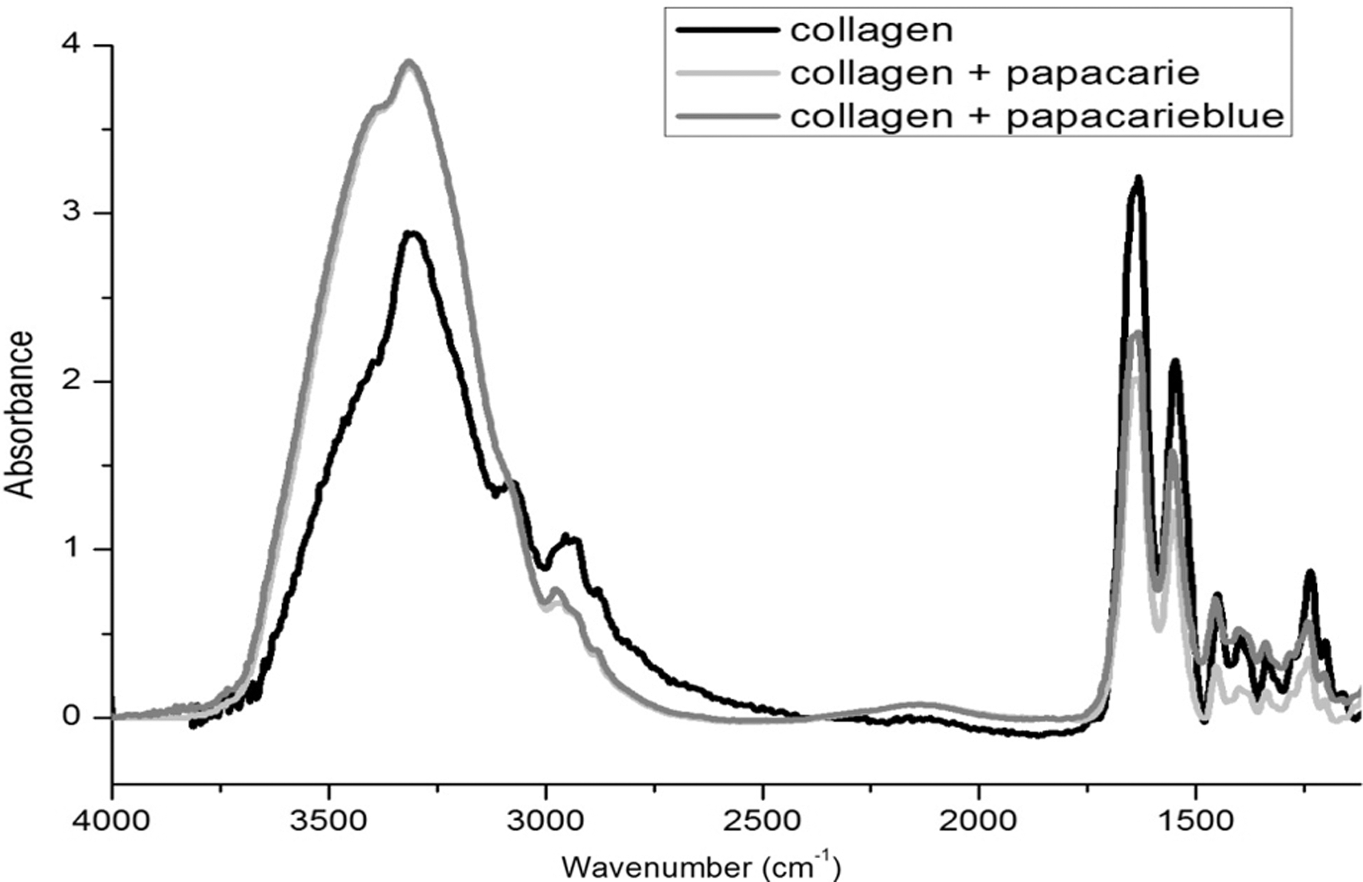
Normalized attenuated total reflectance-Fourier transformed infrared spectroscopy (ATR-FTIR) spectra in infrared range (1000–4000 cm−1) of pure type I collagen membrane and type I collagen after treatment with Papacarie™ and PapaMBlue™.
The effects of red laser irradiation on the collagen microstructure after treatment with PapaMBlue are shown in Fig. 3.
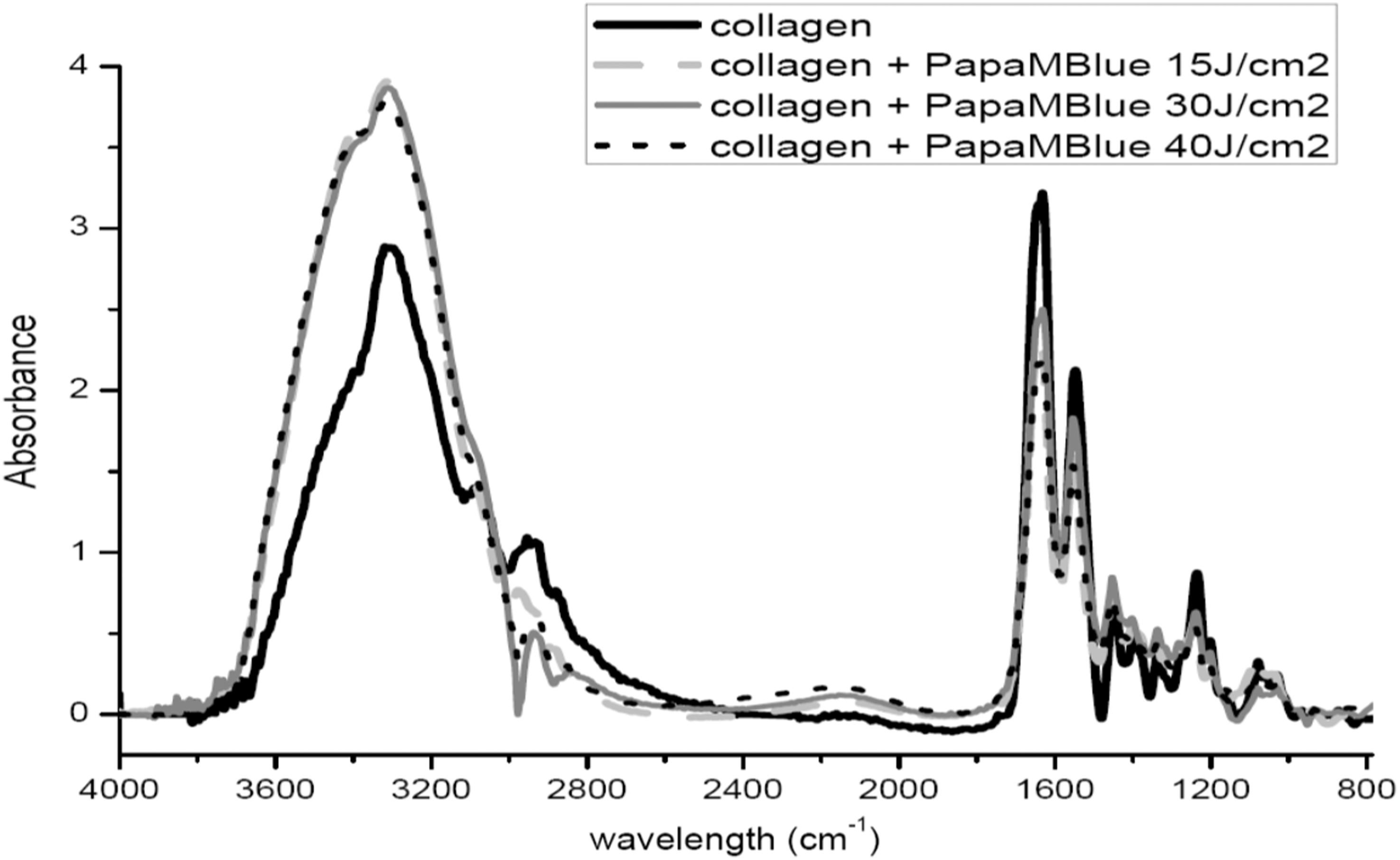
Normalized attenuated total reflectance-Fourier transformed infrared spectroscopy (ATR-FTIR) spectra in infrared range (800–4000 cm−1) of pure type I collagen membrane and type I collagen after treatment with PapaMBlue submitted to different energy densities.
On Fig. 4, we did not identificate after treatment, the disappearance of bands no new bands. However, a decrease in absorbance of the 1630, 1550, and 1235 cm−1 bands was found, corresponding to amide I, II, and III, respectively.
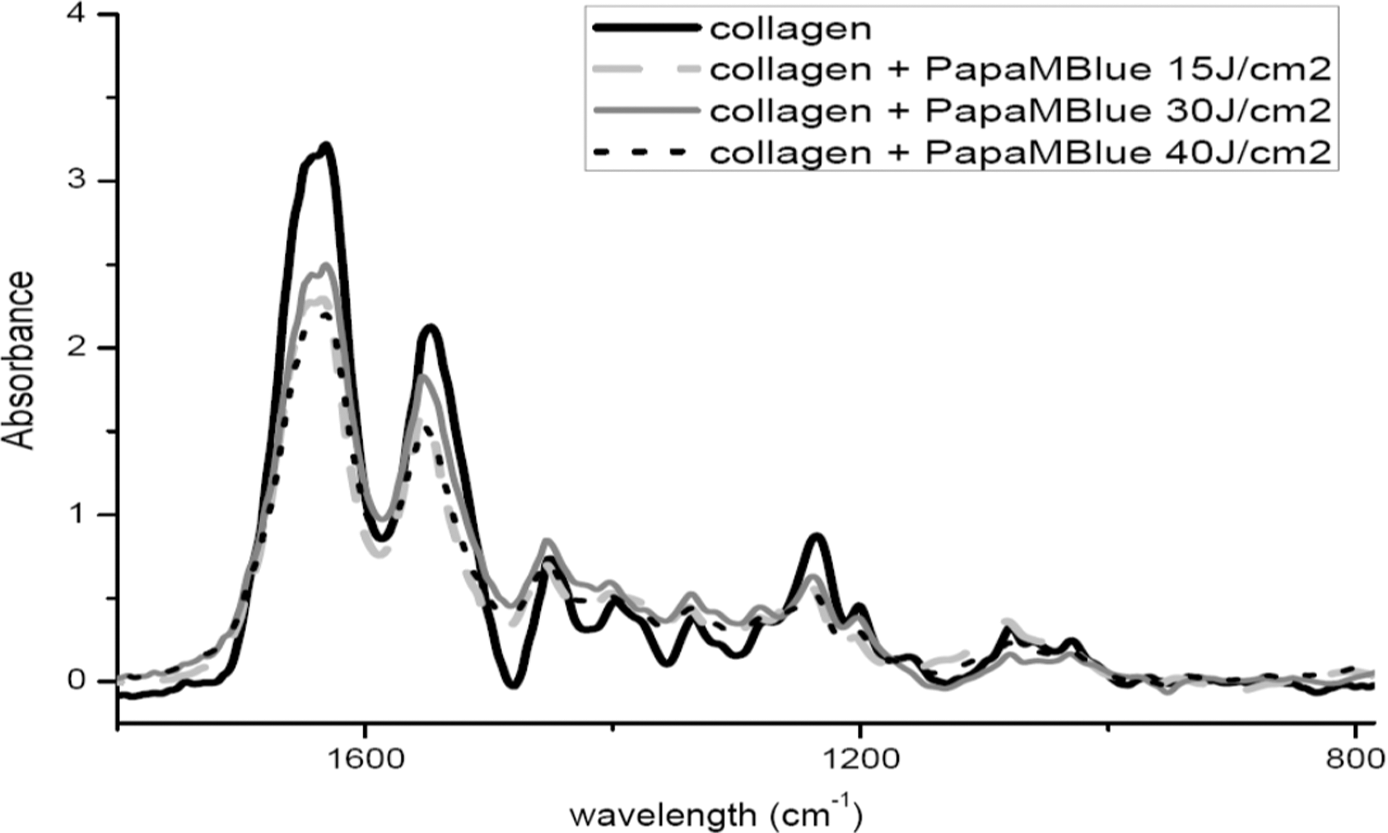
Normalized attenuated total reflectance-Fourier transformed infrared spectroscopy (ATR-FTIR) spectra in infrared range (800–1800 cm−1) of pure type I collagen membrane and type I collagen after treatment with PapaMBlue submitted to different energy densities.
Discussion
For the characterization of organic compounds such as collagen chemical changes, we can use ATR-FTIR in the midinfrared region (4000 and 400 cm−1). Each molecule has its own characteristic infrared spectrum with several bands that enable one to obtain useful structural information. 20 The natural frequency of vibrations and the wavelength absorbed are unique to each chemical group in the molecule and as described by Karoui et al. (2010), 21 “depends on the bond type (C = C, C–H, C = O, N–H, and O–H).”
Amide I and II are the main infrared absorption bands in spectroscopic measurements of biological proteins, such as collagen. The intensities of amide I and II are associated with their helical structure. The intensity of the amide I band can be used to characterize the secondary structure of proteins. 22,23 Amide I and II bands absorb in different regions depending on whether or not they participate in hydrogen bonding interactions. The presence of hydrogen bonds in collagen fiber is an indication of polypeptide chains forming a regular secondary structure. If amide I and II bands appear as α-helix or β-sheets, they absorb in slightly different regions. Thus, the collagen structure can be used to probe amide bands. 22
The bovine Achilles tendon is structurally similar to type I collagen fibers from human dentin when analyzed by ATR-FTIR spectroscopy 18 and can therefore be used to compare the degradation of chemical substances on the helical structure of collagen fibers. The absorbance quotient between the bands of amide III (1235 cm−1) and the pyrrolidine ring (1450 cm−1) determines the whole configuration of the collagen triple helix (Fig. 1). While the pyrrolidine ring is independent of collagen structure, the secondary structure of collagen determines the sheet of the amide band. 24
The absorbance ratios >0.8 confirmed the lack of significant collagen denaturation (Fig. 1), as “only values close to 0.5 denote a change in the three-dimensional structure of the type I collagen triple helix” (Sylvester et al.). 24 The results demonstrate that none of the substances applied over the collagen membrane exerted a negative effect on the collagen triple helix structural disposition.
In the spectra obtained (Fig. 2), absorption bands are observed corresponding to the “fingerprint region of collagen composed by amide I (1600–1660 cm−1), amide II (1500–1550 cm−1), amide III (1320–1220 cm−1), and pyrrolidine rings (1450 cm−1)” as described by Botta et al. (2012). 18 Figure 2 displays the mean infrared spectra of Papacarie, pure collagen membrane, and collagen membranes treated with Papacarie and PapaMBlue. After treatment, no new bands or the disappearance of bands were evident. However, an increase in absorbance of the 3000–3750 cm−1 band was found after the application of Papacarie and PapaMBlue, which indicates the incorporation of water to the collagen structure.
The mean infrared spectrum of the papain-based gel Papacarie shows corresponding infrared absorption bands, such as C–O, C–OH, C = C, N–H, and C–H3. The mean infrared spectra of untreated collagen membranes and membranes treated with Papacarie and PapaMBlue show increased absorbance of the N–H. Moreover, amide A bands are seen, along with a slight decrease in the absorbance of amide bands I, II, and III. However, neither the dissipation of bands nor the formation of new bands was evident.
The addition of methylene blue to the papain gel (PapaMBlue) did not modify the chemical composition of Papacarie (Fig. 2), as demonstrated by the unaltered bands of the chemical structure in the respective positions. Moreover, activation with red laser (660 nm) reduced the intensity of the amide I, II, and III bands of collagen covered with PapaMBlue, indicating a change in the collagen triple helix conformation (Fig. 4) independently of the energy used for irradiation. Thus, irradiation with red laser reduced the intensity of these bands because changes in the amide I band are determined from predominantly C = O stretching, whereas the amide III band results from a combination of C–N stretching and N–H in plane bending as well as C–C stretching. However, the addition of the pigments reduced the intensity of the bands corresponding to the C–O, ν(C–O), and ν(C–C) bonds as well as polysaccharides.
This study demonstrates that methylene blue added to Papacarie does not degrade collagen fibrils (Figs. 1 –4). The findings indicate that this product represents an innovation in dentistry and is viable for use in the removal of carious tissue and the disinfection of cavities, which could assist in improving quality of life, especially for children who live in areas without access to complete dental offices.
Conclusions
The findings of the present study demonstrate that papain gel with and without the addition of methylene blue does not promote the alteration of the chemical structure of type I collagen. The irradiation of methylene blue added to the papain gel with red light also did not alter the chemical structure of type I collagen.
Footnotes
Author Disclosure Statement
No competing financial interests exist.