
Abstract
Introduction
T
Laser energy is absorbed by specific components of the dental enamel and directly converted into heat. 8 As heat increases, enamel can change composition or suffer melting, cracks, or ablation. The first reports showed increased dental enamel acid resistance without enamel melting using the CO2 (carbon dioxide; λ = 9.6 and 10.6 μm) laser parameters. 9,10
Some authors studied Nd:YAG (neodymium:yttrium-aluminum-garnet; λ = 1.064 μm) laser, showing that it can enhance enamel acid resistance to artificial caries-like formation 11 and reported that the Nd:YAG laser did not increase acid resistance of subsurface enamel. 12,13
Erbium lasers have been suggested as a prevention tool to achieve enamel acid resistance to prevent mineral loss without excessive increase of heat. The Er:YAG (erbium:yttrium-aluminum-garnet; λ = 2.94 μm) laser increases the temperature on irradiated enamel, and it has been shown to induce chemical and morphological changes in the substrate, promoting a more acid resistant surface. However, in vitro studies showed that Er:YAG lasers were not able to reduce enamel demineralization during erosive challenges. 14 –16
The Er,Cr:YSGG (erbium,chromium:yttrium, scandium, gallium, garnet; λ = 2.78 μm) laser is one of the most recent in dentistry. Although it was introduced for cavity preparation, since the wavelength produced is well absorbed by water and hydroxyl radical in the hydroxyapatite, some studies have shown their potential to improve enamel acid resistance. 5,17 –21 In most studies Er,Cr:YSGG laser applied in lower energy densities is related to caries prevention and hardness loss reduction on enamel caries lesion, showing that it can be an alternative for the enhancement of the enamel's resistance to acid. In addition, these lasers could produce a cariostatic potential comparable to the use of fluoridated dentifrice. 3,5,18 –21
The chemical analysis of postirradiated dental structure with Er,Cr:YSGG lasers demonstrated a calcium ion increase and hydroxyapatite crystal reorganization after laser irradiation, due to enamel thermal effect that could explain it. 22 Abad-Gallegos et al. 28 concluded the Er,Cr:YSGG laser irradiation with 1 or 2 W during 30 sec achieved insignificant radicular superficial temperature increase and it was insufficient to produce lead surrounding tissue injuries (periodontal ligament and alveolar bone). 23
Jorge et al. evaluated the in situ influence of cavity preparations with Er,Cr:YSGG laser and restorative materials containing fluoride on caries lesion prevention and observed a positive influence promoting acid resistance to adjacent enamel surface after Er,Cr:YSGG laser dental preparation. 5
The exact mechanism leading to enamel gain acid resistance is not entirely explained; some authors attributed this to organic matrix evaporation, leading to less soluble enamel, structural changes that reduced permeability, or dental enamel melting. Depending on the temperature following laser irradiation, different effects on dental enamel are produced that could result in decreased enamel solubility and increased acid resistance. 2,3,24
However, the ideal parameters of Er,Cr:YSGG laser to inhibit demineralization have not been determined. The laser parameters vary among studies and there is no consensus about the best one; 25 low-energy densities applied for a short time can cause only water heating, vaporization, and pressure inside the enamel, without promoting melting or recrystallization on enamel structure. The laser effects on dental enamel have become more evident as power increases and a high power leads to the undesired ablation. Parameters should be tested because irradiation conditions have already been shown to cause greater protective effects against erosion, and the increase in the resistance of enamel to acid demineralization is highly dependent on laser parameters such as pulse duration, energy density, and the number of overlapped pulses. 26 Thus, it is necessary to establish the ideal parameter to the clinical use of the laser that promotes the highest acid resistance with the lowest possible power.
The objective of the present study was to evaluate the effect of Er,Cr:YSGG laser irradiation pulse frequency and power on enamel surface and erosive resistance.
Methods
Ethics statement
The study protocol was approved by the Guarulhos University Ethics Committee with Animal Research (CEUA-UnG, process No 017/2015).
Experimental design
The experimental units consisted of 63 bovine dental blocks, divided into 21 groups. Groups were composed of one nonirradiated control group, and the combination of 20 power and pulse frequencies of low radiance Er,Cr:YSGG laser energy delivery parameters was selected to compose experimental groups, with three repetitions per group (Table 1). Moreover, a nonirradiated control group was added. The response variables were superficial microhardness (KHN) and surface roughness (Ra). They were measured in three phases: baseline, after laser irradiation, and after erosive challenge.
Ø, combination not available.
Sample preparation
From 26 fresh bovine lower incisors, 63 enamel blocks of 4 × 4 × 2 mm were sectioned with double-faced diamond disks (n. 7020; KG Sorensen, Barueri, SP, Brazil) used at low speed (Kavo, Joinville, SC, Brazil) and under water irrigation. Enamel surfaces were wet polished with 400-, 600-, and 1200-grit silicon carbide paper (Carburundum Abrasivos Ltda., Vinhedo, Brazil) using a polishing machine (Teclago PL02 RB LAB Com. Tecnica Ltda, Sao Paulo, Brazil). Next, the specimens were polished with diamond pastes (6, 3, 1 μm; Arotec SA Ind e Com., Cotia, Sao Paulo, Brazil) on cloths, under mineral oil lubrication. The 63 enamel blocks were randomly assigned into 21 groups (n = 3 per group).
Enamel surface microhardness test
Microhardness test was carried out by a single blinded examiner, calculated by the average of five central indentations, with 100 μm distance from one other. Indentations were performed with a microhardness tester (HVS 100; Panambra, Sao Paulo, Brazil), and a Knoop diamond indenter with 50-g was applied for 5 sec. After irradiation, and after erosive challenge phase, the indentations were performed either on left or right side of baseline indentations from randomized each experimental unit.
Enamel surface roughness test
Surface roughness was evaluated by a single blinded examiner using a profilometer (TR200; Time Group, Inc., Beijing, China). A microneedle was used to obtain three profiles. Three points were initially marked to ensure repeatable measurements of the profiles. From these points, two perpendicular and one transversal profiles were obtained on the surface of each specimen, with a cutoff of 0.25 mm (λc), and a speed of 0.1 mm/sec. The surface roughness was recorded, and the mean value of roughness (Ra expressed in μm) of the three profiles was determined for each unit in three phases: baseline, after irradiation, and after erosive challenge.
Enamel laser irradiation
The Er,Cr:YSGG laser (iPlus, Waterlase, Biolase, CA) has a wavelength of 2780 nm, with a power output range from 0.10 to 10.0 W with pulse frequency repetition range from 5 to 100 Hz. Twenty combinations were selected to treat bovine enamel of experimental groups as described on Table 1. Each enamel block was irradiated during 30 sec according to experimental group, with wiping movement using a MZ8 tip on the entire area. The tip was positioned 1 mm from the enamel surface (focused mode). The beam diameter at the focal area used was 0.75 mm. The control group (G0) received no laser irradiation.
Erosive challenge
Following the laser treatments, all enamel blocks were immersed in 80 mL of 0.01 M HCl (Merck, Darmstadt, Germany), pH 2.0 at 37°C for 2 min. They were then stored in a supersaturated mineral solution (1.5 mmol/L CaCl2, 1.0 mmol/L KH2 PO4, 50 mmol/L NaCl, pH 7.0) for 3 h. This protocol was performed four times.
Statistical analysis
Levene test was performed to verify data normality. Mean differences from baseline phase and after irradiation were plotted as function of pulse frequency (Hz) and power (W) in graphs to demonstrate the effect on enamel. The Pearson correlation test was performed to observe the correlation between study variables and laser treatment responses. Analysis of variance three-way ANOVA, considering as factors: the repeated measurements from the phases, pulse frequency (Hz), and power (W) and Tukey's test were performed for the statistical analysis (p < 0.05). The statistical analysis of surface roughness considered the three phases: baseline, after irradiation, and after erosive challenge. The statistical analysis of microhardness test considered only two phases: baseline and after irradiation, since after erosive challenge most specimens could not be measured.
Results
Enamel surface microhardness test
After laser irradiation, the surface of specimens from G9; G13; G17; and G18 was altered and microhardness could not be measured. These groups were excluded from microhardness analysis. The surface microhardness results showed a not statistically significant triple interaction between the factors phase × power × pulse (p = 0.130). No statistically significant double interaction was observed to phase × power (p = 0.49) or to power × pulse (p = 0.49). Statistically significant differences were observed in phase × pulse double interaction (0.01); the main factor power showed statistically significant differences (p = 0.03) with 0.25 W (362.2 ± 15.7) statistically significantly different from 0.75 W (293.4 ± 19.6); 0.1 W (298.8 ± 15.7); 0 W (302.3 ± 31.4); 0.5 W (323.3 ± 18.1); and 1.0 W (302.2 ± 22.2). All groups showed no statistical differences in the baseline phase; groups irradiated with pulse frequency of 10 and 15 Hz showed a decrease in surface microhardness after erosive challenge (Table 2).
Uppercase letters indicate differences between pulse frequency (lines) and lowercase letters indicate differences between experimental phases (columns)
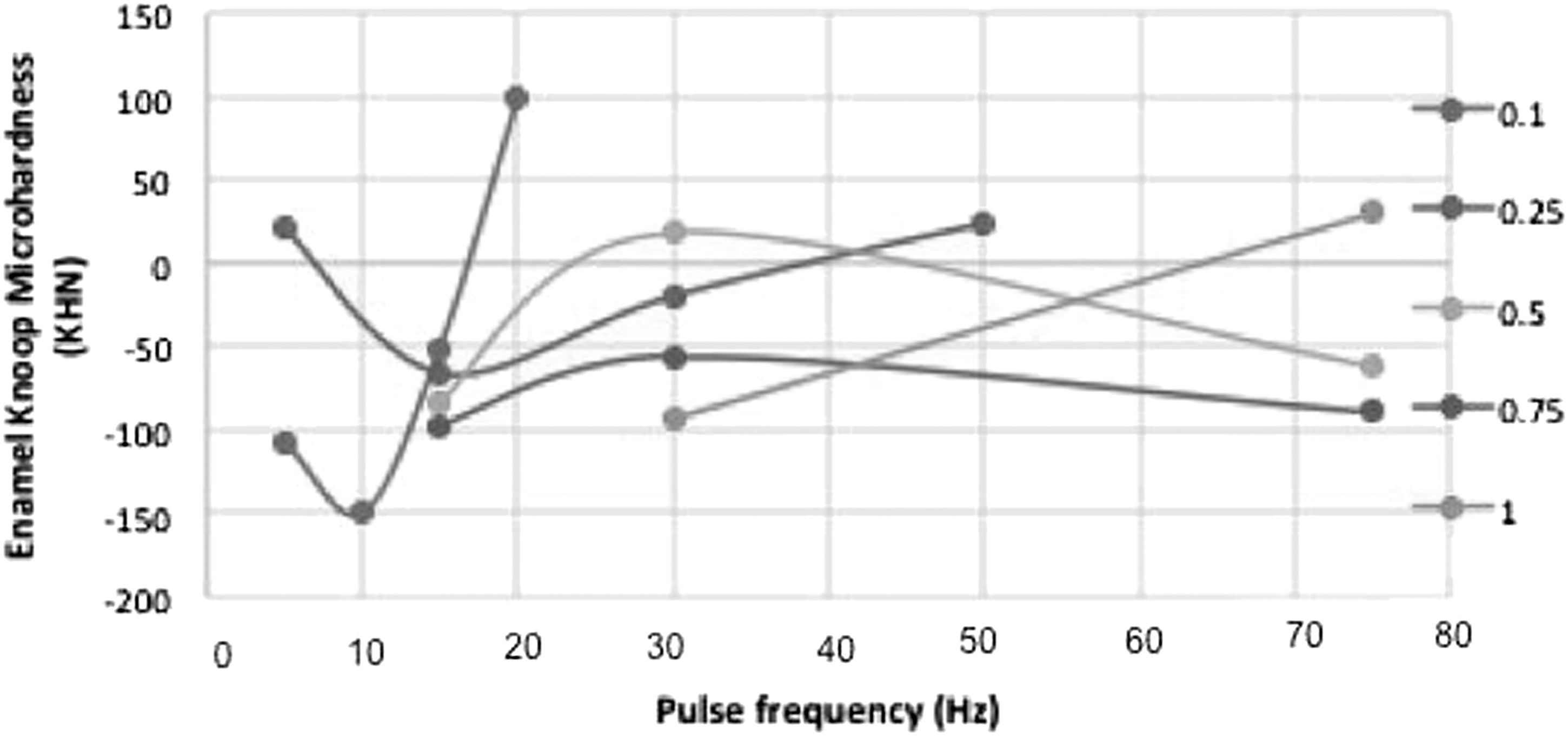
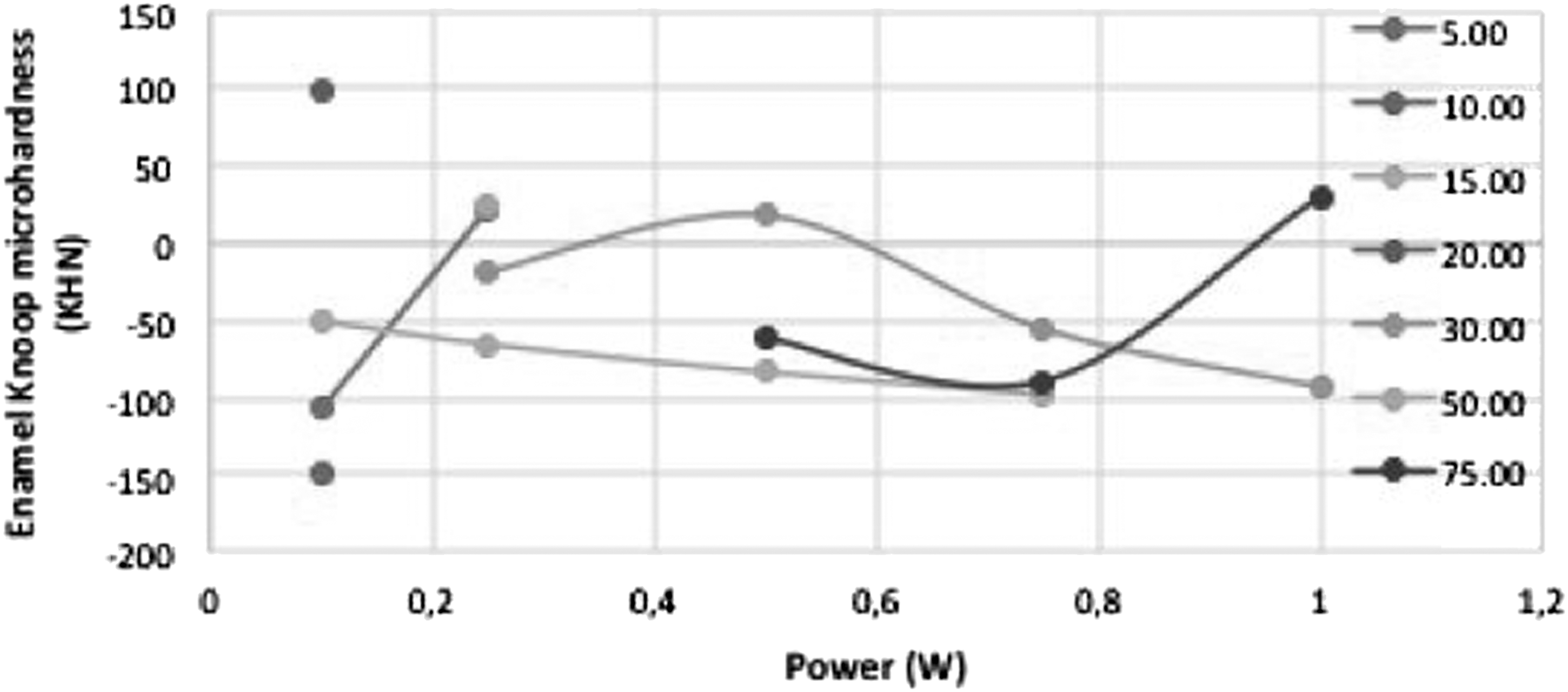
In Figs. 1 and 2, the effect of Er,Cr:YSGG on laser power or pulse frequency on enamel surface microhardness are shown. It was observed that the increase in the pulse frequency using power of 0.10 and 0.25 W resulted in a reduction of enamel surface microhardness followed by an increase (Fig. 1). In contrast, power of 0.50 and 0.75 W showed an initial increase followed by a decrease. Power of 0.75 W showed only an increase in the microhardness pattern. In Fig. 2, it was observed that increase of power using pulse frequency of 15 Hz resulted in enamel microhardness decrease. Pulse frequency of 30 Hz resulted in a discrete increase, followed by a decrease in enamel microhardness. Both power of 5 and 75 W seem to result in enamel microhardness increase as function of pulse frequency increases. Due to absence of more than one combination of power and pulse frequency of 10 and 20 Hz the effect pulse frequency was not described. Moreover, because of the loss of G9; G13; G17; and G18, the effect of 10; 20; and 50 W power could not be observed. Being so, only the combination used in G4, G5, G8, G11, and G20 resulted in higher microhardness enamel values after laser irradiation. In addition, only two samples of G11 (506.6 ± 104.5), G16 (240.4 ± 141.7), G20 (24.95 ± 3.46) and one sample from G4 (56.72), G5 (21.4), G7 (195.4), G10 (581.4) could be measured after erosive challenge.
Effect of Er,Cr:YSGG laser power in function of pulse frequency on enamel surface microhardness change after laser irradiation. Er,Cr:YSGG, erbium,chromium:yttrium, scandium, gallium, garnet.
Effect of Er,Cr:YSGG laser pulse frequency in function of power on enamel surface microhardness change after laser irradiation.
Enamel surface roughness test
The surface roughness results showed statistically significant triple interaction between the factors phase × power × pulse (p < 0.01). Tukey test showed no statistically significant difference among specimens surface roughness in the baseline phase (Table 3). After laser irradiation and erosive challenge statistically significant differences were observed in almost all groups with increase in the surface roughness (Tables 4 and 5). Comparing the phases, only the irradiation with low power (0.10 and 0.25 W) did not result in surface roughness increase.
Uppercase letters indicate differences between Frequency (columns) and lowercase letters indicate differences between Power (line).
Uppercase letters indicate differences between Frequency (columns) and lowercase letters indicate differences between Power (line).
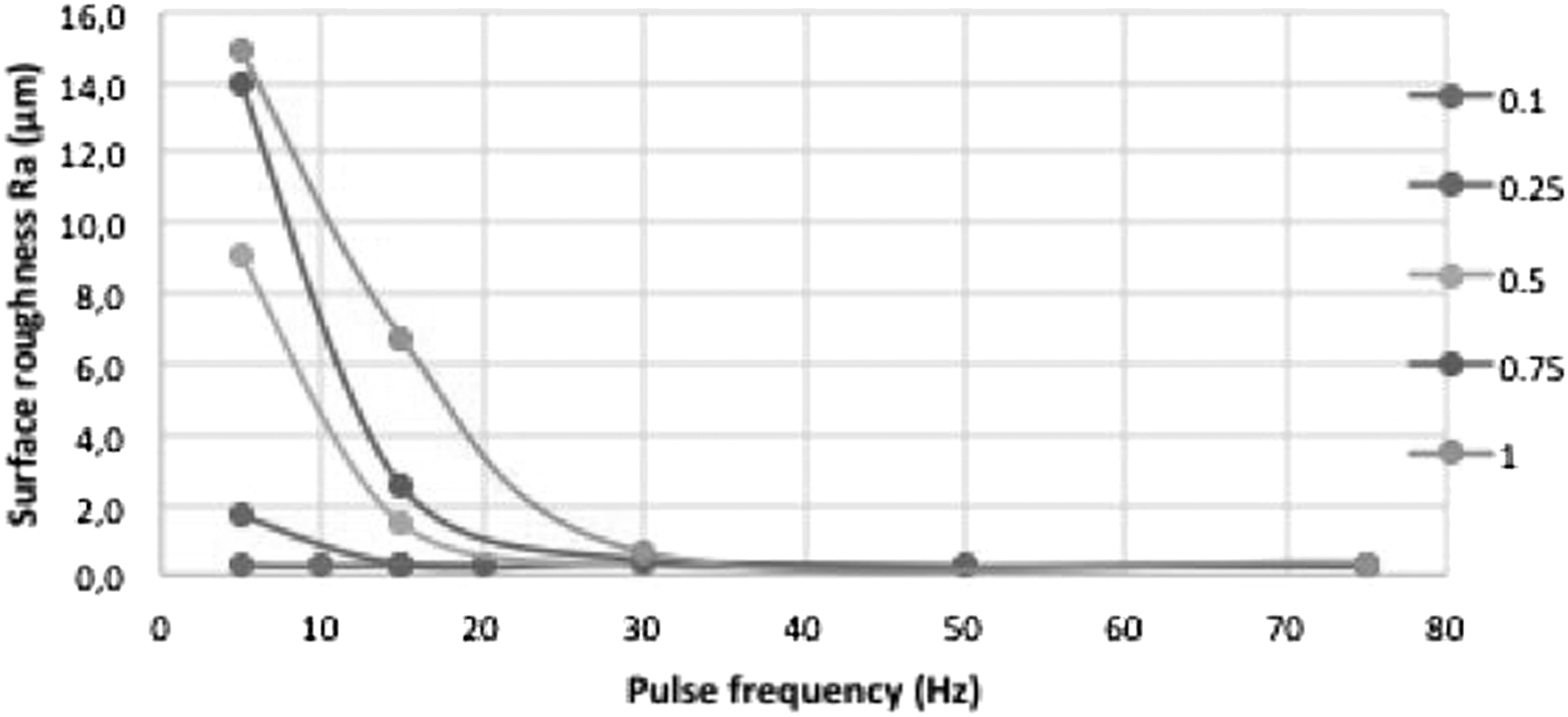
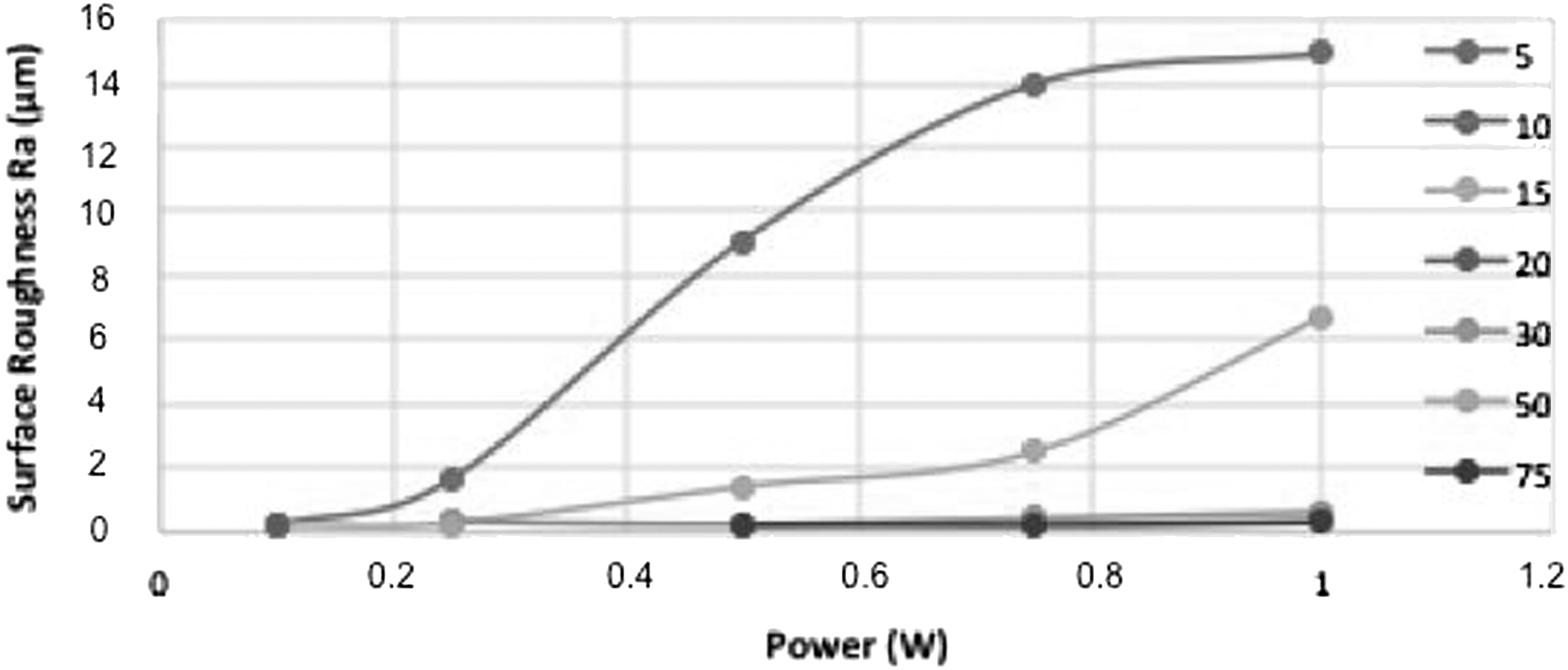
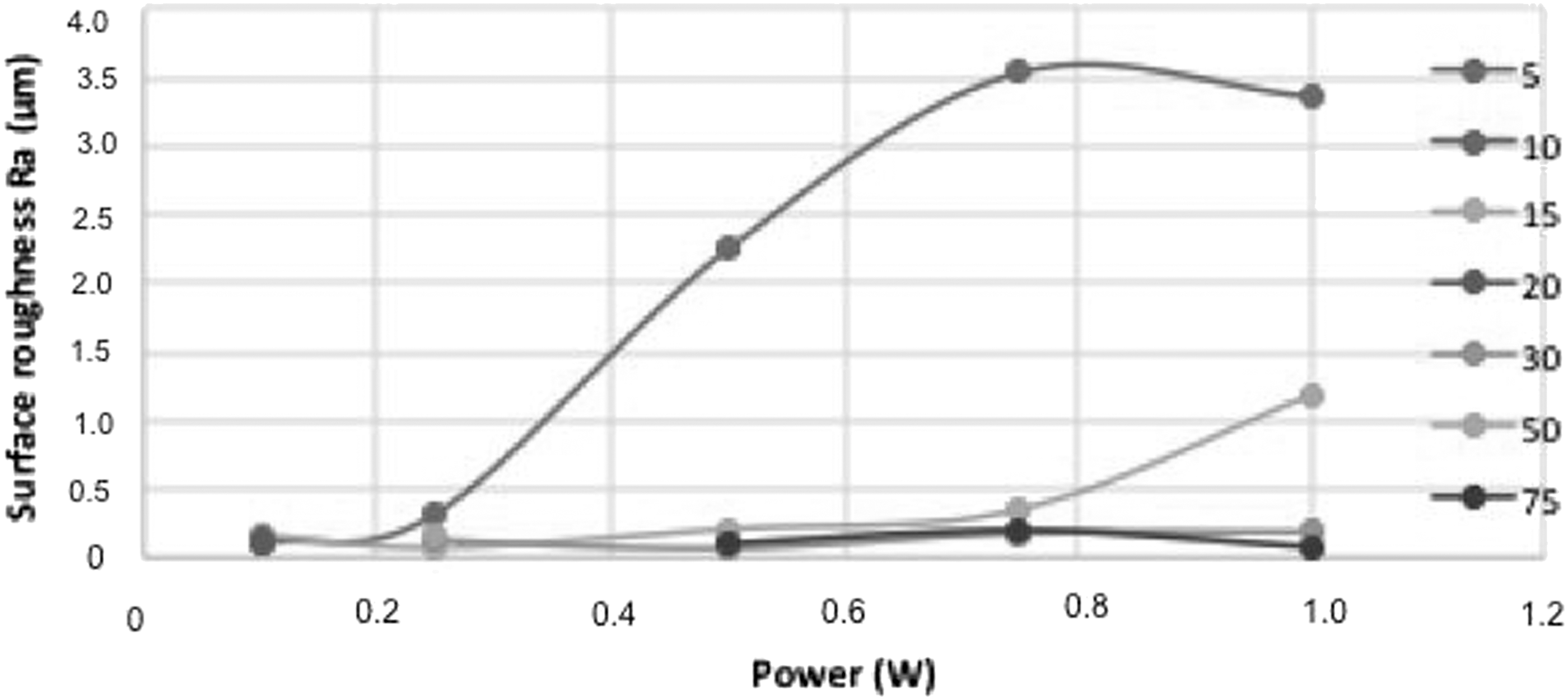
In Figs. 3 and 4, the effect of Er,Cr:YSGG on laser power or pulse frequency on enamel surface roughness is shown. It was observed that the increase in the pulse frequency using power of 0.25, 0.50, 0.75, and 1.0 W resulted in lower surface roughness (Fig. 4). The power of 0.10 W seems to cause fewer alterations on enamel surface roughness. Figs. 5 and 6 showed that increase of power using pulse frequency of 5 and 15 Hz resulted in an increase in enamel surface roughness. Due to absence of more than one combination of power and pulse frequency of 10 and 20 Hz the effect pulse frequency was not described. Both power of 5 and 75 W seem to result in enamel microhardness increase as function of pulse frequency increases.
Effect of Er,Cr:YSGG laser power in function of pulse frequency on enamel surface roughness change after laser irradiation.

Effect of Er,Cr:YSGG laser power in function of pulse frequency on enamel surface roughness change after erosive challenge.
Effect of Er,Cr:YSGG laser pulse frequency in function of power on enamel surface roughness change after laser irradiation.
Effect of Er,Cr:YSGG laser pulse frequency in function of power on enamel surface roughness change after erosive challenge.
The combination used in G1, G2, G3, G4, G8, G11, G12, G15, G16, G19, and G20 resulted in no surface roughness alteration after laser irradiation. After erosive challenge, the same behavior could be observed, but the alterations of surface roughness were lower than in the laser irradiation phase.
The Pearson correlation of microhardness and roughness was weak, but statistically significant (p = 0.007; r = 0.037). The Pearson correlation of power and microhardness (p < 0.001; r = −0.44) and roughness results (p < 0.001; r = 0.45) and the Pearson correlation of pulse frequency and microhardness (p < 0.001; r = 0.43) and roughness results (p < 0.001; r = −0.47) were statistically significant.
Discussion
The present study systematically evaluated the effect of low-energy densities produced by Er,Cr:YSGG laser with different setting parameters, including laser pulse frequency and power, on bovine enamel surface microhardness and roughness. Previous studies showed that differences between human and bovine enamel are not significant, thus enabling the substitution of human enamel by bovine enamel, even for erosive models of in vitro studies. 27
Surface microhardness, surface profilometry (roughness), microradiography, chemical analysis, and scanning electron microscopy (SEM) have been considered the most established laboratory assessments for enamel erosion. 28 Among them, surface microhardness has been reported to have sensitivity for measuring the very initial stages of erosion, when enamel softening starts, 28 and the further stages are better measured by surface roughness. Both tests were used in the present study to establish the best combination of laser parameters to supposedly alter the composition or solubility of the enamel to improve acid resistance without causing ablation or fusion.
This study was composed of three measurement times: baseline, after laser irradiation, and after erosive challenge. It was observed that baseline values were similar and differences in the next studied time must be attributed only to laser irradiation. In the best scenario, the ideal parameter for laser irradiation should not alter surface roughness or enamel hardness. The pulse frequencies of 5, 20, 30, 50, and 70 Hz and all studied power, except for 0.75 W, did not alter the enamel microhardness. On surface roughness, the combination of pulse frequency and power of groups G1, G2, G3, G4, G8, G11, G12, G15, G16, G19, and G20 showed no statistically significant differences. Crossing the surface microhardness and roughness results after laser irradiation, the safe combination of parameters was observed in groups G1, G4, G8, G11, G12, G15, G16, G19, and G20.
After erosive challenge, almost any groups could have its surface microhardness evaluated, and the only group that maintained normal microhardness was G11. This group also showed no alterations on surface roughness after erosive challenge. Thus, the use of Er,Cr:YSGG laser with pulse frequency of 30 Hz and power of 0.50 W seems to be the safest parameter to improve enamel erosive challenge.
Since a pattern of pulse frequency and power was not clear, the correlation of the parameters was tested, and the Pearson test suggested a positive correlation between microhardness and roughness of combinations tested. However, the correlation of response variables was very weak. It could be explained observing the correlation of each parameter and response variable. The increase in pulse frequency increased microhardness and decreased roughness and the increase in power decreased microhardness and increased surface roughness. This way the best parameter should be the highest pulse frequency and lowest power. Although the laser equipment used has a power output range of 0.10–10.0 W with pulse frequency repetition range of 5–100 Hz, the equipment does not allow the combination of highest pulse frequency and lowest power, and intermediate combinations in the G11 group seems to be the best option. In addition, the absence of these parameters is a limitation to the experimental design, since the behavior of these combinations in graphic representations and statistical analysis was jeopardized, and more studies must be developed testing the combination of highest pulse frequency and lowest power parameters.
The erosive challenge performed in this research used hydrochloridric acid. This treatment was designed to simulate the clinical conditions present during the early stages of dental erosion due to gastroesophageal reflux disease, since their treatment is often difficult to manage. 29,30 It can be observed that citric acid is the acid more frequently used in literature to promote erosive challenge. 31 Citric acid is commonly found in sparkling and fruit-based drinks, and it can provide a strong erosive challenge. It has been used to study initial erosion, and it is considered ideal when testing the potential for fluorides to prevent enamel erosion. 31,32 However, the treatment of dental erosion consists on removal or restriction of acid in oral cavity. Erosion produced by extrinsic acids derived from sparkling and fruit-based drink consumption must be demonstrated to patients. Intrinsic acid exposition of enamel by gastric acid requires medical referral and management of the patient as the primary method for its prevention and control. The focus of our study was to evaluate the efficacy of Er,Cr:YSGG laser parameters in incipient erosive lesions, which were created by a single exposure simulating gastric acid.
To the best of our knowledge there are no studies with Er,Cr:YSGG laser to prevent acid erosion prevention focused on gastric acid. Moslemi et al. (2009) 22 evaluated the effect of the Er,Cr:YSGG laser on the enamel acid resistance. By determining the calcium ion using atomic absorption spectrometry they observed no improvement in acid resistance. Due to the variety of parameters and methodologies used in the literature, it is difficult to make comparisons with different lasers and parameters. 26
Er:YAG lasers have a wavelength and enamel physical interaction close to Er,Cr:YSGG lasers. In a study with Er:YAG laser irradiation on surface roughness of eroded dental enamel with citric acid was observed that at any given pulse repetition rate, the surface topography of the laser treated substrates was similar following post erosive like lesion formation and that both showed more changes than in the control group (1, 2, 3, and 4 Hz), but atomic force microscopy images showed that the specimens irradiated by the Er:YAG laser at 1 Hz presented a less rough surface than those irradiated at 2, 3, and 4 Hz. 8 Dos Reis Derceli et al. (2015) 20 showed that Er:YAG laser setting of 60 mJ, 3.92 J/cm2, 2 Hz failed to inhibit dental erosive wear submitted to erosive challenge with citric acid. It has been reported that Er:YAG laser irradiation settings of 0.15 W, 85 mJ, an energy density of 5.2 J/cm2, and frequency of 2 Hz did not reduce the progression of erosive lesions on enamel submitted to in situ erosion with citric acid. They also showed that demineralization of eroded enamel after erosive challenge is approximately two thirds higher than that observed in sound surface and is higher than demineralization of initial carious lesions. 16
Conclusions
The interaction of Er,Cr:YSGG laser with different setting parameters of power and pulse frequency may differently alter enamel surface. Highest pulse frequency and lowest power combination next to 30 Hz and 0.50 W was considered the best combination to prevent enamel acid erosion.
Footnotes
Acknowledgment
This study was supported by CAPES.
Author Disclosure Statement
No competing financial interests exist.