
Abstract
Introduction
V
The pathophysiology of primary VVs is complex, 5 not clearly understood, 3 and has been relatively underinvestigated. 1 Several theories have been proposed to explain the development of primary VVs such as primary incompetence of the saphenous vein valves, inherent weakness of the vein wall, and the presence of arteriovenous fistulas. 6
Recent studies demonstrated that a disturbance in the regulatory mechanisms of vessel wall homeostasis is a potential factor in initiating VV development. 4 Altered angiogenesis could be one of the key factors in the pathogenesis of this disease. 4 Angiogenesis, the process of new blood vessel formation in normal and pathological conditions, is a multi-step process governed by positive and negative endogenous regulators. 7 In normal tissues, there is equilibrium between endogenous stimulators and inhibitors of angiogenesis. 7 Many growth factors have been identified that might be responsible for the angiogenic response in chronic venous insufficiency. 8 Although the molecular mechanisms of angiogenesis are relatively well characterized, the successive intervention and relationship between these growth factors during the angiogenic cascade are less understood. 7 Based on this, several investigators have suggested the potential benefits of studying the plasma concentration of some growth factors (such as VEGF, PDGF, EGF, ANG1, and ANG2) in relation to the pathophysiology of this disease.
Vascular endothelial growth factor (VEGF) is the most widely investigated and most efficient angiogenic molecule, characterized 20 years ago. 7 It is a highly specific potent mitogen of vascular endothelial cells (ECs). 9 It promotes EC survival and migration and increases microvascular permeability. 9 This will play an important role in maintaining vascular reactivity and integrity. 10 Platelet-derived growth factor (PDGF) is a potent mitogen and chemotactic factor for a variety of mesenchyme-derived cells. 11 It plays an important role in wound healing, stimulating cell proliferation, migration, and angiogenesis. 7 Epidermal growth factor (EGF) is a small polypeptide that stimulates cell proliferation. 12 Angiopoietin 1 (ANG1) and angiopoietin 2 (ANG2) are proangiogenic factors secreted by activated ECs that bind the tyrosine kinase (Tie2) receptor with similar affinity, but opposing activity. Numerous investigations have revealed the significance of Tie2 in normal and pathological conditions that destabilize the blood vessel and sustain angiogenesis. 13,14 For many years, the traditional treatment of greater saphenous vein (GSV) insufficiency was high ligation with or without stripping of the vein. Today, endovenous laser ablation (EVLA) is a common procedure in daily practice that can be performed in an outpatient setting. 15
Many studies have explored the changing plasma concentration levels of growth factors in patients with primary VVs, both before and after treatment by traditional surgery. 16 The purpose of our research was to measure plasma VEGF, PDGF, EGF, ANG1, and ANG2 levels among patients with VVs before and after treatment with EVLA to investigate the role of these growth factors in the development of VVs.
We could find no previous similar studies. The main initial hypothesis is that the altered plasma concentrations of these growth factors in pretreatment patients with primary VVs would be normalized (compared with a control group) after treatment with EVLA.
Materials and Methods
This is a prospective nonrandomized study. Between March and August 2016 inclusive, thirty patients with lower extremity primary VVs (who were planned to undergo EVLA) were enrolled. Twenty healthy adults were used as a control group. The study was approved by the Institutional Review Board (IRB) at King Abdulla Teaching Hospital (Registration No.: 31/94/2016). A written informed consent was obtained for each participant.
Patients
After a full history and physical examination suggestive of primary lower limb VVs, patients underwent preoperative duplex ultrasonography for both deep and superficial venous systems to evaluate the patency of the deep veins and look for saphenofemoral junction (SFJ) incompetence.
Inclusion criteria
Patients should be 18 years of age and above with primary lower extremity VVs, judged by a single vascular surgeon to be candidates for EVLA. Patients should exhibit SFJ incompetence with reflux in the GSV, in the absence of any evidence of acute or chronic deep venous thrombosis, as proven by preoperative duplex ultrasonography.
Exclusion criteria
Patients with a concurrent acute illnesses, chronic disease process, cancer, arteriovenous malformation, and atherosclerosis were excluded. Patients receiving medications and patients not candidates for EVLA (severe tortuosity, very superficial vein <1 cm deep in the skin, and aneurysmal vein >2.5 cm in diameter) were also excluded from the study.
After applying the abovementioned inclusion and exclusion criteria, thirty patients were enrolled.
Patients were classified according to the CEAP classification of American Venous Forum for chronic, lower extremity venous disease. 2
Technique of EVLA
The procedure was performed under tumescent anesthesia. The GSV was cannulated just below the knee by ultrasound-guided micropuncture. The tip of the laser fiber (diode laser features a wavelength of 1470 nm) was positioned 2 cm distal to the SFJ, and then the pullback technique was used to deliver the energy. Patients were discharged on the same or next day according to patient preference. They were given paracetamol 1000 mg four times per day if they had pain and advised to wear elastic stockings for 1 week. Follow-up Duplex U/S was done 1 week after EVLA to evaluate the adequacy of surgery by demonstrating the complete obliteration of the GSV and to rule out major thrombotic effects.
Control group
Twenty healthy persons coming to the blood bank for blood donation (14 males and 6 females with a mean age of 37.21 years) were selected as a control group. They did not have any clinical evidence of chronic venous insufficiency or lower extremity VVs and they were not on chronic medications or used any type of medications during the last 2 weeks.
Sample collection and processing
Blood specimens were collected from the brachial vein using venipuncture into two plain tubes on the morning of surgery and 1 week after the EVLA. The serum was obtained by incubating the samples on ice for 30 min. The samples were then centrifuged at 4000 rpm for 7–10 min. The resulting supernatant was transferred to multiple Eppendorf tubes and stored at −80°C in freezers for analysis.
Determination of serum growth factor levels
Assessment of plasma growth factor levels was done using the sandwich enzyme-linked immunosorbent assay (ELISA) technique according to manufacturer's recommendations. Human ELISA kits for ANG1, ANG2, PDGF, VEGF, and PDGF (Abcam, UK) were used in this study.
Prepared standards and samples were added into 96-well plates and incubated for 2 h at room temperature. After washing with a buffer, preprepared biotin conjugate was added and incubated for 2 h at room temperature. Then, preprepared streptavidin-HRP was added and incubated for 1 h at room temperature, followed by incubation with TMB substrate solution for 30 min at room temperature, avoiding exposure to direct light. The reaction was subsequently stopped by the provided stop solution. Plates were immediately read on a microwell ELISA reader spectrophotometer using 450 nm as the primary wave length. Then, absorbance readings underwent a series of mathematical equations to generate a standard curve to calculate sample results.
Statistical analyses
Data are expressed as mean ± standard deviation. GraphPad Prism software (company and country) was used for statistical analyses. Unpaired nonparametric Student's t-test was used to compare between the means. Differences were considered statistically significant at p < 0.05.
Results
Thirty patients (17 men and 13 women) with primary VVs were enrolled. The mean age was 38.11 years (range, 21–57). The control group consisted of 20 persons (14 men and 6 women) at a mean age of 37.21 years (range, 20–55). Therefore, the mean age and gender distribution between the study group and the control group were comparable.
Postoperative complications
Complications that could potentially interfere with the results were not encountered. Doppler US performed 1 week after EVLA revealed no evidence of deep vein thrombosis or superficial thrombophlebitis. No wound infection occurred.
The patients were categorized according to CEAP classification of American Venous Forum for chronic lower extremity venous disease. Twenty-eight patients were in class C2 (VVs) and only two patients were in class C6 (VVs and active ulcer). All patients had EPAS2-3PR CEAP classification.
A significant difference was obtained between pre-EVLA, post-EVLA, and control group for each studied growth factor.
The mean plasma concentrations of (VEGF, PDGF, EGF, ANG1, and ANG2) in preoperative, postoperative, and control group are shown in Table 1. A significant difference was obtained between pre- and postoperative values and between preoperative and control group with p = 0.001 and p = 0.004, respectively. On the other hand, no significant difference was found between postoperative and control groups, especially in EGF and VEGF (p = 0.564, 0.515, respectively) (Fig. 1).
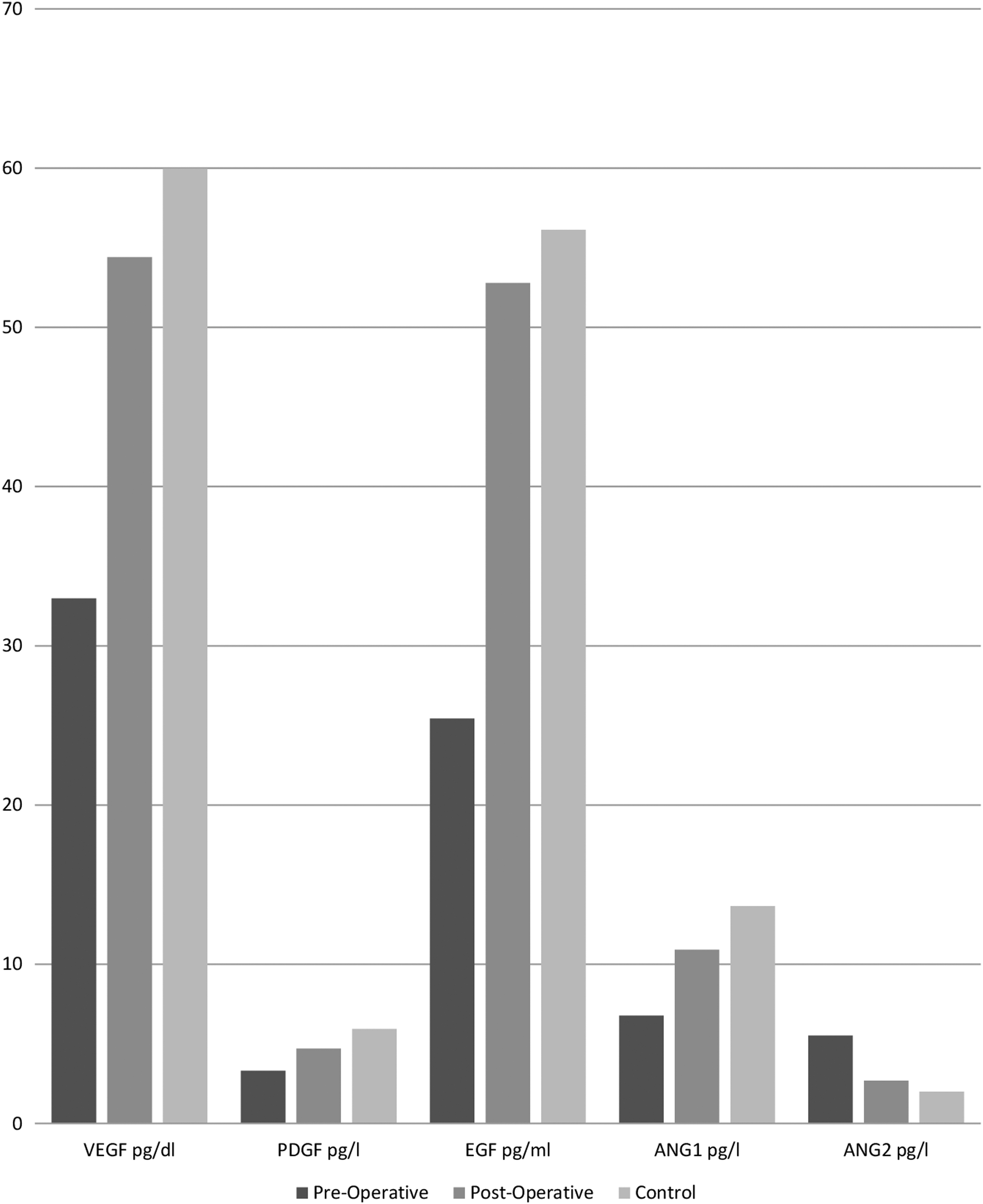
Plasma VEGF, PDGF, EGF, ANG1, and ANG2 levels before and 1 week after treatment with endovenous laser ablation compared with the control group.
Discussion
Many studies have explored the potential relationship between primary VVs and growth factors. 16 During the process of angiogenesis, growth factors might become altered either as a causative mechanism or simply as a result of the pathological process. 5 Previous published data in the literature demonstrated changes in plasma concentration of patients with primary VVs, but not of patients with VVs post-treatment with EVLA. Shoab et al. demonstrated elevating plasma VEGF levels in patients with venous disease compared with the control group. 17 Howlader and Coleridge Smith showed a trend toward raised VEGF in all stages of chronic venous disease, but this only reached statistical significance in those with healed ulceration. 8 Similarly, Tisato et al. studied the plasma concentration of VEGF, PDGF, and EGF before and after hemodynamic correction by conservative hemodynamic correction of venous insufficiency (CHIVA) and found significant increase in preoperative results compared with the control group, which were significantly decreased post-treatment. 16 In contrast, Hollingsworth et al. suggested that loss of VEGF release with experimentally induced venous stasis may be an important mechanism in the development of primary VVs. 10
This is the first study to monitor changes in the plasma concentration of some growth factors after treatment of primary VVs with EVLA. Our results demonstrated a statistically significant trend toward normalization of the concentration of VEGF, PDGF, EGF, ANG1, and ANG2 1 week after treatment with EVLA compared with the control group. The significance was not only in the mean values but also in the interference of distributions. The interference of distributions of growth factors (especially ANG1 and ANG2) was relatively small preoperatively versus controls and preoperatively versus postoperatively. Such trends in the concentration changes might indicate a pivotal role in the pathophysiology of primary VVs.
The timing of the post-EVLA sample collection was determined according to the half-life of the studied factors. The half-lives of VEGF, PDGF, EGF, and ANG1 and ANG2 are 30–45 min, 8–13 h, 1.4 h, and 18 h, respectively. 9,11,12,18,19 Therefore, postoperative blood samples were collected 1 week after EVLA to minimize the effect of surgery and immediate postoperative recovery period on the concentrations of these growth factors. The numbers of both patients and control group were determined according to a limited budget of this study. Future studies may calculate and recruit accordingly by increasing the number of patients, considering patients with both bilateral and unilateral VVs treated surgically, and increasing the 1-week period for blood sampling to 1 month postprocedure to minimize the effect of the procedure on levels of plasma growth factors.
This study demonstrated a statistically significant decrease in plasma concentrations of VEGF, PDGF, and EGF in patients with primary VVs, which were significantly increased to reach the concentration of the control group 1 week after treatment with EVLA. These findings may suggest the presence of a negative feedback mechanism triggered by VVs that regulates systemic VEGF, PDGF, and EGF expression levels. Previous studies demonstrated changes in messenger RNA expression and altered transcription of growth factor receptors in the wall of the excised VVs. 4,5 Based on this, we can explain our results by suggesting that there is an inhibitory feedback mechanism that downregulates systemic production of these growth factors. In addition, treatment of VVs with EVLA would eliminate the inhibitory mechanism and thereby allow systemic production of VEGF to resume, over time, to nearly normal concentrations as shown in our results.
Plasma concentrations of ANG1 and ANG2 in patients with VVs have not been fully elucidated in the literature. Agonistic ANG1 functions are antagonized by ANG2, which is believed to inhibit ANG1–Tie2 signaling. ANG1 is required to maintain the quiescent resting state of the endothelium, while ANG2 destabilizes the quiescent endothelium. 20 Assuming that the endothelium is destabilized in the wall of VVs, this could explain the decreased plasma concentrations of ANG1 in preoperative results. The increased level of ANG2 is most likely reflecting the post-ELVA inflammatory activity. On the other hand, the increased plasma concentration of ANG1 (which is dominant in stable activity) would be consistent with the assumption that the endothelium is stabilized postoperatively.
Growth factors play a key role in the process of wound healing. Thus, activation of the inflammatory cascade in the postoperative period 20 is another possible explanation for the elevated plasma concentration of these growth factors in the post-treatment group. However, the fact that the tissue damage induced by EVLA is minimal, when compared with the traditional open ligation and stripping surgery, favors the explanation of the difference in concentrations on the basis of the pathophysiology of VVs and their treatment. EVLA induces thermal energy that damages the endothelial and intimal layers, the internal elastic lamina, and the media to some degree. 21 This is far less than the amount of damage induced by formal surgery. However, future studies may compare the effect of EVLA versus open surgery on growth factors.
Post-EVLA thrombotic events might affect the growth factor concentration. Although major thrombotic events were not encountered in our patients, as evident by follow-up Duplex U/S after 1 week, occult thrombi in the wall of treated veins might be another explanation for the change in concentration. Further detailed studies should take this into consideration. The complexity of the pathogenesis of primary VVs indicates that further investigations are needed to understand the interactions between growth factors and angiogenesis.
Conclusions
The altered plasma concentrations of growth factors (VEGF, PDGF, EGF, ANG1, and ANG2) among patients with primary VVs normalized 1 week after treatment with EVLA compared with the control group. This may support their role in the pathogeneses of the disease. Future studies may evaluate if these changes can play a prognostic and/or predictive value regarding the adequacy of treatment and the possibility of recurrence.
Footnotes
Author Disclosure Statement
No competing financial interests exist.