
Abstract
Introduction
L
Many previous studies have been conducted to investigate effects of low-power laser irradiation on cells and biological systems. However, the molecular mechanism of changes in cell composition and its functions induced by laser irradiation still have not been fully understood so far. 1,3,13 –16 Indeed, in the last few years, several laser-induced cellular function changes have been proposed, measured, and explained by Wang et al. 7,17,18 and Farivar et al. 19 They assumed that cytochrome c oxidase, a multicomponent membrane protein, is the main optical absorber of red and infrared light that activates cellular functions. However, the dose control and side effects of laser irradiation/stimulation are still relatively unknown. 7,17 –19
Red blood cells (RBCs) are considered to be one of the simple biological systems because they do not have a nucleus. 20 “The RBC membrane is a composite structure, consisting of a membrane skeleton protein lattice and a lipid bilayer to which the membrane skeleton is attached by way of interactions with trans-bilayer proteins. The major components of the membrane skeleton are α- and β-spectrin, ankyrin, F-actin in the form of short filaments (protofilaments), protein 4.1R, adducin, dematin, tropomyosin, tropomodulin, and proteins 4.2. Spectrin is the most plentiful skeletal protein.” 21 Many scientists have studied the effects of low-power laser on RBC membrane proteins. 1,3 On one hand, several studies showed a significant effect of laser irradiation on RBC membrane functional state. 1,3,16 On the other hand, another team of researchers found no noticeable effects of laser irradiation on these cells. 22,23
The current study was carried out to investigate the influence from a low-power diode laser (50 mW) irradiation on the structure of membrane proteins of human RBCs over time.
Materials and Methods
Sample collection
The blood samples of 5 mL were drawn from 12 apparently healthy participants through a venipuncture into ethylenediaminetetraacetic acid-containing tubes (1 mg/mL of blood) as an anticoagulant. The samples were then processed immediately after blood collection.
In this work, the experiments were performed in full compliance with the ethical guidelines of the University of Baghdad-Iraq, strictly prohibiting any disclosure, sharing, or referring to the identities, private information or personal records of any donors or volunteers who provided written consent to have their data used.
Suspending media
Three suspending media were used in this study. Each medium is denoted by M carrying a number as a subscript that refers to its osmolarity in mOsm. The isotonic solution (M300) consists of mM KCl 150 and Tris-HCl 10. The hypotonic solution (M200) consists of mM KCl 100 and Tris-HCl 10. The hypertonic solution (M600) consists of mM KCl 150, Tris-HCl 10, and sucrose 300. The three solutions were brought to PH 7.4 to simulate the PH of the blood.
Laser setup
The experiments were conducted with a low-power diode laser source with a collimated beam that has an irradiation spot of 5 mm diameter. The wavelength of light that emitted from this laser was 650 nm (red light) with output power equal to 50 mW (mill watt). The power density was calculated by dividing the power (W) to the area of irradiation spot (cm2; 0.05 W/0.2 cm2) = 0.25 W/cm2. This beam was directly used to irradiate blood samples in tubes for 10, 20, 30, and 40 min, respectively.
Preparation and irradiation of RBC suspension
Separation of blood components was performed by centrifuging the blood at × 1500g for 5 min. After that, the plasma and the buffy coat (leukocytes and platelets) were removed via gentle aspiration. Then, the packed RBCs were washed two times in M300 and the supernatant was neatly discarded after each wash. Next, the washed packed RBCs were suspended in M600 at an approximate hematocrit of 2%, and the suspension was divided into five equal aliquots. One aliquot was left without irradiation and served as control, whereas the remaining four aliquots were exposed to the laser for 10, 20, 30, and 40 min, respectively. After each specific irradiation time, the control and the irradiated tubes were centrifuged. After centrifuging, the packed RBCs were washed one time in M200 and resuspended in M200 at an approximate hematocrit of 0.2%. The percentage of cells that suffered from denaturation of membrane proteins was calculated in each sample as described below.
Counting the denatured or damaged cells
The technique that was used in the current study for counting the denatured or damaged cells was previously mentioned by Fischer, 24 –26 in which opposing regions of the inner membrane surface touch each other in the dimple area of the membrane when RBCs were suspended in a hypertonic solution. In normal RBCs, such a mechanical contact is reversible by swelling the cells in hypotonic solution. However, this mechanical contact can be made permanent and form membrane cross bonding known as bridges. This is due to the denaturation of membrane proteins when the cells are treated with chemical compounds such as H2O2, glutaraldehyde, SH, amino, and carboxyl reagents 26 –29 or simply heated to temperatures between 42°C and 48°C. 30 When cross-bonding formations take place, referring to the denaturation or damage in membrane proteins, single or several hollows (white dots) can be seen under the microscope on suspending the cells in a hypotonic solution. As seen in Fig. 1, the white dots represent the sites of the cross-bonded regions between the two sides of membrane proteins; these cross-bonds are strong enough to resist the membrane tension that occurs at osmotic swelling.
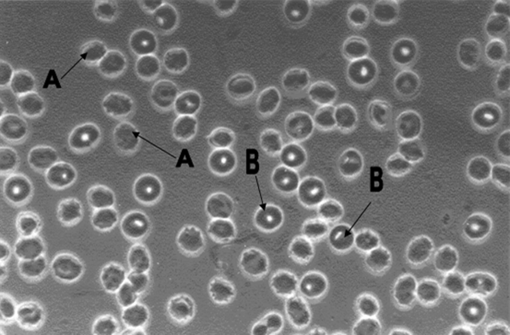
Red blood cells (RBCs) in M200 solution under the microscope (40 × objective) after they were exposed to laser irradiation for 30 min. A, normal cells. B, denatured or cross-bonded cells (the white dots inside the RBCs represent the sites of the cross-bonded regions between the two sides of membrane proteins).
In the current study, a Neubauer counting chamber was utilized, and random samples of RBC suspension were selected to count the damaged cells by positioning and filing according to a fixed scheme. For counting the damaged RBCs after laser irradiation, a drop of the RBCs in M200 solution was placed on the counting chamber and examined by phase-contrast microscopy (40 × objective). Two medium-sized squares on the counting chamber were selected for counting the cross-bonded or damaged cells in all the samples studied. Then, the percentage of cross-bonded cells was calculated (Fig. 1).
Statistical analyses
Student's t-test for paired data was used to determine the differences among experimental groups. Results are expressed as mean ± standard deviation. p Value <0.05 was considered to be statistically significant.
Results
The present study was performed to investigate the effect of low-power diode laser (with an output power of 50 mW) on RBC membrane protein structure during the exposure of RBCs to the laser for different periods of time. The membrane cross-bonding technique previously mentioned by Fischer 24 –26 has been used in the current study to detect denaturation in membrane proteins. In this study, RBCs were suspended in a hypertonic solution (M600) to induce contact between the two inner faces of the membrane, and exposed to the laser for different time periods. After irradiation, the cells were resuspended in a hypotonic solution (M200) and examined microscopically. If the laser irradiation causes denaturation in membrane proteins, cross-bonding or bridges will form permanently and could be seen as single or multiple hollows (white dots) inside the cells. In contrast, unaffected RBCs appear under the microscope without any hollows (Fig. 1).
The results indicated that the percentage of denatured or cross-bonded cells in two irradiated aliquots for 10 and 20 min, respectively, was nearly similar to the percentage of denatured cells in the nonirradiated control aliquot. These results clearly show that the irradiation of RBCs by low-power diode laser (50 mW) up to 20 min did not cause any change in membrane protein composition, whereas increasing the irradiation time to 30 min caused denaturation of membrane proteins, resulting in the formation of membrane cross-bonding in a considerable number of RBCs (9.45 ± 2.3%, n = 12) compared to control aliquot. Further, the percentage of denatured cells increased significantly when the irradiation time was extended to 40 min (28.71 ± 4.58%, n = 12) compared to control aliquot (Fig. 2). The current results showed microscopically that there is no hemolysis or membrane rupture after irradiation of RBC suspension by the low-power diode laser for all time periods of irradiation in comparison with control sample.
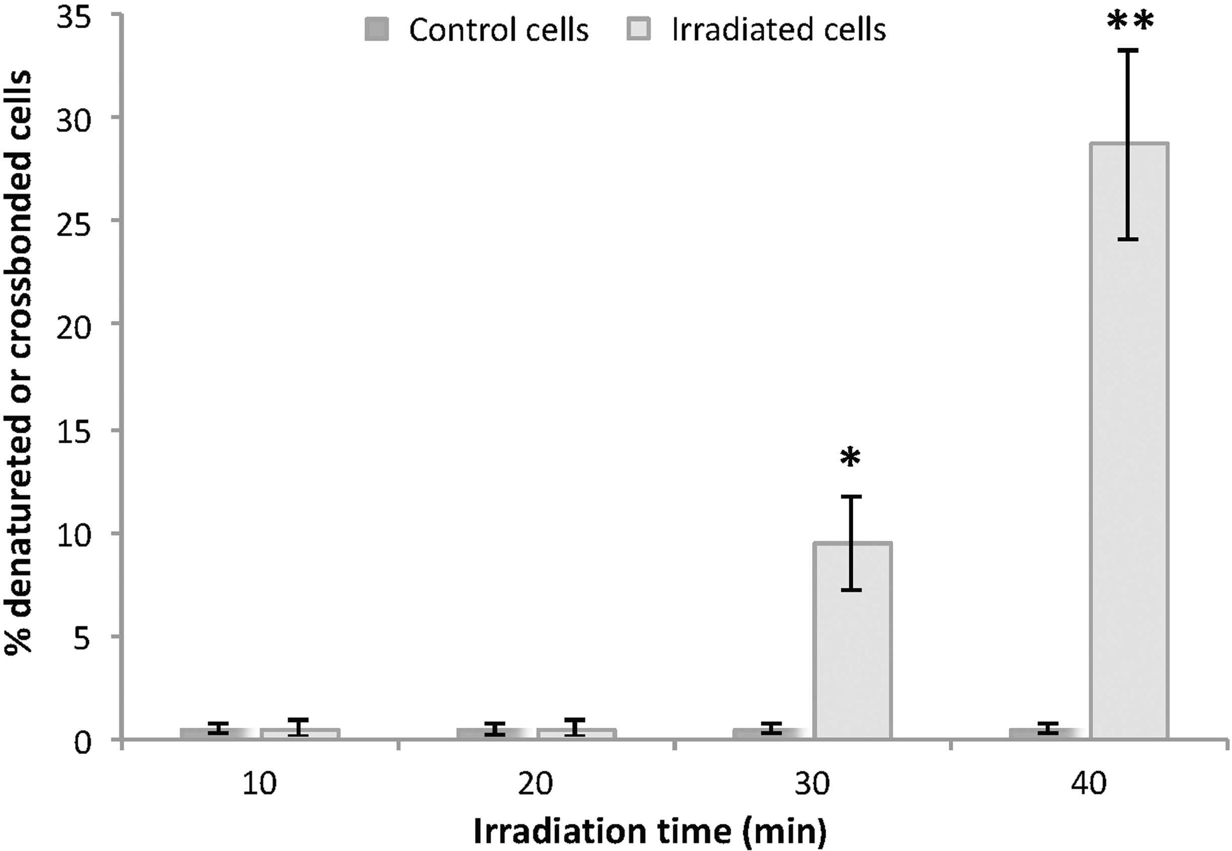
Influence of irradiation time on the percentage of denatured red blood cells (RBCs) that were exposed to low-power diode laser (50 mW). Each value represents mean ± standard deviation of 12 experiments for 10, 20, 30, and 40 min of irradiation time. *p < 0.0001, **p < 0.0001 (by paired t-test) in comparison with nonirradiated RBCs (control).
Discussion
Many studies were conducted to investigate the influence of low-power laser irradiation on RBCs. For instance, several researchers have detected that the deformability of erythrocytes was decreased 13 and their osmotic fragility was increased after exposure to low-power laser. 13,31 Further, other researchers found that low-power laser irradiation induced long-term conformational transitions of RBC membrane due to changes in membrane proteins and lipid bilayer. 16 In contrast, other group of researchers did not find any difference between the irradiated and control cells in hemoglobin content, the absorbance of single cell, and in cell shape after an exposure of these cells to the low-power laser. 23
The present research work was performed to investigate the influence from a low-power diode laser (50 mW) irradiation on the structure of membrane proteins of human RBCs over time. The results of this study indicated that an irradiation of RBCs by low-power diode laser for 20 min did not cause any change in membrane protein composition, whereas increasing the irradiation time to 30 min caused denaturation of membrane proteins, resulting in the formation of membrane cross-bonding in a considerable number of RBCs, and the percentage of denatured cells increased in a dose-dependent manner to the irradiation.
Several studies indicated that when the RBCs are heated at about 45°C, some denaturation of membrane proteins begins, and a substantial amount of RBC membrane proteins are denatured at ∼50°C due to dissociation of the spectrin molecules into α and β subunits. 25,26,30,32 According to the present results, the laser–RBC interaction mechanism that resulted in the denaturing/cross-bonding of RBCs could be interpreted as follows “the laser photons are absorbed in RBC membrane and hemoglobin by electronic absorption bands called chromophores, the energy of these absorbed photons converts to heat. When the irradiation time by low-power laser was 20 min or less, it seems that the heat energy was not enough to increase the temperature of membrane proteins above 45°C, which is required to induce denaturation of membrane proteins. As the time of irradiation increased to 30 min, there were a significant number of cell membranes starting to change and denature, and the denaturation process increased with increasing irradiation time; it is obvious that the prolongation of the irradiation time leads to an increase in the numbers of absorbed photons. The photon energy is converted to heat that leads to an increase in RBC temperature above 45°C (and may even reach 50°C), which causes denaturation of membrane proteins in most cells. As a consequence of the denaturation of membrane proteins especially spectrin, cross-bonding or bridges will form between the opposite faces of the membrane. These cross-bonded areas appear as white dots inside cells under the microscope, which is considered a good indication of the denaturation in membrane proteins.”
The calculated results of the present study show the maximum safe dosage from a low-power diode laser (650 nm) at 0.05 W and a power density 0.25 W/cm2 for an irradiation time of 20 min is 300 J/cm2. As mentioned previously, a 20-min irradiation time did not cause any denaturation or damage in the RBC membrane protein with a corresponding dose of 300 J/cm2. In comparison, the effective dose that is used for wound healing is 57.3 J/cm2, 10 which is much lower than the maximum safe dosage that was determined in this study. This finding can be beneficial in determining an optimized dosage level in medical applications.
Conclusions
It can be concluded that the effect of a low-power diode laser (50 mW) on the RBC membrane proteins depends on irradiation time. When irradiation time is 20 min or less, the laser has no detectable effect on RBCs and does not cause any denaturation in membrane proteins. However, it could cause damage and denaturation of membrane proteins when the time of irradiation was increased to 30 min or more. Therefore, a low-power diode laser with an output power 0.05 W and power density of 0.25 W/cm2 can be used safely in medical applications that include blood irradiation because it will not “denature” or “cross-bond” RBC membrane proteins during medical applications when controlling the irradiation time to 20 min or less.
Footnotes
Acknowledgments
This study was carried out at the Laser Institute for graduate studies and College of Dentistry-University of Baghdad. Many thanks to Eben Allen for help in editing this article for grammar.
Author Disclosure Statement
No competing financial interests exist.