
Abstract
Introduction
PMMA
However, some researchers report rates of 22–30% for tooth debonding as a result of denture repairs, especially in the anterior region 2,10 and 33% as a result of accidents or mechanical fatigue. Consani et al. 11 reported that microwave irradiation for prosthesis disinfection significantly decreased the shear bond strength values in ridge lap area used for the denture tooth/resin adhesion. Despite this, grinding and monomer application on the bonding surface did not significantly improve the bond strength. Krishna et al. 12 also compared the bond strength of cross-linked acrylic teeth to three types of denture base resins after different chemical surface treatments and found that the bonding ability altered according to these variables. 11,12 Wax contamination of the ridge lap area of the teeth, 13 different polymerization techniques, or the careless application of the separating medium were carried out to test their negative effect on SBS. 10
An effective bonding relies on the chemical and mechanical preparation of the PMMA and the acrylic resin teeth, a very important factor for the long-term success of prosthesis. Researchers have described using different techniques, including sandblasting, 7,9,14,15 bur grooving, 9,16,17 wetting with methyl methacrylate (MMA) monomer, 9,18 laser irradiation, 9,19 or a combination of these, on the ridge lap area to improve the adhesion and increase the surface area. Although some of them found that the SBS was reduced with mechanical treatments like sandblasting and bur grooving, 7,16,17 some surface treatments enhanced the SBS between the acrylic teeth and the PMMA.
In the existing literature, lasers have been shown to provide a relatively safe and easy means for altering the surface of materials. Lasers' ability to change the surface topography and wettability characteristics are crucial factors on ceramics and metals for improved adhesion and bonding. 20 Akyıl et al. 21 observed that different type of lasers (Nd:YAG, CO2 and Er:YAG) is effective to modifying on the yttria stabilized zirconia surface. In addition to this Akin et al. 22 reported that Nd:YAG laser is able to alter zirconia postsurface causing scratch-like traces and shallow pits on the surfaces.
Recently, some studies 9,14,15 have evaluated the effects of laser irradiation on the SBS of acrylic teeth to PMMA, and laser irradiation of acrylic teeth using an erbium: yttrium aluminum garnet (Er:YAG) laser with 10 Hz/300 mJ for 20 sec was effective in obtaining better SBS, 9 and these studies showed that surface treatments produced irregularities in the PMMA. However, a literature investigation showed that no study has evaluated the effect of the Er,Cr:YSGG laser, in different powers, on the acrylic teeth bonding.
As such, the purpose of this study was to evaluate the effects of different pretreatment techniques (air abrasion, grinding with a tungsten carbide bur, and laser irradiation with different powers of 1–4 W) for enhancing the SBS values between acrylic teeth and PMMA. The null hypothesis was that there is no difference in the SBS of heat-cured denture base resin (PMMA) to acrylic resin teeth treated with different pretreatments, especially laser irradiation of different powers (1–4 W).
Materials and Methods
For this study, 70 central incisor denture teeth (Eray Delux, Turkey) were selected and ground on the ridge lap area with a tungsten carbide bur (H129FSQ; Brasseler USA) to remove the glaze layer and create similar angulations, then steam cleaned with distilled water to remove any residue. Abrasion of the teeth surfaces was carried out to a depth of less than 0.5 mm using a medium grit rotary abrasive. A digital caliper (Mitutoyo, Tokyo, Japan) was used to arrange in a 6 mm diameter the bonding area of each tooth, rounded to the nearest ± 0.03 mm. Then seven pretreatments were used on seven groups (n = 10).
G1- Control Group: These specimens were not treated.
G2- Ground with a carbide bur: The bonding surfaces were roughened with a tungsten carbide bur (Edenta AG, Hauptstrasse 7, Switzerland) using a speed of 15,000 rpm (K9, KaVo EWL, Leutkirch, Germany) for 10 sec by the same operator. The bur was moved on the surface in a scan at 1 mm/sec for a long specimen diameter.
G3- Air abrasion group: The bonding surfaces were sandblasted by alumina particles with a diameter of 120 μm at a pressure of 2 bar for 20 sec and from a distance of 10 mm.
G4-7 Laser groups: The bonding surfaces were irradiated with an Er,Cr:YSGG laser (Millennium; Biolase Technology, San Clemente, CA) that produces laser pulses at a 2.78 μm wavelength. The optical fiber of the laser was 6 mm in diameter and was placed 10 mm from the tooth surface and applied for 20 sec with water/air flow of 50% in each case. The laser parameters included a pulse energy of 100 mJ, a repetition rate of 10 Hz, a power setting of 1–4 W, and a pulse duration of 140–200 μs.
After the surface treatment protocols and before the SBS tests, rectangular wax specimens (36 × 12 × 5 mm) were prepared, and the heat-polymerized PMMA (Meliodent, Heraeus Kulzer GmbH Grüner Weg 11 63450 Hanau Germany, Lot Number: 12JAN143) was cured following the manufacturer's recommendation after the teeth were fixed and stored at 37°C for 24 h in distilled water. A universal testing machine (Lloyd LF Plus; Ametek, Inc., Lloyd Instruments, Leicester, United Kingdom) was used for the SBS tests, which were conducted with a crosshead speed of 1 mm/min. Figure 1 presents a schematic representation of the SBS test. The maximum force was recorded, and the SBS values were measured in megapascals (MPa), and the data were analyzed with a one-way ANOVA and a post hoc Tukey's test to determine the differences among the groups (p = 0.05).
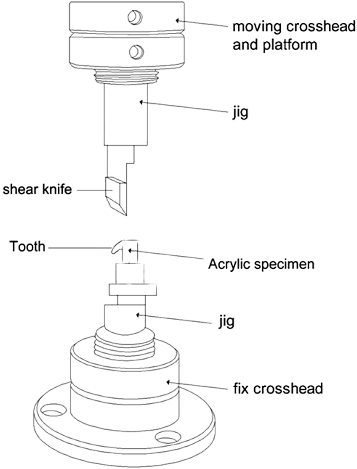
Schematic representation of test protocol (Akin et al. 9 ).
Analyses were performed using an SPSS 13.0 (SPSS for Windows; SPSS, Inc., Chicago, IL) software package with a p < 0.05 significance level. SEM and stereomicroscopy analyses have gained popularity for evaluating roughness area in the dental material surface after various pretreatments. 23,24 To evaluate the surface morphology, four additional specimens of the acrylic denture teeth were prepared in each group and evaluated for a surface analysis with a scanning electron microscope (SEM; JSM-6060LV, JEOL, Tokyo, Japan) at magnifications of 35 × and 150 × (Fig. 2) after covering the surface with a thin layer of gold. To evaluate the fracture pattern (Fig. 3), a stereomicroscope (Stemi DV4; Gottingen, Germany) was used at 32 × magnification, and the patterns were classified as adhesive failure, in which the acrylic denture teeth had completely separated from the PMMA surface; cohesive failure, in which the acrylic denture teeth were completely fractured; or mixed failure, in which both failure types were observed (adhesive and cohesive).
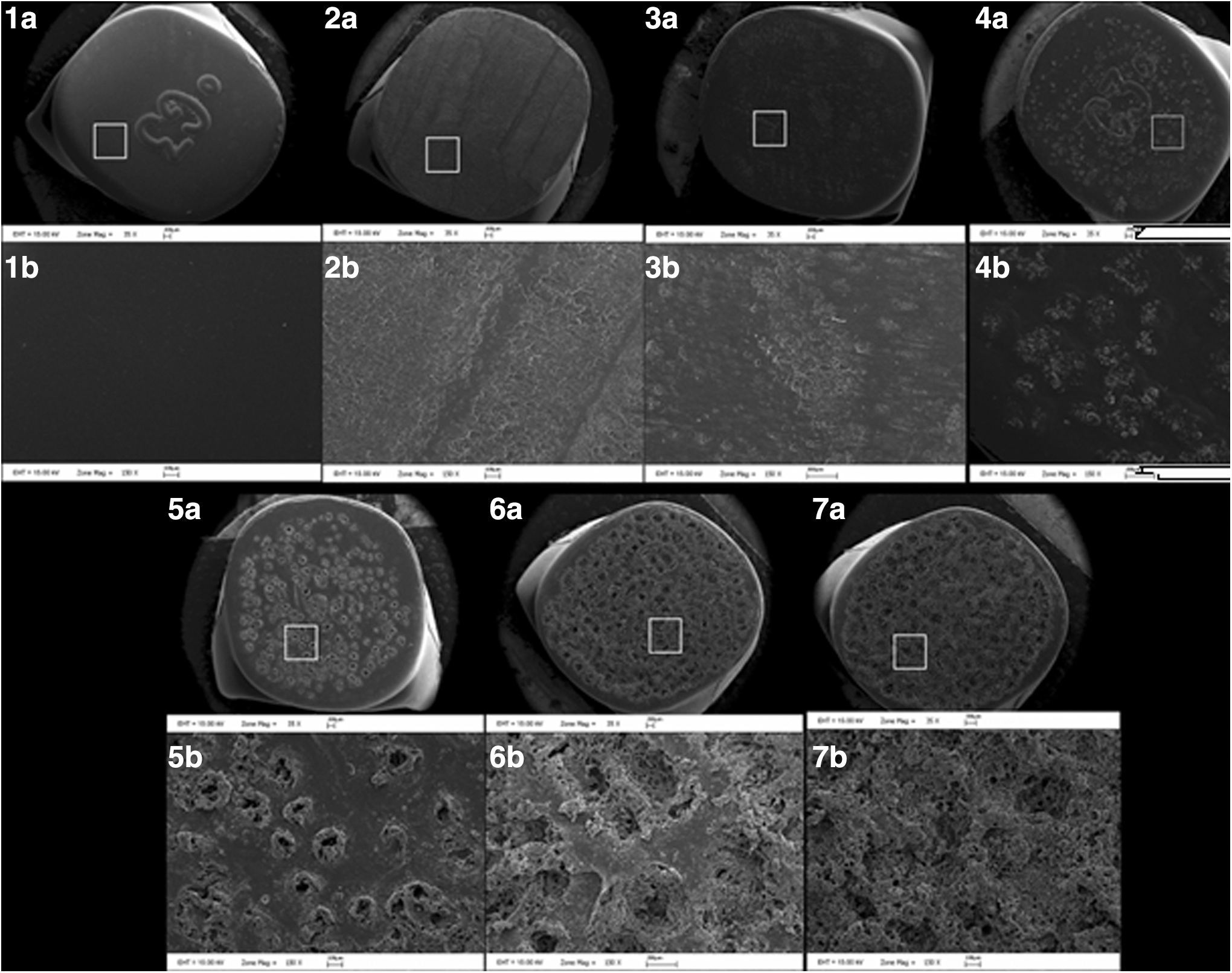
SEM images of the specimens ( × 3000). Surfaces:
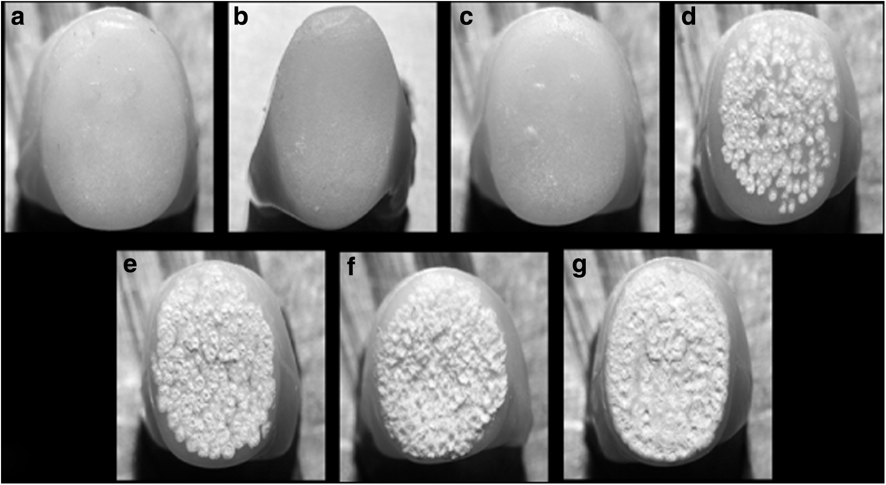
Stereomicroscope images of the specimens after surface treatments:
Results
The groups' mean, standard deviation, and statistical significance are presented in Table 1. The group of specimens that received air abrasion exhibited the highest SBS value (15.94 [2.1] MPa), and the lowest SBS values were observed in the control group (14.34 [1.8] MPa). All the surface treatments increased the surface roughness (Fig. 2, 2a/b–7a/b) and SBS values of all the specimens, but no significant differences were found between the control group and the surface treated groups (p = 0.521 by ANOVA) (Table 2). SEM images of the control, carbide bur, air abraded, and Er,Cr:YSGG laser-treated groups are presented in Fig. 2. Stereomicroscopy view of the groups is introduced in the Fig. 3. The control group specimen presented smooth and glossy surface in the stereomicroscopy evaluation (Fig. 3a), and these findings are compatible with the SEM evaluations (Fig. 2, 1a and 1b).
p > 0.05.
Mean with the same letters was not significantly different.
SD, standard deviation (p = 0.521).
When evaluating SEM images, it could be seen that surface treatments resulted in damage in varying rates. Carbide bur group demonstrated some scratch-like traces extending along on the surface (Fig. 2, 2a and 2b).
When the stereomicroscopy image was evaluated, it could be seen that bur traces and slightly roughened flat areas existed between these deeper traces with dull appearance on the surface due to structural loss (Fig. 3b). Air abraded group showed irregularities with porosities and shallow pits compared to the untreated specimen; also, remnants of the alumina particles are viewed on the surface (Fig. 2, 3a and 3b). Stereomicroscopy view of the air abrasion groups presented diffuse and intensive irregularities in harmony with the SEM images (Fig. 3c). Representative patterns differ from each other with their surface characterization in both bur and air abraded groups. While the linear irregularities are seen perpendicular to the direction of bur movement, circumferential and closer irregular areas are viewed in air abraded group (Fig. 3b, c).
The surface textures of the laser groups gradually deteriorated with similar characteristics according to lasers' output energy (Fig. 2, 4a/b–7a/b). Melted areas, deeper crevices, and many small pits were observed with the 1–4 W laser irradiations (Fig. 2, 4b–7b). In 3 and 4 W group, deeper and large cavities were also detected in the SEM images. Stereomicroscopy views revealed microstructure irregularities on the surfaces in all laser pretreatment groups. While clear rough surface was seen in air abraded group, excessive thermally induced surface fractures and porosities were observed in laser treated groups. Stereomicroscopy evaluations of laser groups represented affected area hollowed by intensive pulsation of laser beam. And melted resin polymer debris is seen as foamy. Depending on the increase in power, foamy areas are seen larger (Fig. 3d–g). Destruction characterization appearance due to pulsation and laser's heat effect was also compatible with SEM images. As the laser's output power rises melting areas expand and get converged (Fig. 3d–g). Further, higher laser power settings may cause heat damage to the acrylic resin teeth because of local temperature changes. All the specimens mostly showed adhesive failures, as opposed to cohesive or mixed failures between the acrylic denture teeth and the PMMA surface.
Discussion
The SBS of acrylic resin teeth to a PMMA denture base material is essential for rehabilitation success. Acrylic resin teeth debonding mostly occurs in the maxillary anterior teeth, and there are several reports in the literature to evaluate the bond strength between denture base resin and acrylic resin teeth. 20 Cunningham et al. 17 reported different mechanical tests available to perform bond strength of acrylic resin teeth to denture base resin. These are ISO 3336 (1977), BS 3990 (1980), ANSI/ADA 15 (1985), DIN 13907 (1983), and JIST 6506 (1989). In many, researchers 25 –29 used SBS test for single maxillary or mandibular anterior tooth.
In this study shear load was performed to teeth to evaluate the effect of surface treatments. However, an effective bonding is impossible because of tooth and resin types and brands, 11 different polymerization techniques, stress distribution, etc. Therefore, it is also necessary to improve the SBS by enhancing the surface topography on the PMMA using different mechanical and chemical treatments. Sandblasting 7,9 and grinding with a carbide bur 9,30,31 have been established as the most frequently used ridge lap modifications for acrylic resin teeth; dental laboratory technicians use them due to convention and because of their easy processes. Some studies 9,30 –32 have evaluated the influence of grinding grooves on the SBS of PMMA, and according to the researchers, the SBS of the PMMA increased with the modification of the ridge lap area through grinding.
This study also showed higher SBS values after grinding with a tungsten carbide bur compared with an untreated surface, but the differences were statistically insignificant. These results contradicted the studies of Huggett et al. 31 and Cardash et al. 16 Some studies have suggested using sandblasting 7,9 and laser irradiation 9,14,15 to improve the adhesion of PMMA and its adaptation to the ridge lap area of acrylic resin teeth by producing micro-retentive areas in which more wettability may occur on the surface, which may cause better flow due to the roughness obtained through them. Chung et al. 7 examined untreated, ground, and ground and sandblasted acrylic teeth surfaces and stated that sandblasting significantly affected the SBS compared with the untreated surfaces. Storer, 33 Usumez et al., 14 and Akin et al. 15 reported similar results. In addition, another study 34 has shown that air abrasion with 120-μm Al2O3 particles significantly affects the SBS between PMMA and a lining material in comparison with other particle sizes.
Nevertheless, in the current study, sandblasting did not provide bond strength compared with untreated surface. This result is contradicted by the present study. Previous studies 11,32,35,36 have shown that mechanical changes are as effective as chemical treatments for improving the SBS of acrylic resin teeth and PMMA. Chung et al. 7 indicate that the free surface energy of the newly sandblasted resin surface created by sandblasting with 250 μm Al2O3 is undoubtedly higher compared with the untreated surface, which may be a reason why roughening improves bonding. However, in contrast to these results, Cardash et al., 16 Cunningham and Benington, 17 and Nishigawa et al. 35 reported that adhesion does not improve with mechanical treatments or chemical treatments, which damage the ridge lap area of the teeth, as the reason why the resin mass often does not penetrate into the groove made on the ridge lap of the tooth, confirmed by the results of the present study (Fig. 2, 5a/b–7a/b).
Recently, different laser applications (Er:YAG, 15,20 Nd:YAG, and KTP laser 15 ) have been used to increase the SBS between PMMA and a soft liner. Only the Er:YAG laser has been shown to be significantly more effective than the other lasers. Another study 20 showed that an Er:YAG laser treatment (using 300 mJ, 10 Hz, 3 W, and a 700 μs pulse duration) achieved higher SBS between a soft liner and PMMA. Akin et al. 9 also examined untreated acrylic resin teeth and different monomer applications and pretreatments (air abrasion, grinding with a carbide bur, and Er:YAG laser irradiation) on the ridge lap area of acrylic resin teeth and reported that laser etching using 3 W, 10 Hz, and 300 mJ significantly increased the SBS (16.03 ± 1.98 MPa) compared with the other treatments. There have been no studies on the effects of different laser powers on SBS teeth bonding, and conflicting results have been observed in the other related studies. Therefore, the present study aimed to study the effect that different laser powers (1–4 W) have on the ridge lap area of acrylic resin teeth.
The current study is in agreement with the previous studies in that similar SBS outcomes were obtained, but these statistical differences were not significant. Some researchers 37 –40 also reported that high laser power settings may cause damage to various dental materials (ceramic and zirconia), so these researchers suggested selecting the dental lasers' lower power settings. Insight into the details in the microstructure of substance is another basic interest in research. 41 In the present investigation, SEM and stereomicroscopy evaluations were done, as well as in many studies. 42 –44 SEM and stereomicroscopy observation findings of this study are consistent with some researchers' studies. Er,Cr:YSGG laser energy pulse causes instant vaporization of water with a massive volumetric expansion. This expansion causes the surrounding material to ablate increasing the surface area and alterations on the surface texture. 14,20,40
The present study is aimed to produce observations of the effects that various pretreatments, especially those with varied power and frequency, have on SBS. Thus, thermo cycling's effects on SBS were not evaluated. The limitations of the present study include that the aging of the specimens (through thermal cycling and long-term storage) should be described so as to state the real influence that these protocols have on the durability of the bonding-surface union.
Conclusions
There are many conflicting results that mechanical treatments are efficient methods for enhancing the surface topography of acrylic teeth in the literature. Laser irradiation of different powers increased SBS values just as well as did the air abrasion and grinding treatments, but the differences were not statistically significant. So our results validated the null hypothesis. In SEM images, the laser groups, especially those with high laser power and frequency, exhibited deeper crevices and a larger area of rough melting surfaces.
Footnotes
Acknowledgments
This study was previously presented at the 39th Annual Congress of the European Prosthodontics Association on 3–5 September, 2015.
Author Disclosure Statement
The authors have no declared financial interests in any company manufacturing the types of products mentioned in this article.