
Abstract
Introduction
S
Bacteria within biofilms remain viable in areas of necrotic pulp tissue that cannot be accessed using endodontic instruments. 4 In addition, bacteria within competitive biofilms generally have low metabolic rates and tend to be more resistant to antimicrobial substances than their planktonic counterparts. 5 Therefore, disruption of bacterial biofilms via irrigation is a crucial step in endodontic therapy 6 and significant effort has been expended in the development of irrigation activation techniques to improve the efficacy of antimicrobial agents. 7
Conventional needle irrigation remains widely accepted as a basic irrigation technique. Nevertheless, the mechanical flushing action created by conventional needle irrigation is relatively weak, and complete debridement of the root canal system is not achieved using this technique. Therefore, inaccessible canal extensions and irregularities are likely to contain debris and bacteria. 4,8,9 To overcome these limitations, various irrigation activation and irrigant delivery techniques have been developed. 10 –13 The Vibringe sonic irrigation system (VSS) (Vibringe B.V. Corp., Amsterdam, Netherlands) and the EndoActivator sonic irrigation system (EA) (Dentsply; Tulsa Dental Specialties, Tulsa, OK) are irrigation activation devices that can be used to sonically activate endodontic irrigants. 10,14 Passive ultrasonic irrigation (PUI) utilizes the transmission of acoustic energy from an oscillating file or a smooth wire to the irrigant in the root canal to generate acoustic streaming and cavitation. 15 –17
Recently, laser-activated irrigation has emerged as a novel approach in endodontic practice. 7,11 –13,18,19 A new method known as photon-induced photoacoustic streaming (PIPS) (Fotona, Ljubljana, Slovenia), which utilizes an erbium–yttrium–aluminum–garnet (Er:YAG) laser with a 2940 nm wavelength, has recently become available for endodontic use. PIPS method uses a radial stripped quartz tip to transfer laser energy into the irrigation solution. The inventors claim that the transferred laser energy increases the action kinetics of standard sodium hypochlorite (NaOCl) solutions. 20 Thus, smear layer and debris can be removed from the apical third of the root canal system using this device. 11,18 The critical difference between this method and other activation techniques is that the quartz tip is placed only into the pulp chamber and does not extend into the root canal. 12,13
The effectiveness of irrigation activation techniques has been previously evaluated using an ex vivo biomolecular film model. 21,22 In the previous studies, the amount of postirrigation residual collagen has been determined using stereomicroscopic analysis. 21,22 Although these techniques have been effective in determining the amount of residual collagen after irrigation, their application is limited to single-rooted teeth with straight root canals, as multi-rooted or curved teeth cannot be split into two regular halves. To overcome this challenge, Metin et al. 23 have designed a new protein testing model that can be used to detect the amount of protein (residual protein) that remains in the NaOCl irrigant solution immediately after the final irrigation. In this method, the determination of protein in the NaOCl irrigant solution is performed using sodium thiosulfate to rapidly neutralize the mixture of NaOCl and bovine serum albumin. Spectrophotometric analysis can then be performed using the Bradford method. 24
The efficacy of debridement using the PIPS method has not been extensively compared with the efficacies of debridement by other common techniques. The main purpose of this study was to compare the debridement efficacy of conventional needle irrigation and those of the VSS, EA, PUI techniques and PIPS method and on an ex vivo collagen biomolecular film model, using the new residual protein testing method.
Materials and Methods
Specimen preparation
Freshly extracted human mandibular premolars were collected after obtaining patient consent. The study protocol was approved by the Adnan Menderes University Human Assurance Committee (2015/527). The premolars were stored in 0.2% thymol solution until use. Soft tissue remnants and calculus fragments were lightly curetted from the external root surfaces using a periodontal scaler. Periapical radiographs were obtained from the buccolingual and mesiodistal directions to identify teeth with straight single canals and mature roots. Teeth with signs of resorption, root defects, or cracks, as well as those having undergone previous root canal interventions, were excluded. Fifty mandibular premolar teeth were selected with the similar root length according to these criteria. After the preparation of an access cavity, a #10 stainless steel K-File (Mani, Inc., Tochigi, Japan) was inserted into the root canal until it was slightly visible at the apical foramen. Endodontic working length was set to 1 mm short of the initial length. Root canal shaping was completed by the same operator in a crown–down manner using the ProTaper Next rotary system (Dentsply; Maillefer, Ballaigues, Switzerland) up to X3 (0.30 mm tip with 7% taper). Root canals were irrigated with 2 mL of 5.25% NaOCl using a 27-gauge notched-tip needle (Ultradent, South Jordan, UT) between each instrument. Finally, the root canals were irrigated with 2 mL of distilled water and dried using paper points.
Following the root canal procedure, freshly prepared collagen solutions were applied into the root canals to simulate an ex vivo bacterial biofilm. Briefly, the collagen solution was prepared by dissolving 10 mg/mL collagen (type I rat tail collagen; Sigma-Aldrich, St. Louis, MO) in 1 mL of 0.1 M acetic acid (Sigma-Aldrich). This was followed by overnight crosslinking in 1 mL of 5% glutaraldehyde solution (Sigma-Aldrich), which was used to enhance the solution's mechanical and enzymatic resistance properties. The collagen solution (2 mL) was applied into the root canals using a peristaltic pump (Minipuls 3; Gilson, France) (Fig. 1.) to ensure continuous flow through the root canal, as follows. Initially, the collagen solution was drawn into the tubing system of the peristaltic pump. Next, the crown of the tooth was inserted into one end of the tubing system and the apex of the same tooth was inserted into the other end. Finally, the tubing system of the pump was connected back to the pump to complete the loop. Thus, using this method, a continuous flow system was achieved in which the collagen solution was transferred into the root canal five times for 3 min (see Supplementary Video S1 at
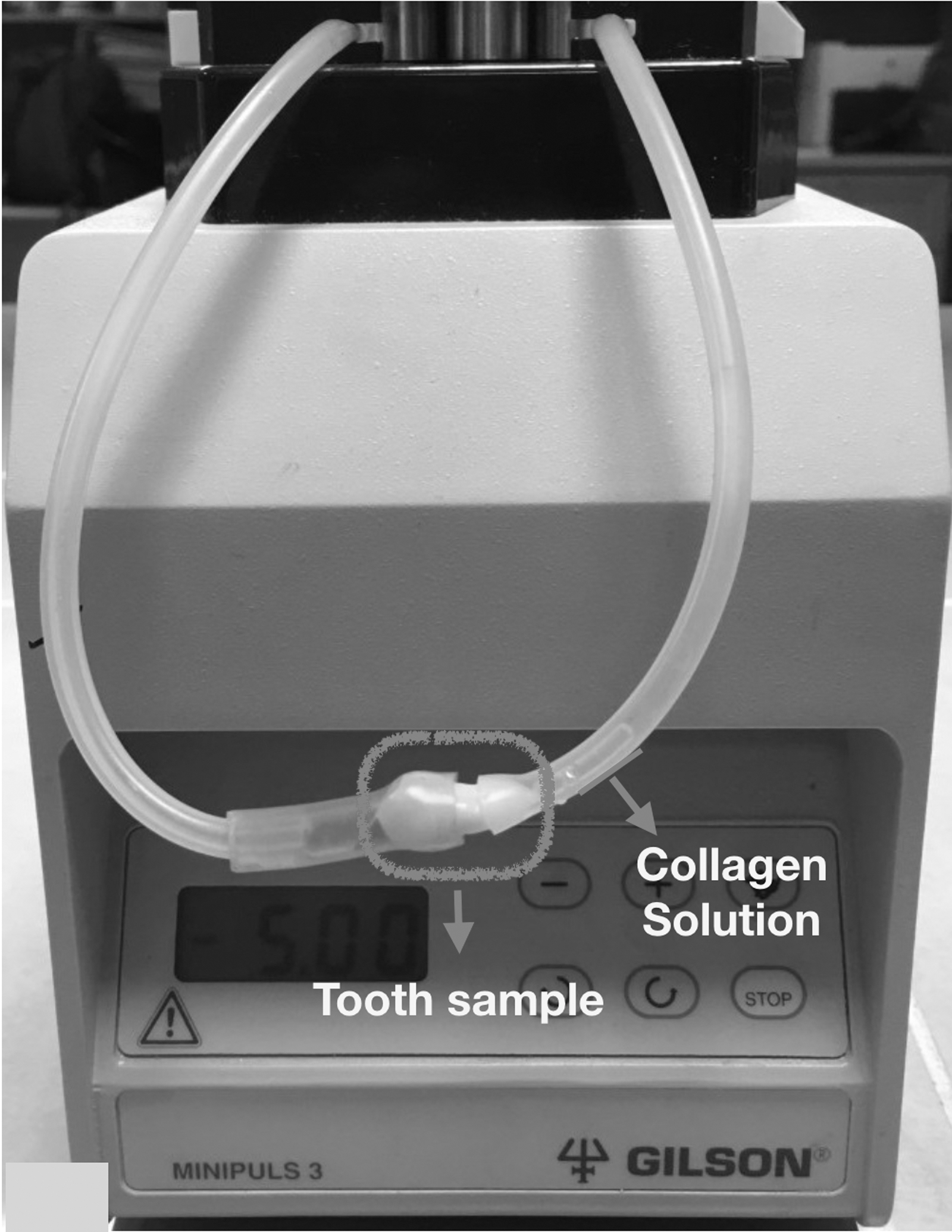
The application of the collagen solution into the root canals via peristaltic pump.
On the following day, two layers of nail polish (Flormar, Istanbul, Turkey) were applied to the entire root surfaces, and then, the apices of the specimens were sealed using glass ionomer cement (Kavitan™ Plus; Pentron, SpofaDental, Czech Republic) to create a closed-end canal creating a vapor lock effect according to the Tay et al. 25
Specimen irrigation
The specimens were randomly divided into five groups (n = 10 specimens per group), which were categorized according to the irrigation activation treatment that each group would receive. In all groups, the total irrigation time was 90 sec, and the total irrigant volume was 3 mL of 5.25% NaOCl. The total irrigation activation time was 60 sec in all groups, except for group 1. Activation was not performed in group 1, which was defined as the control group. The specimens were irrigated using the following procedures:
Group 1 [conventional needle irrigation group (control group)]: a 27-gauge notched-tip irrigation needle (Endo-Eze; Ultradent) was used for 90 sec with 5.25% NaOCl solution. The needle was inserted into the root canal 2 mm short of the working length and was moved 4 mm vertically within the root canal.
Group 2 (Vibringe sonic irrigation group): the irrigation procedures were performed using the same needle as that used for group 1. The notched needle was attached to the tip of the Vibringe sonic irrigation device. During the first 10 sec, the device was switched off and needle irrigation was performed using 1 mL of 5.25% NaOCl. This was followed by 20 sec of activation with the device switched on. The activation was performed without irrigation to obtain a standard activation period comparable with those of the other groups. This procedure was repeated three times.
Group 3 (EndoActivator sonic irrigation group): root canals were irrigated with 1 mL of 5.25% NaOCl using the same needle as that used for group 1 for 10 sec. The activation protocol was then performed using the EA sonic device. Irrigation solution within the root canal was activated using the EA for 20 sec (10,000 cycles/min using the red tip, 25, 0.4%). The sonic tip was inserted into the root canal 2 mm short of the working length. This procedure was repeated three times.
Group 4 (PUI group): the irrigation procedures were performed using the same needle as that used for group 1. After irrigation with 1 mL of 5.25% NaOCl for 10 sec, an ultrasonic tip (ESI, size 15, 0.02 taper; EMS, Nyon, Sweden) was inserted into the root canal 2 mm short of the working length. The tip was activated at a frequency cycle of 28–32 kHz for 20 sec. This procedure was repeated three times.
Group 5 (PIPS group): the laser-activated irrigation protocol was performed using an Er:YAG laser with a wavelength of 2.940 nm (Fotona). A 14-mm-long 300-μm quartz laser tip was used at 0.3 W, 15 Hz, and 20 mJ per pulse, while the water and air of the laser system were turned off. First, the root canal was irrigated with 0.5 mL of 5.25% NaOCl, as in group 1, for 10 sec. The optical fiber of the PIPS device was then placed into the access cavity and the NaOCl solution was activated for 20 sec. When the irrigating solution volume decreased within the access cavity during the activation procedure, a supplemental volume of 0.5 mL of 5.25% NaOCl was provided by needle irrigation. The laser-activated irrigation was continued during the placement of the irrigant. This irrigation and activation procedure was repeated three times.
Determination of residual protein concentration
We used a method previously described by Metin et al. 23 to assess residual protein levels in the NaOCl irrigant solution. Metin et al. 23 have reported that 3% or 3.5% sodium thiosulfate should be used to neutralize the 5.25% NaOCl solution within 15 sec, and that spectrophotometric evaluation should then be performed within 5 min to determine the residual protein level. In our protocol, 3 mL of total irrigation solution was collected from the coronal part of the tooth into test tubes containing 190 μL of 50% sodium thiosulfate solution to yield a final sodium thiosulfate concentration of 3%. The amount of residual protein was then determined using the Bradford method 24 of spectrophotometric analysis (Shimadzu UV-1700, Japan). The protein content was calculated based on a standard curve prepared using bovine serum albumin (Fig. 2.). All measurements were performed three times and mean values were calculated.
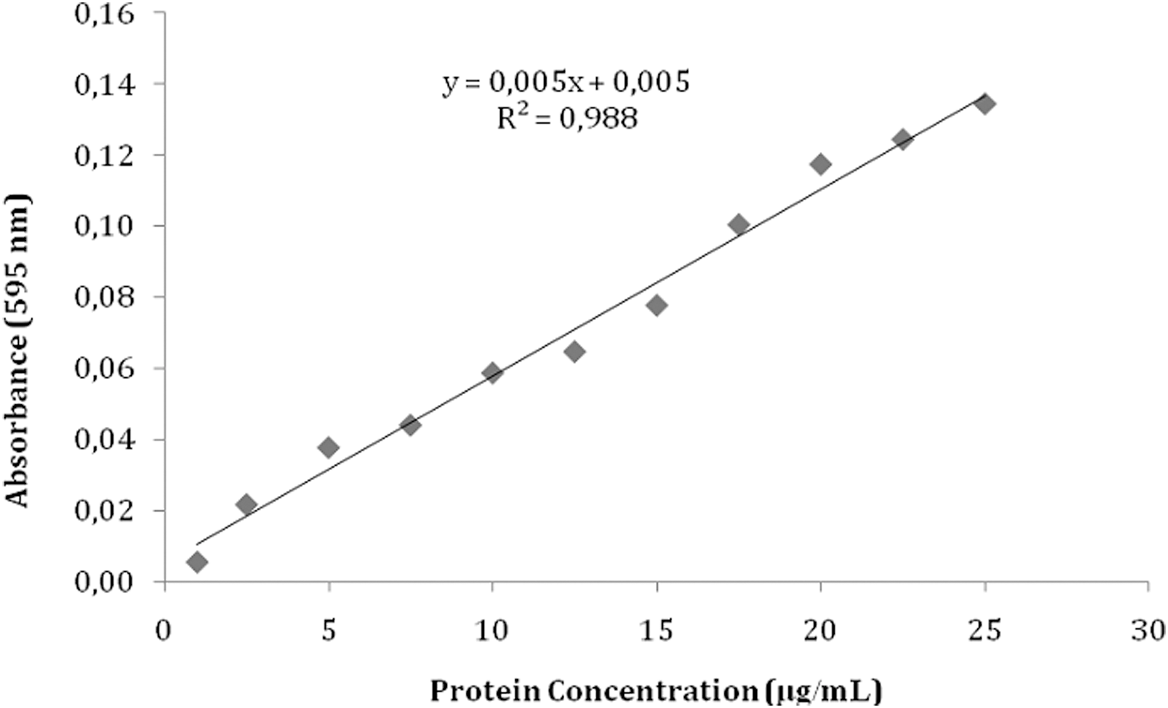
Bradford protein standard curve using bovine serum albumin.
Statistical differences between the control and experimental groups were analyzed using analyses of variance and Duncan post hoc tests. All statistical analyses were performed using STATISTICA 7.0 (2004; StatSoft, Tulsa, OK) at the 95% confidence level (α = 0.05).
Results
Concentrations of residual protein extruded using the NaOCl irrigant solution (mean ± standard deviation) for all groups are presented in Fig. 3. Significantly greater amounts of residual protein were removed in the PIPS group than in all other experimental groups and the control (conventional irrigation) group (p < 0.05). When the other experimental groups were compared with each other, there were no statistically significant differences among the VSS, EA, and PUI groups (p > 0.05). However, the VSS group had the lowest residual protein levels among these experimental groups. The lowest residual protein level was observed in the conventional irrigation (control) group, which was significantly different from all of the experimental groups (p < 0.05), with the exception of the VSS group (p > 0.05). Although there was no statistically significant difference between the control and VSS groups, there was more residual protein removed in the VSS group than in the control group (conventional irrigation).
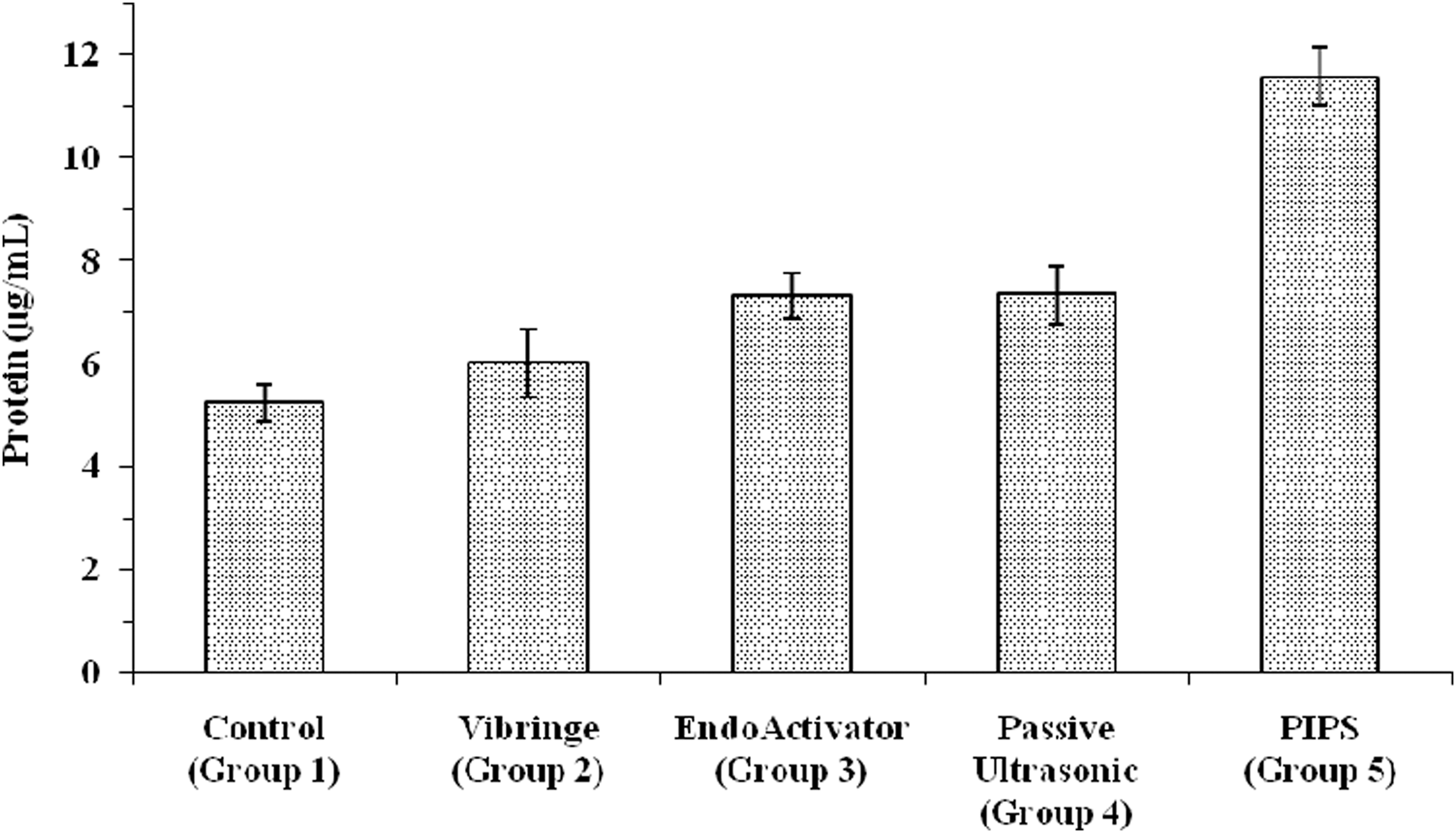
Residual protein levels in the 5.25% NaOCl solution. Bars indicate mean protein concentrations in μg/mL; whiskers indicate standard deviations. NaOCl, sodium hypochlorite; PIPS, photon-induced photoacoustic streaming.
Discussion
Experiments using standardized bacterial biofilms on extracted teeth are not reproducible 26 due to the variable nature of in vivo biofilms. 3 The use of nonstandardized biofilms may generate variations in the clinical results of irrigation tests. For this reason, a collagen ex vivo biomolecular film model was proposed by Huang et al. 21 to improve the standardization and predictability of testing. It is known that certain bacteria may bind cell surface adhesins to type 1 collagen found in dentin, facilitating the formation of biofilm. Importantly, the type 1 collagen model used by Huang et al. 21 mimics an important molecular binding property of bacterial biofilms. In addition, the hydrodynamic properties of biofilm are simulated using a collagen biofilm model in the studies of Huang et al. 21 and McGill et al. 22 Our model allowed simple quantification of, and comparisons between, the efficacies of the different irrigation activation techniques used for biofilm removal. The abovementioned study design was reasonably modified in this study to obtain a continuous flow ex vivo biomolecular biofilm model using a peristaltic pump to apply the collagen solution. The collagen solution was transferred into the root canal five times for 3 min using a peristaltic pump. Although the collagen solution was viscous, it was able to pass through the apical foramen. In previous studies, the collagen solution was applied into the root canal using a small brush 21,22 or was injected into the root canal and drawn apically 27 resulting in a static ex vivo biomolecular biofilm pattern. Using our modified method, we believe that we demonstrated a continuous fluid flow model. Comparing with the other ex vivo biomolecular biofilm models used previously, 21,22,27 the continuous flow model used in present study mimics saliva flow and thus simulates clinical conditions better than static systems. Therefore, continuous flow model may provide more precise information regarding performance evaluation of irrigation activation systems.
Determining the residual protein content in the irrigation solution is a new and practical method to evaluate the efficacy of irrigation activation techniques. NaOCl is the main irrigation solution used in endodontic treatment because of its antibacterial properties. 28 In addition, NaOCl is a powerful organic solvent. Peptide bonds are broken during to dissolution. This leads to rapid breakup of proteins when they come into contact with hypochlorite. 29 Due to the chemical properties of the NaOCl solution, determining the residual protein content in the irrigation solution is not possible. To overcome this problem, Metin et al. 23 developed a method to assess protein content by neutralizing the NaOCl irrigant solution with sodium thiosulfate. Previously, the amount of removed collagen in ex vivo studies was calculated using histological 27 or stereomicroscopic measurements. 21,22 Although histological analysis is quite effective at measuring the removal of collagen, it is time-consuming and expensive when compared with the method developed by Metin et al. 23 The use of stained collagen in these studies 21,22 is another potential problem. Irrigation of stained collagen samples with NaOCl solution might lead only to the removal of the stain, as collagen itself may remain in the biofilm. Collagen within the biofilm cannot be detected using stereomicroscopy without the use of stains. This may also affect the accuracy of the test. In addition, the model design used by the above studies is limited to single-rooted teeth with straight canals, as it required the teeth to be regularly split into two halves to expose the root canal surfaces. 19,21,22,30 –32 This may produce misleading results due to imperfect cohesion of the separated fragments, which would negatively affect the acoustics streaming through the root canal. The design used in our study may be useful for a wide range of root canal and tooth types, as it does not require an additional step to expose root canals after the final irrigation step.
We examined five irrigation activation techniques and methods: conventional irrigation, VSS, EA, PUI, and PIPS. One of the main results of this study was that all experimental techniques, except for VSS, led to the extrusion of a statistically greater concentration of protein than conventional irrigation. Conventional irrigation led to the least amount of residual protein removal when compared with the other irrigation techniques. This is consistent with the findings of previous reports. 13,30 A greater amount of residual protein was removed when using VSS than when using conventional irrigation. However, the evidence for this difference was statistically weak. This is consistent with the findings of Johnson et al. 27 On the contrary, in the present study, VSS was not used for concurrent irrigation and activation according to the manufacturer's instructions, to provide a standardization in irrigation times. It is thought that the utilization of VSS without following the manufacturer's instructions may adversely affect the performance of this device and this is a limitation of this study.
The results of this study were in agreement with the findings of other studies examining the efficacy of the PIPS method in the destruction of bacterial biofilm. 12,31 In this study, we clearly demonstrated the superiority of the PIPS method, as a significantly greater amount of protein was removed when using this method than when using other experimental techniques and the conventional needle irrigation. The PIPS method differs from other activation techniques in that it utilizes photoacoustic and photomechanical phenomena. PIPS is the photomechanical streaming of an irrigant that is activated in the coronal part of the tooth using an Er:YAG laser tip. 13 The subablative parameters used in the PIPS method result in a photomechanical effect that occurs when light energy is pulsed in a fluid. 12,13 In a similar study by Arslan et al., 30 the PIPS method was shown to be superior to other methods of irrigation, including conventional needle irrigation, sonic irrigation, and ultrasonic irrigation, in removing Ca(OH)2 paste. However, in contrast to our findings, Pedullà et al. 33 reported that there is no significant difference in the reduction of Enterococcus faecalis bacterial levels between conventional needle irrigation and PIPS method. In the previous study, PIPS was used for activation for 30 sec, while we used PIPS for activation for 60 sec. The longer activation time used in this study may enhance the debridement efficacy of PIPS. This may explain the difference between the results of the present study and those of Pedullà et al. 33
Consistent with our results, a previous study 19 indicates that the PIPS method is significantly more effective in debris removal than the PUI technique. De Groot et al. 18 have described the superiority of laser-activated irrigation to PUI. Specifically, the high energy transferred to the irrigation solution in a laser-activated irrigation system may lead to superior performance when compared with PUI systems. Further, our results are similar to those of Arslan et al., 32 who concluded that PIPS is more effective than the EA and conventional irrigation in removing both double antibiotic paste and triple antibiotic paste from artificial grooves in root canals. We found no statistically significant differences between the sonic and PUI systems, consistent with the findings of Jensen et al. 34 The results of our study are consistent with those of previously published articles on the efficacy of irrigation activation techniques. 12,18,19,23 This indicates that the residual protein testing model is a practical method to analyze the debris removal efficacy of irrigation activation systems.
Conclusions
The PIPS method was significantly more effective than PUI, EA, VSS, and conventional irrigation in extruding ex vivo biomolecular film in a side-by-side comparison. This suggests that PIPS is a promising adjunctive method to remove bacterial biofilm from root canals in a clinical setting. In addition, the residual protein testing model used in the present study appears to be a practical and useful method to quantify irrigation efficacy and can be used to evaluate newly designed irrigation systems. However, further investigations are recommended to confirm its accuracy.
Footnotes
Acknowledgments
This study was supported by the Adnan Menderes University Research Foundation (DHF-15007). The Foundation had no role in performing the study, including its design or analysis, or in our decision to publish the findings.
Author Disclosure Statement
No competing financial interests exist.
References
Supplementary Material
Please find the following supplemental material available below.
For Open Access articles published under a Creative Commons License, all supplemental material carries the same license as the article it is associated with.
For non-Open Access articles published, all supplemental material carries a non-exclusive license, and permission requests for re-use of supplemental material or any part of supplemental material shall be sent directly to the copyright owner as specified in the copyright notice associated with the article.