
Abstract
Introduction
P
Cell membranes have been identified as an important intracellular cancer treatment target, the reason being that many of the cell membrane natural constitutive macromolecules are readily susceptible to singlet oxygen, which is typically produced during the PDT process. Such membranes include the plasma membrane surrounding the cell, the membranes of endoplasmic reticula distributed throughout the cytoplasm, and the membranes of mitochondria and Golgi apparatus. 4 In addition, the cell surface has glycoconjugates that are involved in numerous functions, including cell proliferation, differentiation, and migration, and cell-cell recognition, among others. 5 –7
Lectins are carbohydrate binding proteins or glycoproteins that are not an antibody or an enzyme, and that can specifically bind to glycans without catalyzing their modification. 8 –11 The interactions of lectins with glycans are pivotal in the regulation of a wide variety of interactions of cells with other cells, the extracellular matrix, and pathogens. 8,9,12 They have been used as recognition tools to differentiate malignant and benign tumors and determine the degree of glycosylation associated with metastasis. 11,13
Evidence indicates that aberrant expression of glycans correlates with pathological conditions, including cancer. 14 Several studies have found that the occurrence, progression, and metastatic behavior of many different tumors are closely related to aberrant protein glycosylation. 11,14 –19 Thus, specific glycan motifs may serve as effective biomarkers for cancers. 7
Studies on human melanoma and murine B16 melanoma cell line have reported significant changes in the glycosylation of cell surface proteins important for the development and progression of melanoma. 20 –23 The occurrence of melanoma, one of the major causes of death in industrialized countries, has increased significantly worldwide over the last decades. According to the World Health Organization (WHO), 132,000 new cases occur globally each year. In the United States, it is considered the fastest growing disease in both sexes. In Brazil, the National Cancer Institute (NCI) estimates 5920 new cases per year, 2950 in men and 2970 in women, with most of these cases in the Southern region, where there is a very clear predominance in fair-skinned individuals. 24 Hence, the need for medical studies of this disease in search for better therapies is pressing.
This study aims to evaluate the action of PDT with two different photosensitizers, 5-aminolevulinic acid (ALA) and Photosan-3, in the inhibition of glycosylation of melanoma cell surface using lectins wheat germ agglutinin (WGA) and concanavalin A (ConA).
Materials and Methods
Cells and tumor
B16-F10 murine melanoma cells were kindly provided by Prof. Dr. Antônio Claudio Tedesco (USP-Ribeirão Preto, Brazil). The cells were maintained with 10% fetal bovine serum, 200 mM L-glutamine, and 1% penicillin-streptomycin-neomycin in Dulbecco's modified Eagle's medium (DMEM; Thermo Fisher Scientific, Waltham, MA) and incubated at 37°C in a 5% CO2 atmosphere.
Isogenic mice were bred from 6-week-old male Swiss mice acquired from the Institute of Biomedical Sciences USP-Biotery. During the experiment, the animals were anesthetized with Dopalen (Agribrands Purina do Brazil, Ltda, Paulinia, SP, Brazil). All procedures were approved by the Ethics Committee on Animal Research (Application no. A32/CEP/2008) and were in accordance with international guidelines on the ethical use of animals in research. The mice were housed under standard conditions with food and water ad libitum.
PDT in tumors
The mice were shaved on the left flank area and injected subcutaneously with 2 × 10⁵ B16 cells in 100 μL of phosphate-buffered saline (PBS). After 7–10 days, a tumor with a diameter of 3–6 mm had been established. The animals were stratified so that the mean tumor sizes in all treatment groups were nearly identical. Tumor volume was calculated according to the formula V = 0.52 × A 2 × b (a, smallest superficial diameter; b, largest superficial diameter). The PDT treatment group was subdivided and injected with either Photosan-3® (n = 5) or 5-aminolevulinic acid (ALA; Sigma-Aldrich Co., St. Louis, MO), n = 5, 40 μg/kg weight. Twenty-four hours after injection, the tumors were exposed to diode laser low-intensity semiconductor Gallium Aluminum Arsenate (GaAIAs) λ = 655 ± 20 nm (Bio Wave LLLT model; Kondortech, São Carlos, Brazil), with continuous emission, power output of p = 30 mW, energy density of 10 J.cm2 and power density of 45 mW.cm2, and probe with 8 mm beam diameter (Table 1). The non-PDT-treated group (n = 5) was not submitted to the photosensitizer and/or laser irradiation. The frequency of treatment occurred every other day and the tumor was measured at each session, were 6 weekly sessions, lasting 960 sec with cumulative dose 60 J.cm2. After 4 weeks of treatment, the mice were euthanized with a lethal dose of anesthetic and the tumors were surgically removed and fixed overnight in 2.5% glutaraldehyde +2% paraformaldehyde (Karnovsky's fixative) in 0.1 M cacodylate buffer (pH 7.2) at 4°C. The cells were postfixed in 2% OsO4 in sodium cacodylate buffer, dehydrated in a graded acetone series, and embedded in Epon 812 (Polysciences, Inc., Warrington). Sections (300 μm) were cut using a Leica Ultracut UCT ultramicrotome (Leica, Wetzlar, Germany) with a diamond knife, mounted on clean coverslips, and allowed to dry for a few minutes on a hot plate (60°C).
Pretreatment of semithin sections
Sections were exposed at room temperature to one of the following agents: 10% NaOH in absolute ethanol for 5–60 min for removal of the epoxy resin; washed in absolute ethanol, dried in air, and stored in a refrigerator at −20°C until use. Before labeling, all sections were treated with trypsin 0.5 mg/mL (Sigma-Aldrich Co.) and with 0.05% sodium borohydride for 4 min to eliminate the autofluorescence of the glutaraldehyde used in fixation.
Lectin labeling procedure
Two types of TRITC-conjugated lectins were used as probes: ConA (specificity: α-D-Mannose and α-D-glucose) and WGA (specificity: N-acetyl-β-D-glucosaminyl and N-acetyl-β-D-glucosamine oligomers), purchased from Sigma-Aldrich Co. (Sigma-Aldrich Co.). The sections were incubated for 30 min with lectin diluted to 20 μg/mL in PBS, followed by three washes in PBS, air drying, and analysis in Leica DMLB with a Leica DFC 310FX CCD camera.
Results and Discussion
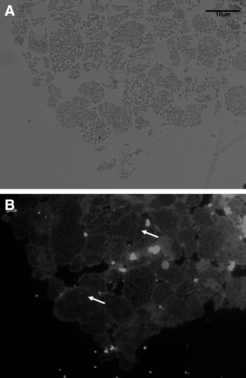
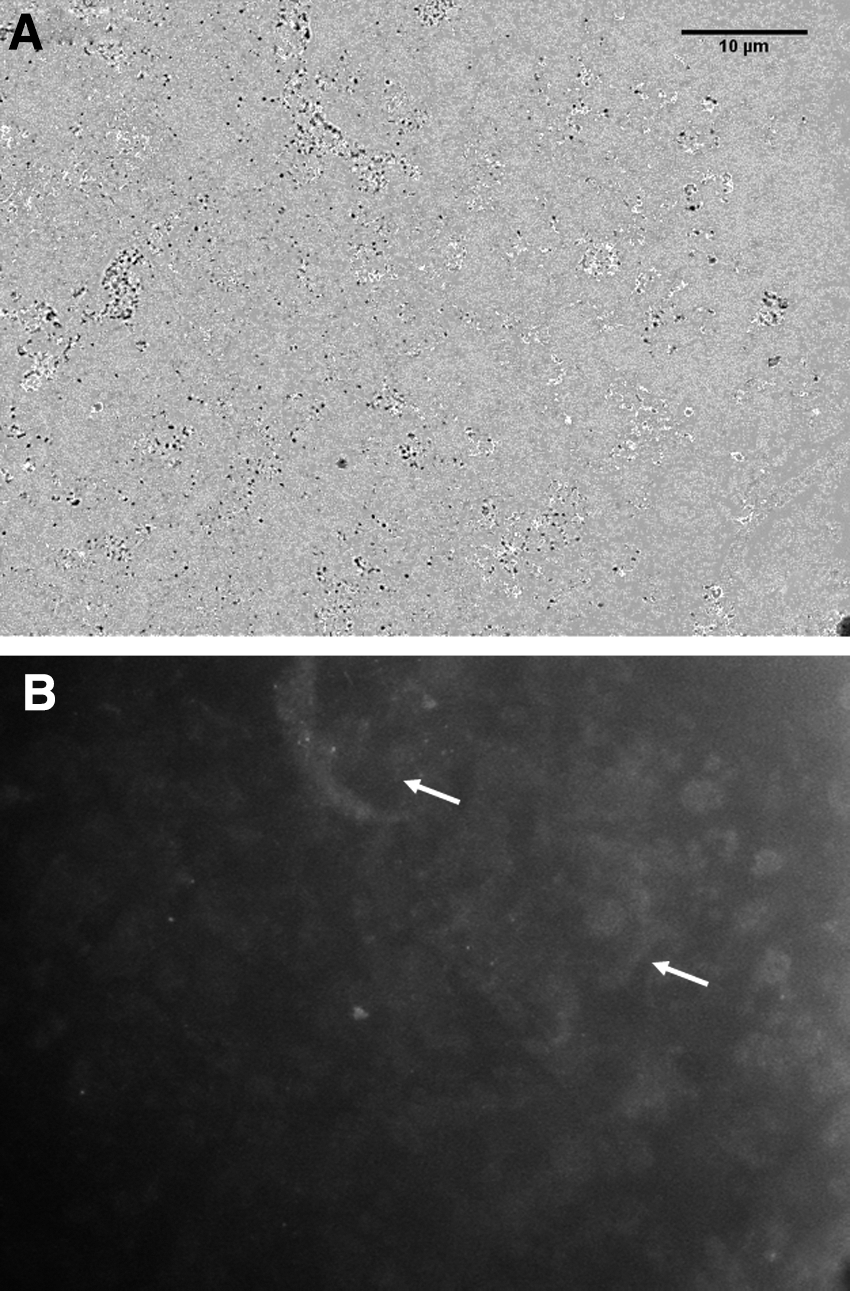
In this study, we showed that PDT alters the glycosylation process of the cell surface in melanoma. Compared to the control group (Fig. 1A, B), both tumors treated with ALA (Fig. 2A, B) and Photosan-3 (Fig. 3A, B) presented positive labeling for ConA. However, when WGA was analyzed, no labeling was observed in either of the two groups of photosensitizers. We hypothesized that PDT can alter the glycosylation process of the cell surface and our results showed glycoprotein alterations with both Photosan-3 and ALA photosensitizers.

Control group not labeled with ConA fluorescence.
PDT with ALA alters the labeling of melanoma surface glycoprotein.
PDT with Photosan-3 alters the labeling of melanoma surface glycoprotein.
Changes in surface glycoprotein are associated with all the cancers described. 7 Studies indicate that aberrant glycosylation is the result of initial oncogenic transformation, as well as a key event in the induction of invasion and metastasis. 25 The presence of carbohydrates on the surface of B16-F10 strain melanoma cells is related to their ability of metastatic colonization. 26 –29 Many authors consider that melanocyte malignancy is associated with changes in surface carbohydrates. 30 In this study, we described that PDT could change the melanoma surface glycoprotein; however, we have not evaluated the principle of the mechanism underlying this change. Nonetheless, since ALA and Photosan-3 kill similar forms of cells, they may cause changes in surface carbohydrates as well.
The glycans present in humans are predominantly bound to transmembrane proteins in two ways: (1) an N-acetyl-glucosamine (GlcNAc) residue is added to an Asn residue in peptide consensus sequence Asn-X-Ser/Thr (where X can be any amino acid, except proline) for N-glycans, or (b) a GalNAc residue is added to the hydroxyl group of Ser or Thr residues in polypeptide O-glycans. Proteins destined for the cell surface undergo glycosylation process by endoplasmic reticulum (ER)-Golgi, where a regulated sequence of events occurs involving both anabolic and catabolic enzymes, such as glycosidases and glycosyltransferases, to structure glycoconjugates. 31 Therefore, the changes found in melanoma surface glycoprotein in this study suggest that Photosan-3 and ALA may interfere with the expression of enzymes involved in protein glycosylation by ER-Golgi or others changes in the system endomembrane by modifying surface glycoconjugates.
In general, rather than a single technique, PDT is a set of related protocols involving photosensitization. Thus, several types of photosensitizers, each with a specific characteristic, are used. 1
Plant lectins have been classified as either polyspecific or monospecific according to their sugar binding specificity, that is, whether they can interact with one or more sugars, such as galactose, mannose, mannose-containing glycans, glucose, and so on. 20,26 WGA is a lectin with binding properties specific to sialic acid and GlcNAc, and Con A recognizes and binds specifically to the residues of α-D-glucomanose of complex carbohydrates. It has high affinity for glycoprotein and glycolipid oligosaccharides on the cell surface, which may connect to several types of cells from different organisms. 32,33
Evaluation with WGA in this study demonstrated that melanoma cells do not express α-galactosyl epitopes after PDT, regardless of the photosensitizer used; however, B16 cells, as well as normal cells, express α-galactosyl epitopes. This can be used to study their ability to metastasize and their capacity to restore surface glycoconjugates. In a comparative analysis of lectin binding in tumor cells, some changes were observed in the binding of Con A, WGA, and Glycine max (SBA) in high and low metastatic cell lines of B16 melanoma cells. 30,34 Studies have demonstrated that lymphoma cells showed a correlation between the reduction of Con A binding sites and metastatic potential; however, the same connection was not observed between lectin WGA and metastatic potential. Nevertheless, these differences in lectin binding can demonstrate some changes in the expression of cell surface carbohydrates in tumor cells with distinct metastatic potentiall. 35 Some lectins able to bind and induce aggregation of normal or malignant cells have been found to be highly cytotoxic, such as Con A, Phaseolus vulgaris (PHA) and WGA, ricin (RIC), Lens culinaris agglutinin (LCA), and Griffonia simplicifolia 1-B4 (GS1B4), which are able to eliminate both normal and malignant cells in relatively low concentrations. 36 Further, pretreatment of cells included in Epon did not affect labeling lectin for fluorescence microscopy. The data reported in this study show that PDT with ALA and Photosan-3 interfered in the expression of glycoprotein in the surface of B16 cells in the presence of lectin Con A (Fig. 1). Further, this study showed that PDT can be an alternative method to alter cell surface carbohydrates; however, further study is needed to clarify how PDT acts on surface carbohydrates.
Footnotes
Acknowledgments
This study was funded by FAPESP (Fundação de Amparo a Pesquisa do Estado de São Paulo—The State of São Paulo Research Foundation, grant contract numbers 2011/05404-7, 2011/05958-2, 2009/15206-8, and 2016/17984-1). Pacheco-Soares, C designed the study. All authors performed the experiments, analyzed the data, and wrote the article.
Author Disclosure Statement
No competing financial interests exist.