
Abstract
Objective:
To determine surface roughness caused by Er:YAG laser irradiation and its effect on the increase in bacterial adhesion.
Background:
Er:YAG laser was proposed as a strategic device to reduce caries by its ability to generate chemical and structural changes in tooth enamel; in turn, it produces undesirable effects on the tooth surface that could increase its roughness and allow a greater accumulation of microorganisms.
Methods:
Eighty-four samples of human enamel were divided into seven groups (n = 12): G1_control (no laser irradiation); G2_100/H2O, G3_200/H2O, and G4_300/H2O were irradiated with Er:YAG laser (12.7, 25.5, and 38.2 J/cm2, respectively) under water irrigation. In addition, G5_100, G6_200, and G7_300 were irradiated with the energy densities described above and no water irrigation. Surface roughness measurements were recorded before and after treatment using a profilometer. Afterward, three samples per group were incubated in a microorganism suspension for the tetrazolium salt (XTT) assay. Biofilm morphology was observed using scanning electron microscopy and confocal laser scanning microscope. One-way analysis of variance and t-tests were performed for statistical analysis (p < 0.05).
Results:
There were no statistically significant differences in roughness values in the G5_100 group before and after treatment, but there were statistically significant differences observed in the other groups evaluated (p < 0.05). No significant differences in adhesion of both strains were detected in irradiated groups compared with G1_control.
Conclusions:
The increase in roughness on dental enamel surfaces was proportional to the irradiation conditions. However, the increase in surface roughness caused by Er:YAG laser irradiation did not affect Streptococcus mutans and S. sanguinis adhesion.
Introduction
I
Interaction between the laser and hard tissue depends on irradiation parameters such as wavelength, emission mode, pulse duration, energy, frequency, spot size, 10 as well as irradiation distance and cooling. 8
A proper laser treatment may prevent enamel demineralization by reducing the enamel diffusion. 3,11 Block enamel diffusion was associated with organic blocking, which preserves and modifies the organic matrix to obliterate diffusion channels in enamel induced by lower energy laser treatment. 5
The use of a wavelength highly absorbed by water (2.94 μm) generates thermal changes in enamel, which may enhance its chemical and/or morphological structure. 1,12 However, there are undesirable morphological changes produced by the photothermal effects that could increase its roughness, such as craters, 4,6,9,13 cracks, 2,3,13,14 exposed enamel prisms, 6,8 ablation, and melted areas, 1,8 in addition to surfaces with a molten lava-like appearance. 1 This lack of smoothness on the treated surfaces seems to be more prone to accumulation of bacteria, which could result in the dental caries process, as suggested by some authors. 4,9,13,14
Streptococcus mutans has been identified as the principal pathogenic agent in caries, 15 while S. sanguinis is one of the most prevalent species in the indigenous oral microbiota, and it can be present in saliva and dental plaque. 16 Both strains are dental plaque initial colonizers with a remarkable affinity for dental enamel hydroxyapatite. 17
However, there is little information related to changes in surface roughness caused by Er:YAG laser irradiation and its possible effect on the increase in bacterial adhesion. This study aims to evaluate the influence of several Er:YAG laser irradiation parameters, which are used in clinical preventive dentistry, on the dental enamel surface roughness and the effect on adhesion of two important first colonizers of dental plaque, S. mutans and S. sanguinis.
Materials and Methods
Tooth selection and sample preparation
The study protocol was reviewed and approved by the institutional research ethics committee. Noncarious human premolars extracted for orthodontic reasons from patients aged 15–17 years were obtained and stored in a 0.2% thymol solution for processing, as described by this research group. 13 Enamel fluorescence was evaluated using a DIAGNOdent® pen (DIAGNOdent; KaVo, Biberach, Germany) and Spectra™ (Air Techniques, Melville, NY), and only teeth with sound enamel were included. 12,13,18 Teeth with caries, restorations, fractures, structure or color defects, or fluorosis were excluded. 13
Each tooth was cut to obtain one enamel block (3 × 3 × 2 mm), from the buccal and lingual surface. The samples were rinsed with deionized water and dried at room temperature for an initial surface roughness evaluation.
Er:YAG laser irradiation
An Er:YAG laser system (Lumenis OPUS DUO™ Er:YAG + CO2, Yokneam, Israel) was used for sample irradiation. The laser parameter settings used were 2.94 μm wavelength emission, 100–300 mJ (based on the experimental groups), 10 Hz, 250–400 μsec, 1.0 mm tip diameter, focused mode, and 18-sec irradiation total time. Eighty-four samples of human dental enamel were randomly assigned to seven groups (n = 12), as described in Table 1.
The tip was positioned perpendicular to the enamel surface, irradiation was performed manually in one direction with a consistent motion, and the tip sample distance was standardized at 1 mm. 13
Surface roughness analysis
The roughness was measured before and after laser treatment using a profilometer (Mitutoyo Surftest SJ-301, Tokyo, Japan). Enamel roughness of each sample was scanned at a length of 0.5 mm with a diamond stylus using a cutoff of 0.08 mm (λc), a velocity of 0.25 mm/sec, and a Gaussian filter. The scanned area was limited to the irradiated surface.
Measurements were performed perpendicular to the samples. For each sample, three measurements were made by the same operator, and mean values were calculated per sample and then for each group. Roughness parameters assessed were average distance from the profile to the mean line over the length of assessment (Ra) and peak-to-valley values of five equal measures within the profile (Rz). All measurements were performed following ISO 4287-1997. 19
Bacterial adhesion test
Streptococcus mutans ATCC 25175 and S. sanguinis ATCC 10556 were used for the experiments. Lyophilized bacterial stocks (American Type Culture Collection, Rockville, MD) were grown on Hemin-Vitamin K (HK)-enriched agar plates [brain/heart infusion agar (BBL; Becton-Dickinson), trypticase soy agar (BBL; Becton, Dickinson), and yeast extract (BBL; Becton, Dickinson), supplemented with 5 μg/mL hemin (Sigma-Aldrich), 0.3 μg/mL menadione (Sigma-Aldrich), and 5% defibrinated sheep blood (Microlab, Mexico City)] at 37°C under anaerobic conditions (80% N2, 10% CO2, and 10% H2) to obtain pure cultures. 20
All experiments were performed using three samples per experimental group. The sterile enamel samples were placed individually into a 96-well cell culture plate with a flat bottom (Costar Cat.# 3599). Bacterial suspensions of S. mutans and S. sanguinis were adjusted to an optical density of 1 at 600 nm in a spectrophotometer (Eppendorf BioPhotometer D30, Germany) and 10 8 cells/mL of each strain were individually added to the samples (200 μL total volume). Plates were incubated at 37°C under anaerobic conditions for 24 h to allow bacterial adhesion on the enamel surfaces. Each sample was then washed three times with the culture broth to remove nonadherent bacteria. The number of viable attached bacteria was determined using the tetrazolium salt (XTT) viability assay.
XTT assay
The XTT cell viability kit assay (Cell Signaling Technology® #9095) was used to evaluate bacterial adhesion on the enamel samples, and 50 μL of the XTT reagent and 100 μL of the enriched culture broth were added to each well and incubated in the dark for 4 h at 37°C. After incubation, 100 μL of the supernatant in each well was transferred to a new 96-well cell culture plate and the absorbance was measured at 450 nm (reference wavelength, 620 nm) using the multi-mode microplate reader FilterMax™ F5 (Molecular Devices). A standard calibration curve was performed on the number of cells/mL to transform absorbance values.
Confocal laser scanning microscopy
An additional sample from each experimental group was prepared to observe bacterial adhesion. Samples were stained using the LIVE/DEAD® BacLight™ (Invitrogen Ltd., Paisley, United Kingdom) Bacterial Viability Kit. The dyes were mixed (1:1 ratio), and 3 μL placed on the surfaces and incubated at room temperature for 15 min in the dark. After incubation, the residual stain was rinsed off using sterile deionized water. Three areas were randomly selected on each tested surface 21,22 and were immediately analyzed using a confocal laser scanning microscope (CLSM, CLSM FV 1000; Olympus, Tokyo, Japan) equipped with a UPLSAPO 100 × O NA: 1.4 and UPLFLN 40 × O numerical aperture (NA): 1.30 lens (Olympus).
Scanning electron microscopy
After the bacterial adhesion test, selected specimens (one per group) were fixed in 2.0% glutaraldehyde for 24 h at room temperature. Then, they were washed three times with phosphate-buffered solution (pH 7.4) and dehydrated through a series of graded ethanol solutions of 20%, 40%, 60%, 80%, and 100%. Samples were subsequently vacuum dried and sputter coated with gold. Subsequently, three fields were randomly selected and evaluated using scanning electron microscopy (SEM, SEM JEOL; JSM-6610LV, Inc., EU) at 20 kV. 20
Statistical analysis
The SPSS 21 statistical package (SPSS IBM, New York, NY) was used. The Kolmogorov–Smirnov test was used to assess data distribution, and thus, a paired t or Wilcoxon test was performed to compare the mean surface roughness Ra and maximum height of profile Rz, before and after irradiation. Subsequently, one-way analysis of variance (ANOVA) was used to compare among the groups; when significant differences were found, the Bonferroni or Tamhane's T2 post hoc test was applied, depending on the Levene's test of variance homogeneity. Significant differences in the number of cells/mL between surfaces tested were determined using ANOVA. To compare both microorganisms per group, a t-test was performed. The significance level for all tests was p < 0.05.
Results
Surface roughness
The Ra and Rz values obtained before and after treatment with Er:YAG laser are presented as mean ± standard deviation (Table 2). Before irradiation, all enamel surfaces showed similar Ra (0.20–0.32 μm) and Rz (1.63–2.51 μm) values, with no significant differences between them. However, after irradiation, significant differences were observed in groups G2_100/H2O, G3_200/H2O, G4_300/H2O, G6_200, and G7_300 compared with the nonirradiated surfaces (p < 0.05). G4_300/H2O showed the highest roughness value after irradiation (1.55 μm), while G5_100 showed the lowest value after irradiation (0.49 μm), which was similar to the control group. No significant differences were observed between G1_control and G5_100.
Mean values and standard deviations.
Capital letters in a column are the comparison between roughness values of different groups. Same capital letters follow means that do not differ statically (one-way analysis of variance, p < 0.05).
Lower case letters in a row are for the comparison of roughness before and after treatment. Same lower case letters follow means that do not differ statistically (paired-samples t-test or *Wilcoxon test, p < 0.05).
AT, after treatment; BT, before treatment.
XTT assay
The number of bacterial cells attached on the enamel samples is presented in Fig. 1. Bacterial adhesion varied depending on the laser Er:YAG irradiation protocol. A significantly higher adhesion of S. sanguinis was observed on the control enamel surfaces compared with adhesion of S. mutans (p < 0.05). All irradiated samples showed increased adhesion of S. mutans and decreased adhesion of S. sanguinis compared with nonirradiated samples, but there were no statistically significant differences among the groups.
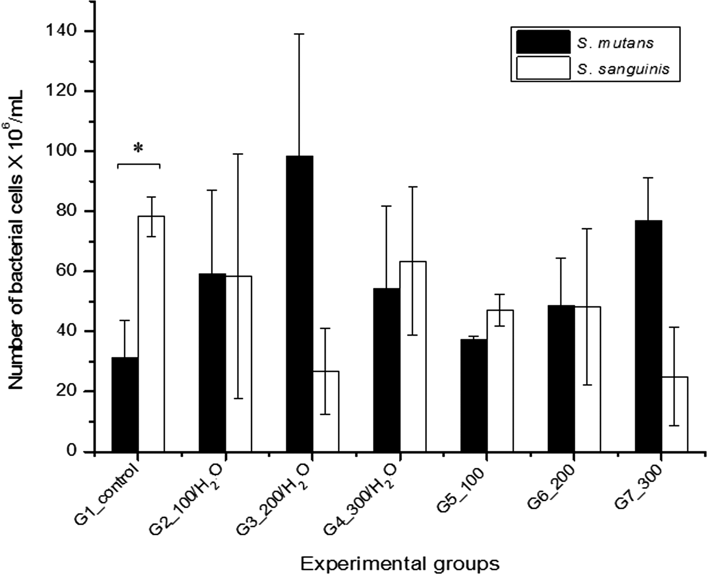
Average bacterial cells of Streptococcus mutans and S. sanguinis adhered to dental enamel irradiated with Er:YAG laser obtained by the tetrazolium salt (XTT) assay. Error bars indicate ± standard error of the mean. Asterisks mean statistical differences in the comparison between bacterial cell values of S. mutans and S. sanguinis under different irradiation parameters (Student's t-test, p < 0.05).
Confocal laser scanning microscopy
Images (xy-views) of biofilm formed on laser-irradiated enamel surfaces are shown in Fig. 2. Sparse bacterial colonies could be observed on all irradiated surfaces after a 24-h incubation. The confocal images confirm the XTT results because S. sanguinis cells can be seen in higher quantities than S. mutans cells on the nonirradiated control surfaces. However, on the irradiated samples, higher amounts of bacterial cells can be observed on the surfaces incubated with S. mutans than on the irradiated enamel surfaces incubated with S. sanguinis. A considerable overlap between the living and dead bacteria can be seen by the yellow color in the G3_200/H2O and G7_300 groups incubated with S. mutans, and in the G4_300/H2O and G6_200 groups incubated with S. sanguinis.
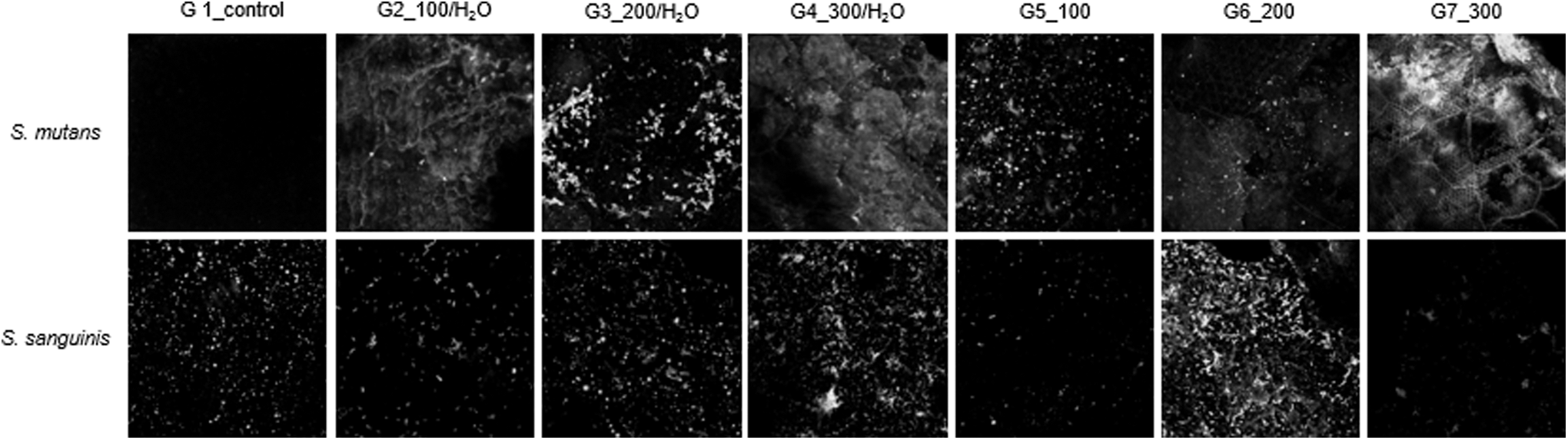
Confocal laser scanning microscopic images (xy-views) of the bacterial cells that adhered to Er:YAG laser-irradiated dental enamel assayed by LIVE/DEAD® BacLight™ Bacterial Viability Kits. Group G4_300/H2O and G7_300 were observed with UPLFLN 40 × O numerical aperture (NA): 1.30 lens.
Scanning electron microscopy
The representative SEM images for laser-irradiated samples are shown in Fig. 3. The control group presented predominantly smooth surfaces with some grooves. Morphological alterations in the irradiated samples include ablation areas with exposed enamel prisms, honeycomb patterns, rough surfaces, and small cracks can be observed. At a higher magnification, melt zones with surface roughening were pronounced, and smooth glaze-like surface layers and fused masses of crystallites were seen. Bacterial cells were scattered into a fused mass of crystallites in irradiated samples. S. mutans could be seen as single, pair, or triplet cells, while S. sanguinis was observed as cell chains. XTT assay results were consistent with the qualitative SEM observations.
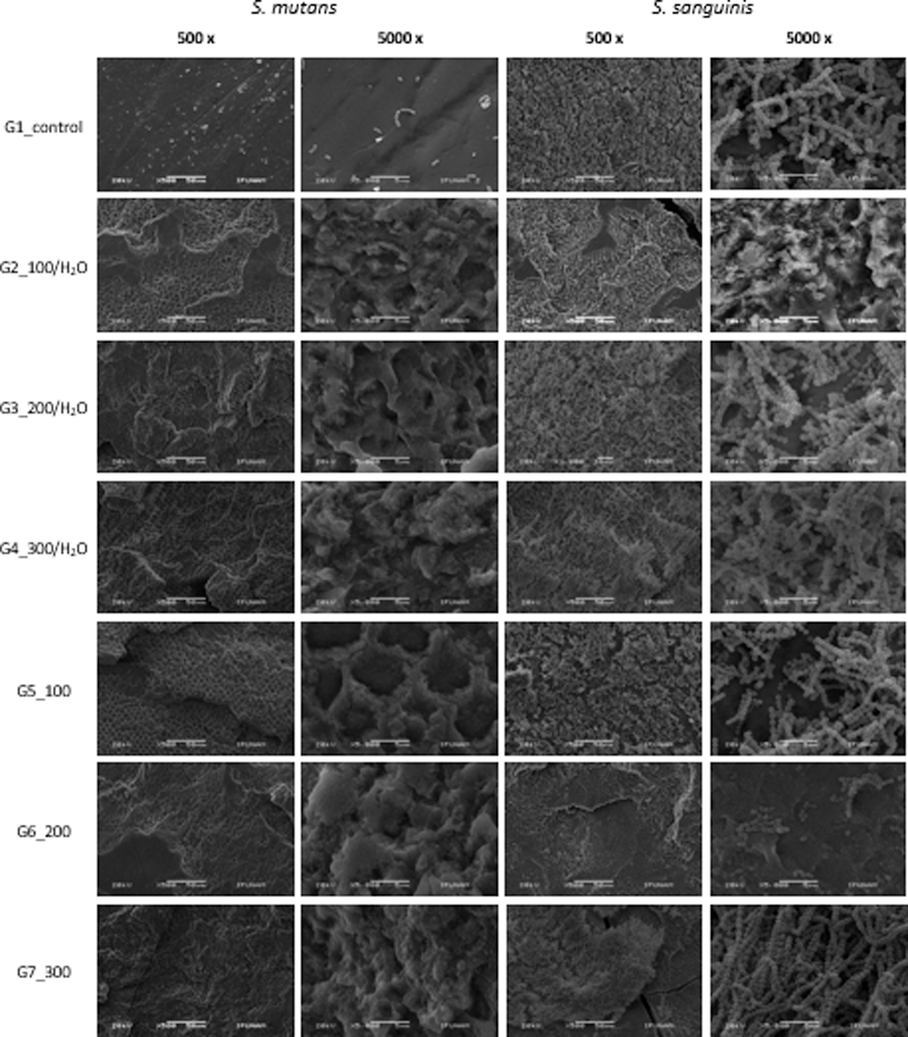
Scanning electron microscopy images of the bacterial adhesion on Er:YAG laser-irradiated and nonirradiated dental enamel surfaces after 24 h of anaerobic incubation with S. mutans and S. sanguinis (magnifications, × 500, × 5000); scale bar = 50 μm and 5 μm.
Discussion
In this study, we evaluated dental enamel surface roughness before and after Er:YAG laser irradiation, and its effect on S. mutans and S. sanguinis adhesion. In addition, different energy pulses 100–300 mJ were applied because they showed positive results for enamel demineralization reduction in previous studies. 4,7,12,23 Irrigation conditions were also evaluated.
Profilometry was used instead of Atomic Force Microscope (AFM) to evaluate roughness, because the high degree of roughness produced by laser irradiation at 200–300 mJ (shown in a pilot study) was not compatible with the z range of the AFM scanner, as reported by McConnell et al. 24 In addition, roughness measurement by AFM depends strongly on the analysis area and the average roughness increases when the scan area increases, 24 while profilometry presents a dimensional scale that is much larger than AFM. 25
For the procedure, enamel samples were not ground flat or mechanically treated before laser treatment to maintain the original morphology, and simulate a real clinical procedure.
Our results showed that baseline surface roughness measurements were homogeneous; control group values were lower than those reported by Barac et al. in the third unerupted molars that have not been exposed to the oral environment, while we used erupted bicuspid teeth. 19 Unerupted teeth have the appearance of less homogeneity with different striations of perikymata and variation in the number of exposed prisms. 26 Conversely, erupted teeth have an absence of big and small crystals, prism boundaries, and the primary cuticle, which were removed by abrasion, wear, and erosion. 27 These characteristics could cause differences in roughness between erupted and nonerupted teeth.
All irradiated surfaces showed an increase in roughness except for G5_100, which was irradiated with the lowest energy per pulse and there was no water irrigation. Roughness values were increased, irrespective of augmented energy per pulse. Morphological changes were proportional to energies per pulse used and were more evident at higher energy densities, as previously reported. 4,9,13,14 The roughness increase observed in laser groups may be related to morphological changes, such as formation of scaly surfaces, small or larger craters, areas of superficial tissue loss, cracks, and blocks. 1,4,6,9,13,14
The irrigated laser groups showed higher surface roughness, which was probably caused by surface ablation that is associated with water spray use. 28,29 The use of cooling water during irradiation has also been discussed, and satisfactory results have been achieved with or without the use of cooling. 1,8
For dental enamel roughness, our results are inconsistent with AFM evaluation in tooth samples treated with an Er:YAG laser at 100–200 mJ with and without water spray. Talu et al. showed that there was greater roughness in irradiated groups without water irrigation. 30 This difference could be because of techniques used and the area in which roughness was evaluated. Other techniques that have been used to evaluate surface roughness included macroscopic observation, SEM, and digital texture analyses based on computer scanning imagery. 31
Although differences in the roughness were observed between irradiated and nonirradiated enamel surfaces, this condition was not enough to produce a significant increase in bacterial adhesion, as we had expected and as had been hypothesized by several authors. 4,9
The amount of S. mutans and S. sanguinis adhered to the Er:YAG laser-irradiated enamel surfaces was determined by an XTT assay, which is a feasible, practical, and straightforward method to evaluate bacterial viability without the need to separate microorganisms from the substrate. This is an advantage to avoid the sonication process, which compromises cellular viability used in classical procedures such as counting colony-forming units, also requiring several days and sometimes multiple procedures. 32
CLSM, a noninvasive and nondestructive method for biofilm observation, was used because fixation and dehydration processes are not required, 33,34 and it is a good way to analyze dental plaque on enamel. 21 When bacterial viability was observed under CLSM, a layer of viable cells (green), up to the entire test surface, was revealed. Dyed S. mutans cells (red) were present in some samples, which may be because of the bacterial growth curve. 35 However, SEM analysis showed that S. mutans does not form classic chains because of anaerobic incubation, as previously reported by Olsson et al. 36
Contrary to our results, bacterial adhesion is caused by an increase in the surface roughness in restorative dental materials because of an increase in the contact area between material and microorganisms, 37 which protects bacteria from the shear force. 38 However, Hu et al. 22 concluded that thermal treatment and photothermal/laser treatments might modulate the physicochemical properties of dental enamel, thereby preventing adhesion by some bacterial species. Specifically, heating reduced the adhesion force of both S. mitis and S. oralis to enamel with or without a saliva coating; however, heating did not affect S. sanguinis adhesion under the same conditions. 22 Our results revealed that S. sanguinis did not show statistical significance when irradiated surfaces were compared with control; however, when adhesion of both Streptococci to an untreated tooth enamel surface was examined, higher adhesion was found for S. sanguinis compared with S. mutans in this study. Previous research demonstrated the antagonistic relationship between S. sanguinis and S. mutans and suggested that S. sanguinis might delay the colonization of S. mutans in oral cavities. 39,40 Er:YAG laser irradiation may produce changes on the enamel surface, which modify bacterial adhesion, although additional research is required to clarify this observation. A decrease in adhesion of the initial colonizer is desirable because this would delay adhesion of cariogenic microorganisms, as well as the formation of biofilm, which is also influenced by the chemical composition of the surface. 37
Conclusions
An increase in the enamel surface roughness was directly related to increased energy per pulse and water spray irradiation parameters. However, the increased roughness of irradiated enamel surfaces did not increase S. mutans and S. sanguinis adhesion.
More studies are needed to determine the physical and chemical properties that are changed in irradiated enamel samples and that modulate adhesion of these initial enamel colonizers.
Footnotes
Acknowledgments
The authors are grateful to the Autonomous University of the State of Mexico (UAEM), University National Autonomous of Mexico (UNAM), and Meritorious Autonomous University of Puebla (BUAP) for supporting this research, and Karina Jiménez Durán, PhD (CLSM), Jacqueline Cañetas Ortega, MS, Manuel Aguilar Franco, MS (SEM), and Ester Luminosa Soberanes de la Fuente, MDent (profilometry).
Author Disclosure Statement
The authors declare that there is no conflict of interest.