




Abstract
Objective:
The aim of the present study was to investigate the clinical treatment effect on oral venous lakes (OVL) treated with neodymium-doped yttrium aluminum garnet (Nd:YAG) laser or a combination of erbium-yttrium aluminum garnet (Er:YAG) laser.
Patients and methods:
Between June 2015 and March 2017, nine patients, suffering from OVL in the mandibular regions, were treated with Nd:YAG laser or combination of Nd:YAG laser and Er:YAG laser in our department. The Nd:YAG laser was mainly performed for the treatment of nine initial lesions. The preset parameters were as follows: average power of 5 W, frequency of 100 Hz, microshort pulse (MSP), tip size of 300 μm, spot size of 3 mm, irradiation distance of 3–4 mm, and speed of 1–2 mm/sec, sequential treatment. The power density at work was 57 W/cm2. If postoperative scars occurred after the Nd:YAG treatment, the Er:YAG laser was used. The parameters were set as follows: power of 3.75 W, energy of 150 mJ, frequency of 25 Hz, very long pulse (VLP), tip size of 0.6 mm, 40% water, and 60% gas. The patients were followed up for 4–8 weeks. The therapeutic results were graded on a 4-point scale system. Adverse effects after laser treatment were evaluated and managed accordingly.
Results:
With single Nd:YAG laser, the therapeutic outcome was excellent in seven patients (77.8%) and good in two patients (22.2%). Scar tissue was encountered in two patients 2 weeks after Nd:YAG laser therapy, and then Er:YAG laser was used for the scar removal. No mucosal necrosis was found in any of the patients.
Conclusions:
The Nd:YAG laser or combined with Er:YAG laser was an effective and safe treatment for patients with OVL in the mandibular region.
Introduction
Oral venous lakes (OVL), referring to violaceous nodules or papules on oral mucosa, consisted of dilated parenchyma veins enclosed by fibrous tissue. 1 OVL were first described in 1956 by Bean and Walsh, and they found that these benign vascular malformations occurred on the papillary dermis. 3 Studies have shown that OVL were caused by local damage to adventitia and elastic tissue due to sun exposure. 4 Lesions often occurred in the elderly, exposed to the sun on the surface (such as lips) or oral mucosa (most commonly in the ventral side of the tongue, buccal mucosa, and lining mucosa). 5 Patients seek treatment because of recurrent bleeding, impact of facial appearance, or oral function caused by OVL. 4,6,7 Various treatment options available for OVL have been documented in the literature, including surgical excision, cryosurgery, sclerotherapy, and electrocoagulation. Each treatment method had its own advantage, but also resulted in the formation of scars, bleeding, and pain. In this study, we presented our experience and clinical results using the neodymium-doped yttrium aluminum garnet (Nd:YAG) laser or combined with the erbium-yttrium aluminum garnet (Er:YAG) laser for the treatment of nine patients with OVL in the mandibular region.
Patients and Methods
Subjects
This study recruited nine patients diagnosed as OVL from the Department of Oral and Maxillofacial Surgery, Yantai Stomatological Hospital (Yantai, China) between June 2015 and March 2017. The ages ranged from 54 to 79 years old. There were six male patients with an average age of 63.43 ± 6.36 years old, and three female patients with an average age of 67.21 ± 6.84 years old. The sizes of the lesions in diameter ranged from 5 to 15 mm. The lesions involved different sites: five were in the lower vermilion border of the lip, three were in the lower vestibule and the inner surface of lower lip, and one was in the buccal surface of the alveolar ridge of the mandible and lower vestibule. Yantai Stomatological Hospital Review Board approved the clinical study (Bioethics Committee Approval Number: ytkq201502006), and patients had been informed the surgical procedure and precaution and signed the consent form.
Treatment
Variable pulse width 1064 nm Nd:YAG laser (Fotona Lasers, Slovenia) and 2940 nm Er:YAG laser (Fotona Lasers) were used. Before surgery, all the patients underwent routine electrocardiogram examinations and blood tests to exclude systemic diseases, and clinical photographs were taken. The procedure was performed under the local anesthesia. Local anesthesia was achieved by infiltration around the lesions using a solution of 4% articaine hydrochloride and epinephrine 1:100.000. The surgeon, assistant, and patients all wear goggles to protect against laser. The parameters were set as follows: average power of 5 W, frequency of 100 Hz, microshort pulse (MSP), tip size of 300 μm, spot size of 3 mm, irradiation distance of 3–4 mm, and speed of 1–2 mm/sec, sequential treatment. The power density and the energy density at work were 57 W/cm2 and 570 J/cm2, respectively. The irradiation would not stop until the surface of lesion shrunk and turned into gray-white plaques (Table 1). The average irradiation time was 10 ± 1.68 sec. Thin slices of ice (The thickness of each slice of ice is about 3 mm) were used intermittently during operation for cooling and reducing thermal damage. After treatment, the patients were told to avoid chewing hard food on the side with the lesion. The therapeutic efficacy was evaluated at 1, 2, and 4 weeks after treatment. At revisit, scars were encountered after treatment, which affected the facial appearance or oral function, and then Er:YAG laser was used for the scar removal. The operation area was anesthetized with Articaine before irradiation. The parameters were set as follows: power of 3.75 W, energy of 150 mJ, frequency of 25 Hz, very long pulse (VLP), tip size of 0.6 mm, 40% water, and 60% gas. Noncontact delicate irradiation was used to remove scar. The distance between the tip and the lesion site was 1–2 mm. After local gums were cut up, hyperplastic scar tissue was excised, and 4-0 absorbable suture was used for suturing tissue.
The Surgery Parameters of Nd:YAG Laser
LED, light emitting diode; Nd:YAG, neodymium-doped yttrium aluminum garnet.
Criteria for evaluating therapeutic efficacy and safety
According to the criteria proposed by Achauer, 2 the therapeutic efficacy of patients with OVL can be classified into four grades: I (poor) grade (lesion reduced by 0–25% in size; evident scars), II (medium) grade (lesion reduced by 26–50% in size; local scars), III (good) grade (lesion reduced by 51–75% in size; few scars), and IV (excellent) grade (lesion reduced by 76–100% in size; no evident scars). In addition, the location of lesion has influence on the therapeutic efficacy.
Results
All nine patients completed the treatment and achieved a good result. Of the nine patients, the single Nd:YAG laser was administered once in seven cases and twice in two cases. According to the follow-up visit, seven (77.8%) showed an excellent response and two (22.2%) showed a good response. The operation area in one patient was swelled, and ulcer pseudomembrane was formed. Scar tissue was formed in two patients 4 weeks after Nd:YAG laser therapy, and Er:YAG laser was used for scar removal. The wound healed well, and no evident scars were observed 10 days after scar removal (Figs. 1 –8).
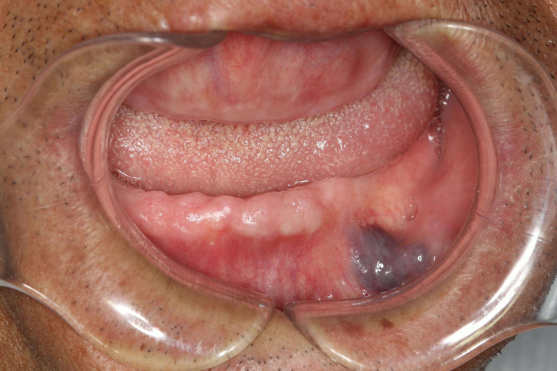
A venous lake could be found at vestibule of the left side lower jaw: before treatment.
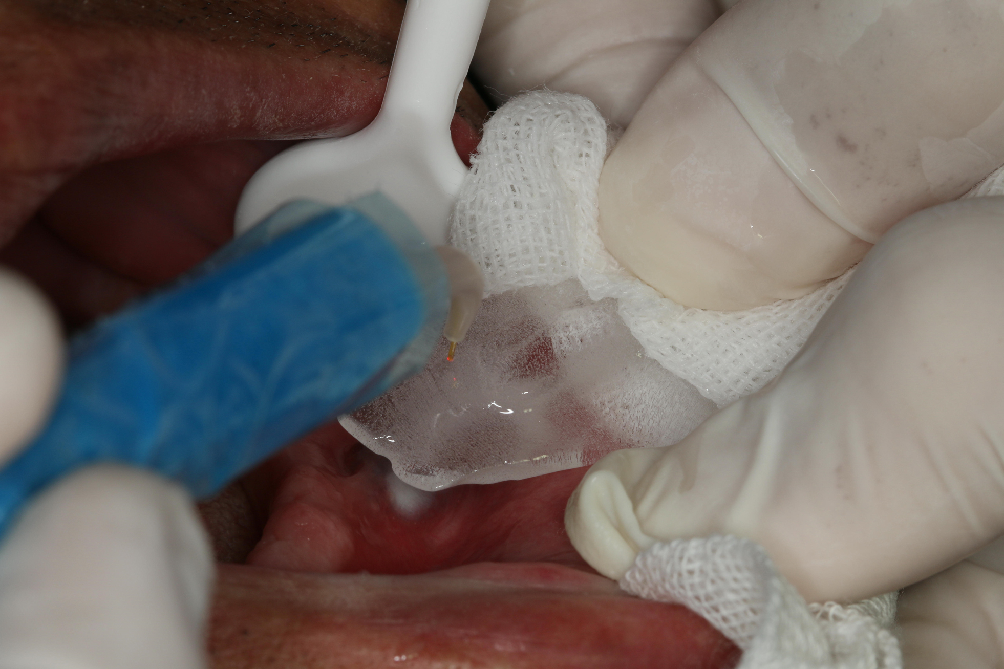
Nd:YAG laser was used, and the parameters were set as follows: average power of 5 W, frequency of 100 Hz, MSP, tip size of 300 μm. Thin slices of ice were used intermittently during operation for cooling and reducing thermal damage. MSP, microshort pulse; Nd:YAG, neodymium-doped yttrium aluminum garnet.
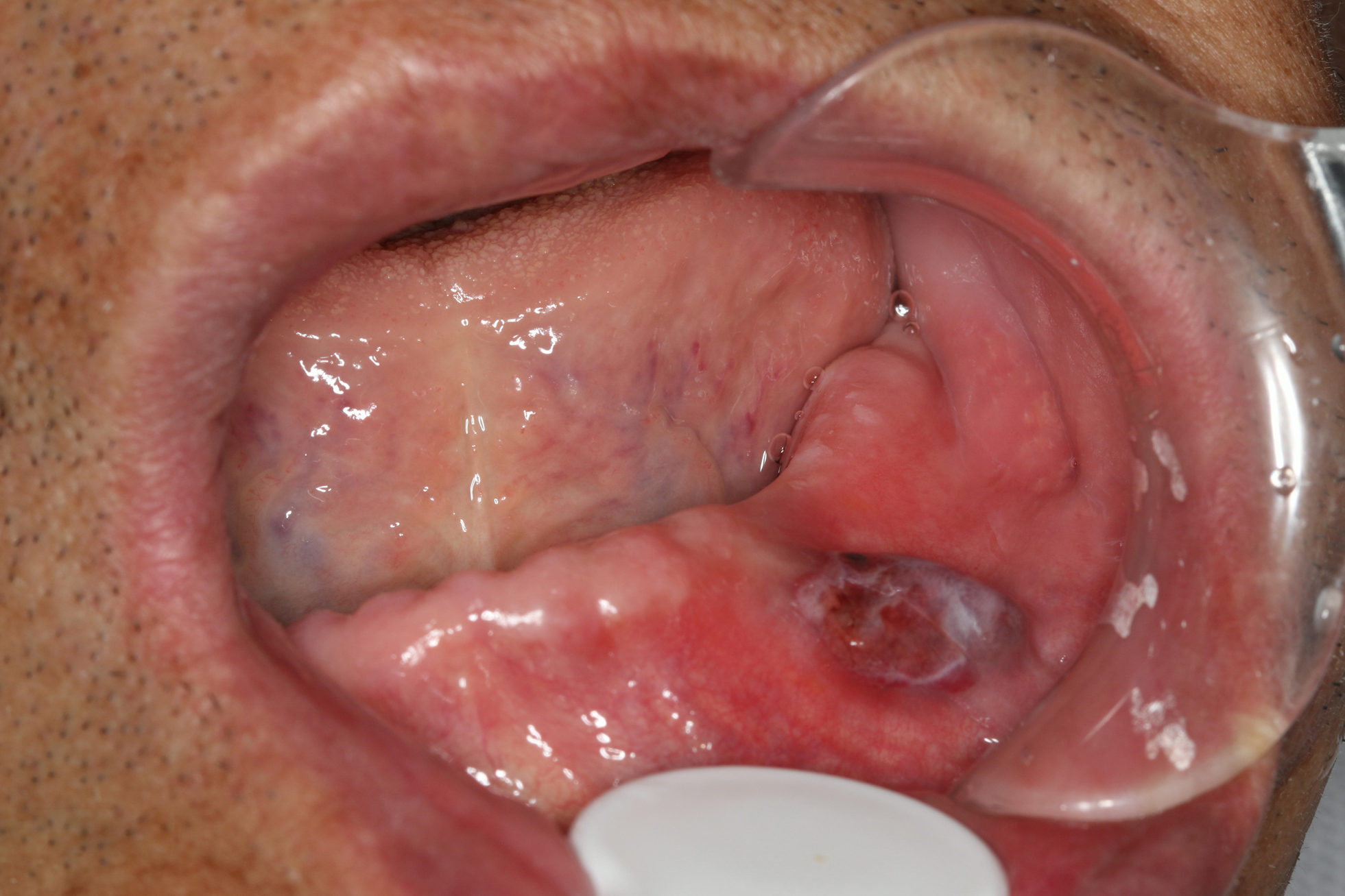
Immediate performance after treatment: the surface of lesion shrunk and turned into gray-white plaques.
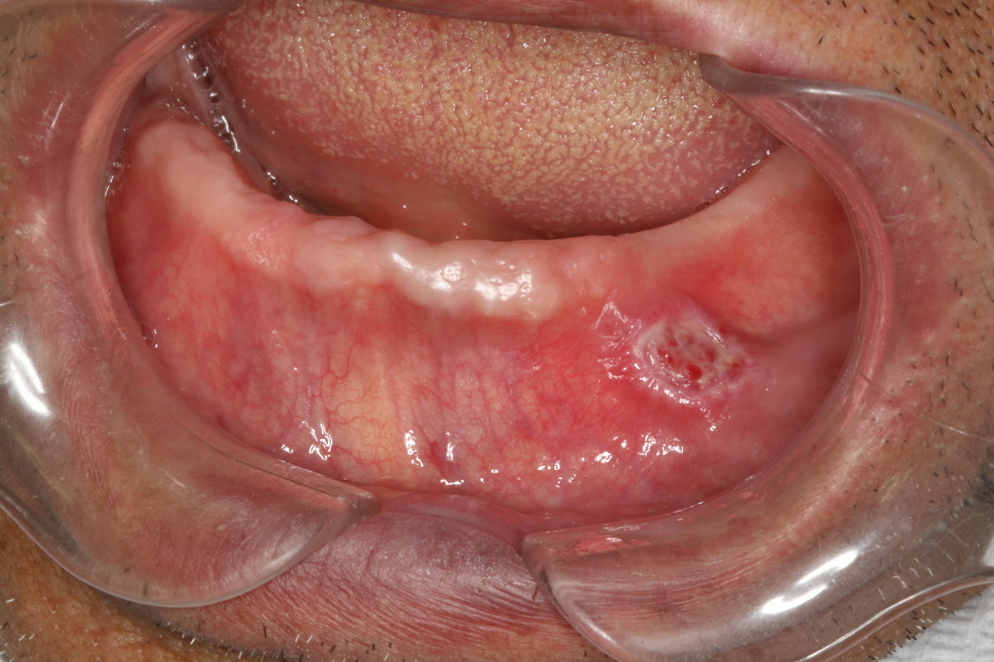
Two weeks after Nd:YAG laser treatment: the operation area mainly covered the surface of white pseudomembrane.
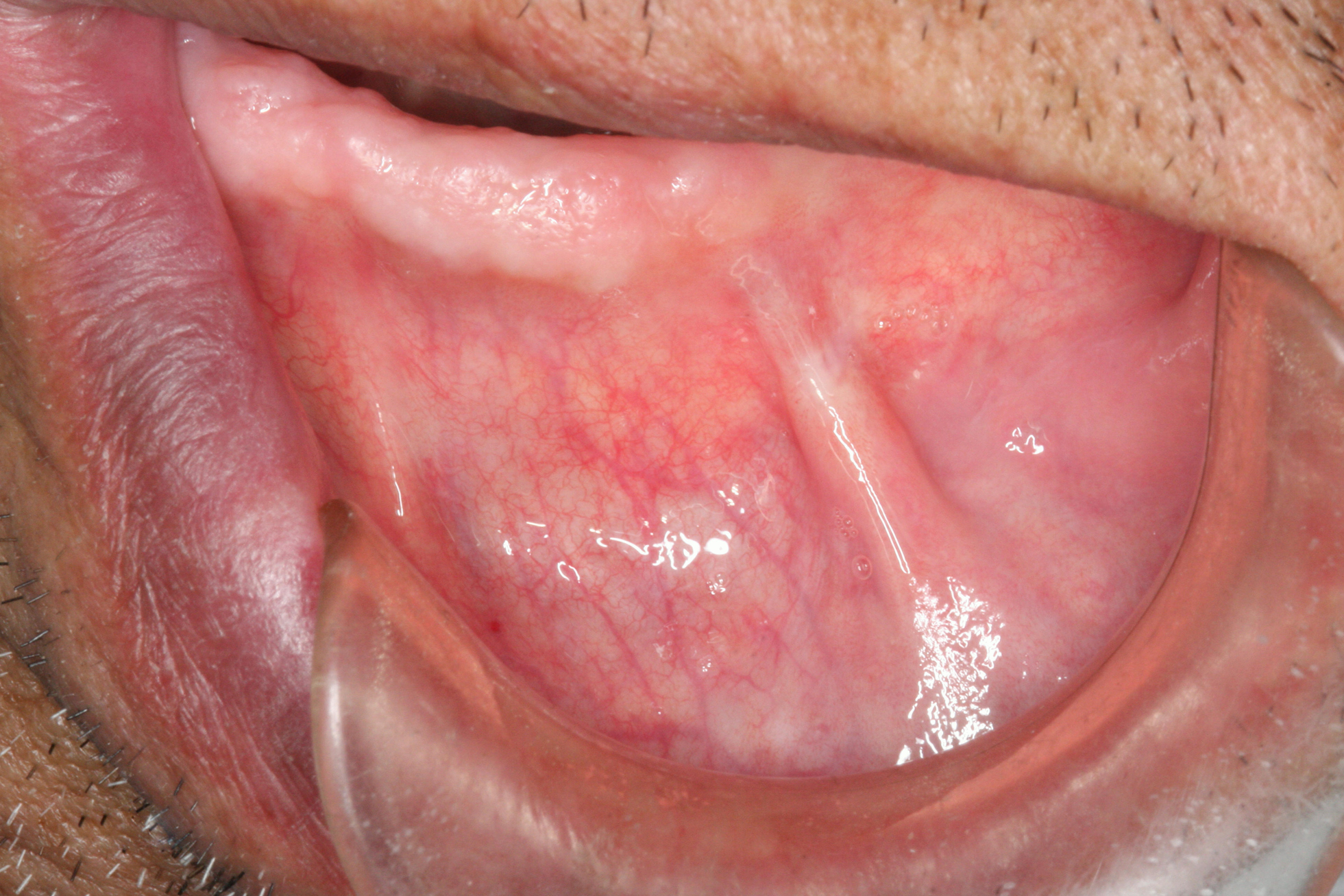
Four weeks after Nd:YAG laser treatment: scar tissue was formed at the operation area and the buccal frenum had a high attachment point.
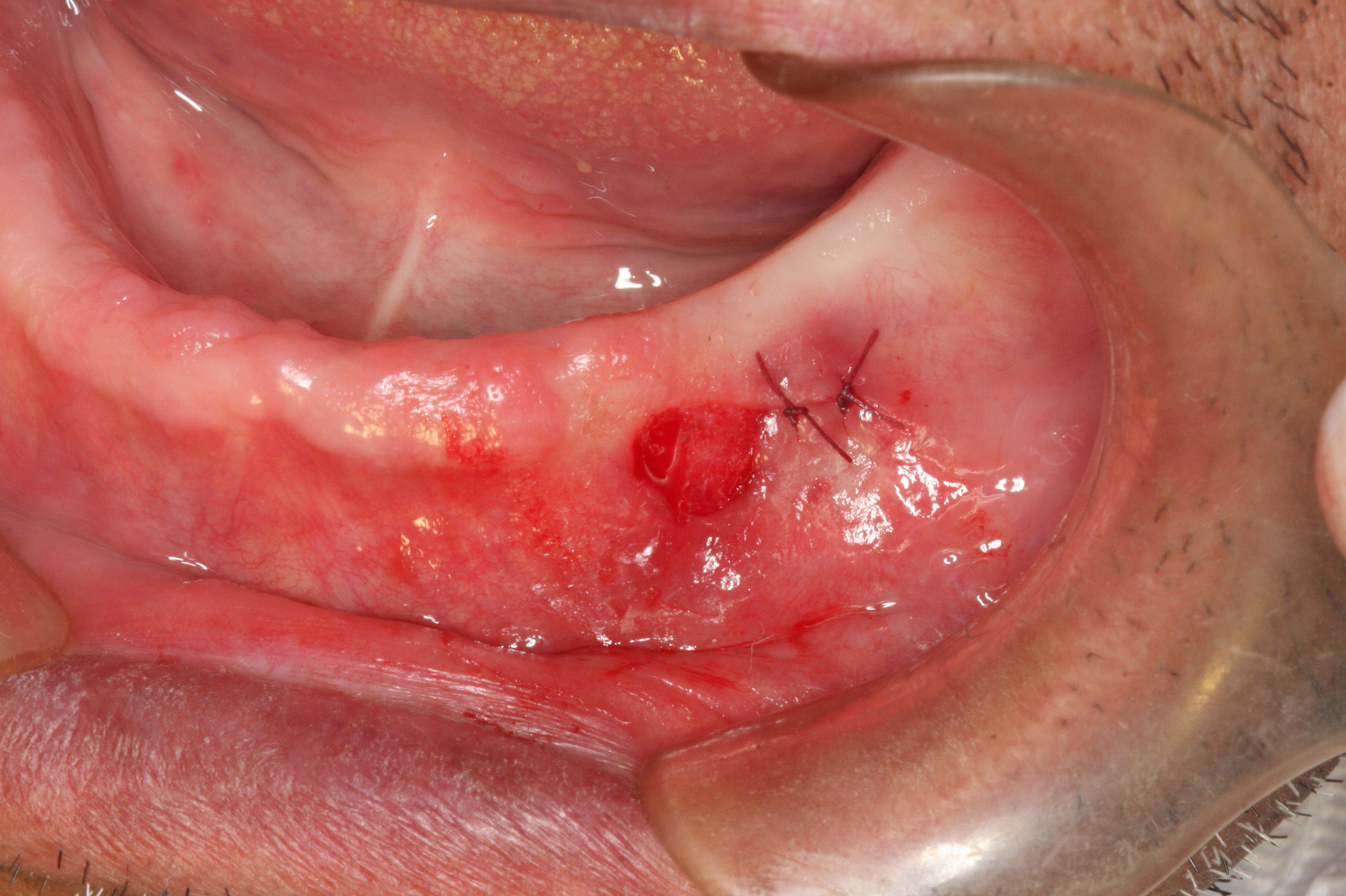
Immediate performance after Er:YAG laser treatment: scar tissue was removed, and the attachment point of buccal frenum was corrected. Er:YAG, erbium-yttrium aluminum garnet.
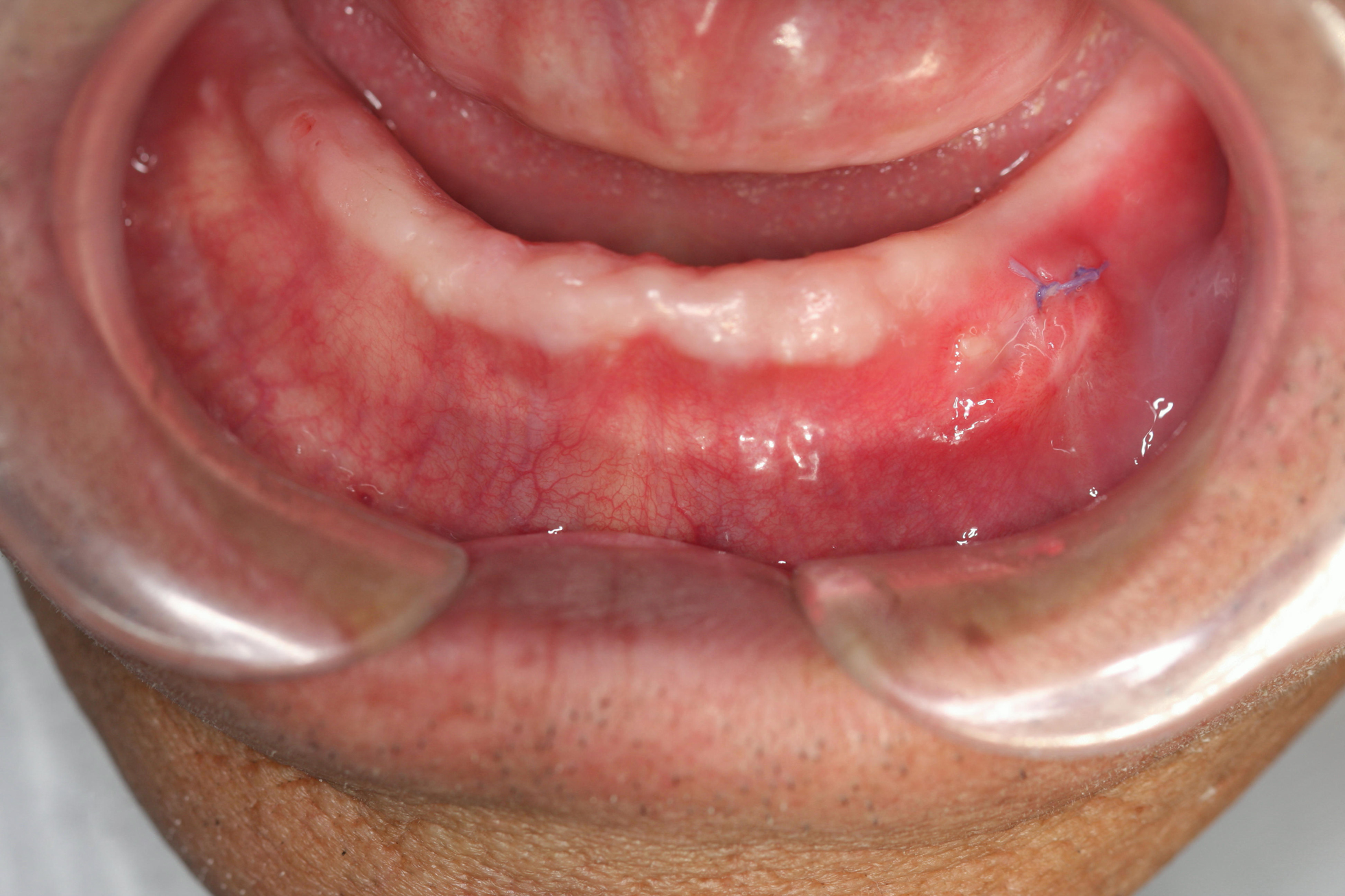
Ten days after Er:YAG laser treatment: the wound healed well.
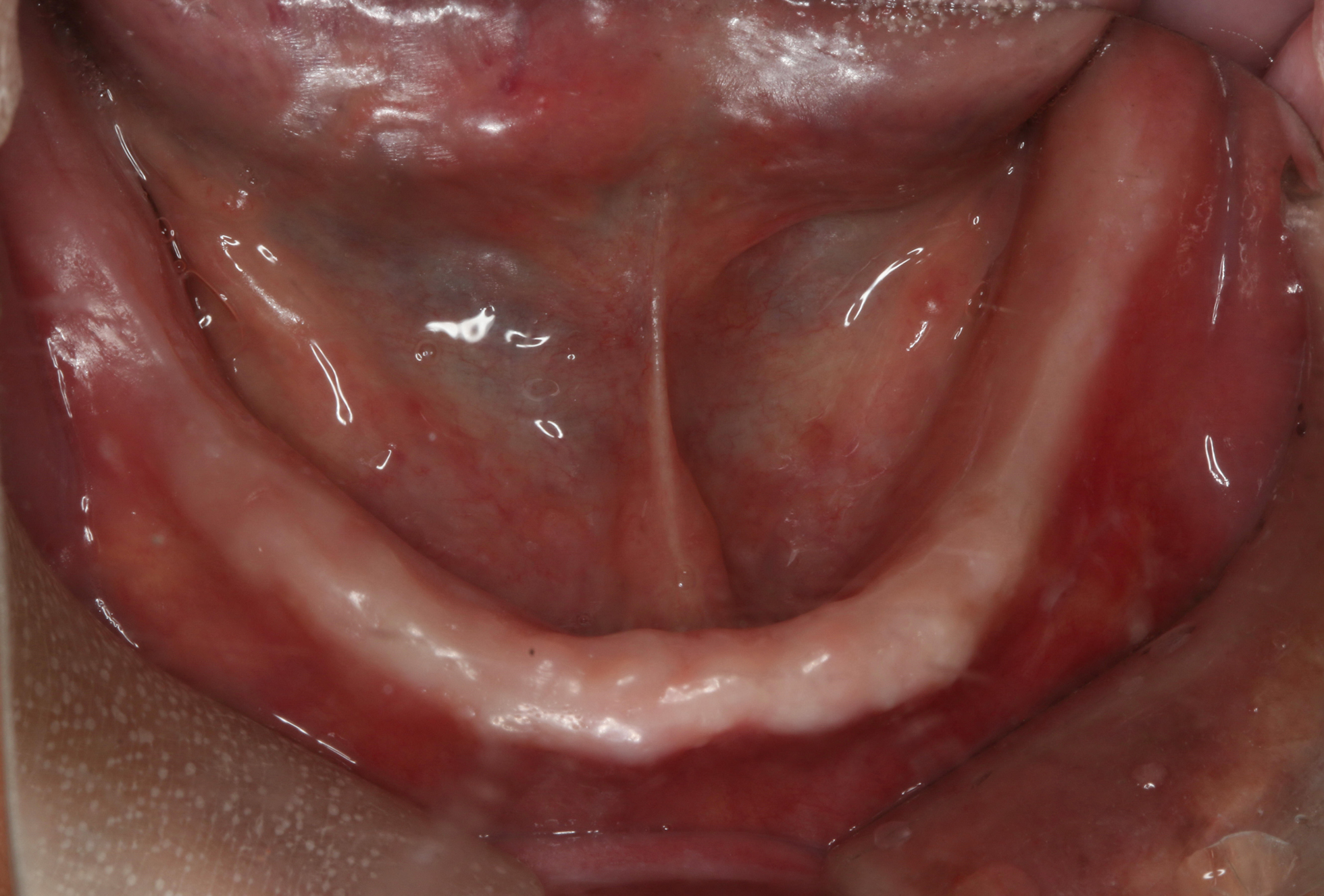
Three weeks after Er:YAG laser treatment: the wound healed well, and no evident scars were observed. Complete denture. Full denture is about to be made.
Discussion
The treatments of OVL include surgical excision, cryosurgery, sclerotherapy, electrosurgical excision, laser therapy, phototherapy, and a combination of these. 4 Although the recurrence rate of surgical resection is low, there is the possibility of bleeding and scar formation. 8 In cryosurgery, no anesthesia is required, and the mucosa changes little in color after treatment. However, complications such as local frostbite and postoperative swelling can occur. 9 Sclerosing therapy is simple and cost effective, but it may require multiple treatments to achieve acceptable therapeutic effects. In addition, studies have shown that sclerosing agent has adverse effects on lung connective tissue. 10 Laser treatment has become a more favored method for the treatment of OVL because of its easy manipulation and less side effects. In 1983, Anderson and Parrish developed a new laser equipment based on the selective photothermolysis theory. They proposed that some colored molecules in the tissue absorb specific wavelengths of light and convert light energy into heat energy. Therefore, local tissue can be selectively damaged. 11 There is evidence that laser with a certain energy, duration, and wavelength can target oxyhemoglobin and remove vascular structure without damaging surrounding tissue. It provides an effective way for treating skin or mucosal vascular abnormalities such as OVL. 12 –14 Lasers used for OVL treatment include argon laser (488–514 nm), pulsed-dye laser (PDL, 577 and 599 nm), alexandrite laser (755 nm), diode laser (800–810 nm), Nd:YAG laser (1064 nm), and CO2 laser (10,600 nm).
The results show that pulse width and optical absorption efficiency are two important factors influencing therapeutic efficacy. 6 PDL is used for treating superficial vascular malformations. Oxyhemoglobin in vascular lesions has three absorption peaks at 418, 524, and 577 nm, respectively. PDL has a wavelength of 577 nm, so it can be readily absorbed by oxyhemoglobin, thus ensuring a high absorption efficiency. However, purpura, bruises, and pigment deposition may occur after PDL therapy. 12 In addition, melanin can mildly absorb the wavelength of PDL light, resulting in the change of skin color, which might become the potential concern of dark-skinned patients. 20 PDL has short pulse width, and the penetration depth of PDL irradiation is ∼1 mm; therefore, PDL tends to be used to treat small OVL on skin surface. 12,15 Compared with PDL, Nd:YAG laser is less readily absorbed by oxyhemoglobin. However, it has longer pulse width (50–60 ms), and the penetration depth of Nd:YAG laser irradiation can reach 7–10 mm. Thus, it has more influence on deep vascular structure than PDL. 16 John et al. first reported that Nd:YAG laser has therapeutic effects on low-flow venous malformations in head and neck region. 7 Bekhor used Nd:YAG laser to treat OVL in 35 cases and adjusted the parameters of laser therapy according to sizes of lesions. 16 They found that OVL was completely removed in 94% of the patients. Becher et al. found that Nd:YAG laser has comparable function to PDL, and as a therapy method for superficial vascular malformations it can be used to completely remove OVL or significantly improve conditions in more than 90% of the patients. 12 Migliari et al. used Nd:YAG laser to treat OVL (<1 cm) on the lips or oral mucosa of 16 patients. 5 They found that the operation area was slightly swelled 1–2 days after treatment and completely healed (epithelialization) by 2–4 weeks. No pigment deposition, scars, shrinkage, and wrinkles were observed. Stephanie Mlacker pointed out that lasers with larger penetration depth such as Nd:YAG laser (wavelength of 1064 nm) would be used to remove OVL, which is a noninvasive and rapid therapy and can maximally reduce the formation of scars. 17 However, the patients should be told that there is still a possibility of the scar formation.
In this study, the therapeutic efficacy in the treatment of OVL with single Nd:YAG laser reached grade IV (excellent) in seven patients (77.8%) and grade III (good) in two patients (22.2%). Scar tissue was formed in two patients 2 weeks after Nd:YAG treatment, which may be attributed to two factors as follows: (1) the surgeon is not much experienced with the laser therapy technique, and (2) the sizes of lesions are larger in these two patients. Nd:YAG laser with variable pulse width (MSP/SP, 100–300 μs) was used in this study. Because of limited pulse energy, short working time can lead to the formation of purpura due to incomplete coagulation of blood vessels. However, if working time was too long or the pulse width was longer than the thermal relaxation time of lesion, the tissues surrounding lesion would be damaged because of heat penetration. 3 Moreover, during the initial stage of laser therapy, we did not use thin slices of ice for cooling. Instead, we used a solid stick of ice for intermittent cooling, so the local cooling was not performed in a timely and complete manner. The literature indicates that plastics or transparent glass sheet should be pushed on OVL during laser treatment, which can improve therapeutic effects. 6 During the later stage of laser therapy, we pushed self-made transparent slices of ice with a size of 3 mm on the lesion site, which resulted in more uniform transmission lines and better cooling effects.
Er:YAG laser has a wavelength of 2940 nm, which is in the infrared region. It can be strongly absorbed by water and photoelectrolysis, resulting in the increase in water temperature. Therefore, vapor is released from target tissue and microexplosions occur. Finally, the target tissue can be removed through microexplosions. 18 The penetration depth of Er:YAG laser irradiation is only 20–30 μm, which has characteristics of high precision, high efficiency, less heat diffusion, and less mechanical damage during excision. 19 Compared with traditional scalpels, Er:YAG laser is more conducive to wound healing and the reduction of local inflammatory response, as well as the formation of scar tissue. 20 In this study, scar tissue was formed in two patients 2 weeks after Nd:YAG laser therapy. Thus, Er:YAG laser was applied for scar removal. Good clinical results were obtained, and no scars and shrinkage were observed.
Conclusions
In this study, we used the Nd:YAG laser or a combination of Er:YAG laser for the treatment of OVL, and good therapeutic efficacy was achieved. This might provide a new choice of OVL treatment. As the number of patients with OVL increases, the parameters of laser treatment should be adjusted accordingly to achieve optimal therapeutic effects and prevent the occurrence of complications.
Footnotes
Author Disclosure Statement
No competing financial interests exist.
Funding Information
There was no funding provided for this article.