




Abstract
Objective:
The aim of this study was to evaluate the effect of photodynamic therapy (PDT) on multispecies oral caries biofilms composed of Streptococcus mutans, Lactobacillus casei, and Candida albicans.
Background:
The abovementioned microorganisms largely cause dental caries, especially early childhood caries (ECC), by synthesizing of acids in the presence of sugar. PDT is considered an effective process to remove oral biofilms, and erythrosine, an oral bacterial disclosing agent, is an ideal dye that can be used as a photosensitizer in PDT. However, until now, there are no studies that have reported the effect of erythrosine-mediated PDT on biofilms, including the three microorganisms.
Methods:
The biofilms were formed on hydroxyapatite discs, and erythrosine was used as the photosensitizer, diluted to a concentration of 40 μM for 3 min. Light was irradiated for 10 and 20 sec using a blue light-emitting diode dental curing light. After the experiment, the colony-forming units of each microbial group cultured on blood agar plates were counted, and a confocal laser-scanning microscope was used to evaluate the effect of PDT.
Results:
The counts of all three microorganisms significantly decreased in the PDT group compared with those in the control group. For S. mutans and L. casei, there was a larger decrease proportional to the amount of energy irradiated.
Conclusions:
Overall, PDT showed a significant antimicrobial effect against oral biofilms composed of the three microorganisms, suggesting its potential clinical application for infants with ECC.
Introduction
S
Candida species are not only the normal commensal microorganisms of the oral cavity but also are the opportunistic pathogens. 2 C. albicans is the most prevalent Candida species in the oral cavity. A previous study analyzed the number of microorganisms in the dental plaque of children aged 2–5 years, classifying them into groups of children with no caries, caries, and ECC, and found that the level of C. albicans was the highest in the ECC group. 1 In addition, another study found a significantly higher level of C. albicans in patients with severe ECC than in those without caries in a study of children aged 12–71 months. 3
There have also been reports of the interaction of C. albicans with other cariogenic microorganisms. For example, Falsetta et al. 4 demonstrated that the exopolysaccharides (EPS) produced by C. albicans resulted in an increased density of S. mutans and promoted the growth of a biofilm, resulting in increased virulence. C. albicans also contributes to the production of an acidic environment, which is favorable for the survival of lactobacilli, thereby enhancing their durability. 5
Biofilms are a conglomerate of several populations of surface-attached microorganisms, comprising either single or multiple species. Biofilms formed in the oral cavity are involved in the etiology of dental caries through the production of copious quantities of EPS. Oral biofilms are also regarded as a significant factor contributing to the resistance of oral bacteria to antibiotics, with several resistance mechanisms reported to date. 6,7 Because of the nature of biofilms, it is difficult to use antibiotics for the prevention of dental caries, as it can result in the destruction of not only the caries-inducing bacteria but also the normal beneficial oral bacterial flora. 8,9 Therefore, the currently used methods for preventing dental caries involve mechanically remove biofilms, such as tooth brushing. 10 However, prognosis with mechanical method depends on patient compliance. Therefore, alternative techniques, or additional therapy other than mechanical or antibiotic therapy, are required to effectively remove oral biofilms. 6
Photodynamic therapy (PDT) is a cell-removal technique involving the combination of a photosensitizer and a light source in the presence of oxygen. Recently, PDT has attracted attention in dentistry to remove the external cell layer of an oral biofilm, as it can reduce the mass size without requiring extensive compliance of the patient or promoting microbiological resistance. 6,11 And according to previous study, PDT also inhibits both biofilm formation and viability of the biofilm too. 12
Erythrosine is a xanthene dye that is characterized by light absorption at wavelengths of 500–550 nm. It is ideal for use as a photosensitizer in PDT compared with other dyes because it has been approved for use in the oral cavity as a plaque disclosing agent and exhibits no direct toxicity to the host. 13
Although S. mutans, Lactobacillus casei, and C. albicans have all been shown to be closely related to the development and/or progression of ECC and have synergistic interactions, no study has yet explored the effects of erythrosine-mediated PDT for a multispecies biofilm containing these three species. In this study, we formed a multispecies biofilm in vitro with S. mutans, L. casei, and C. albicans to more closely reproduce the oral environment, in which ECC occurs and progresses. Moreover, we aimed to evaluate the specific effects of PDT on the biofilm by distinguishing its influence on each of the component species.
Materials and Methods
Bacterial strains and culture conditions
The strains used in this study were S. mutans ATCC 25175, L. casei ATCC 334, and C. albicans KCTC 7270. Bacteria and yeast were inoculated in brain-heart infusion (BHI) broth (Becton, Dickinson and Company, Sparks, MD) and incubated under aerobic conditions, supplemented with 5% CO2 at 37°C for 18 h. Cell dilutions were established by constructing a standard curve relating the bacterial cell numbers with the culture turbidity, measured by a spectrophotometer (Smart Plus 2700; Young-woo Institute, Seoul, Korea). All species of the bacteria were then diluted to 10 9 colony-forming units (CFU)/mL with phosphate-buffered saline (PBS), and this suspension was used as the inoculum for biofilm formation.
Cariogenic biofilm formation
Since there was no previous study of biofilm formation with the three microorganisms based on ECC, pilot studies were conducted with setting various inoculation ratios and culture conditions before this study. The experimental conditions, in which all three microorganisms were well grown and colony counting of three microorganisms was possible, were selected after pilot studies.
S. mutans, L. casei, and C. albicans were cultured in BHI liquid medium supplemented with 1% sucrose at 37°C for 24 h. BHI broth and the diluted bacterial solution were inoculated into a 24-well culture cluster (SPL Life Sciences; Pocheon-si, Gyeonggi-do, Korea) to achieve a final concentration of 1 × 10 7 CFU/mL for all three species. To best reproduce the tooth surface, hydroxyapatite discs (Biosurface Technologies; 1.27 cm diameter) were placed at the bottom of each well. The plates were then cultured at 37°C for 24 h to allow for the formation of biofilms on the disc surface.
Photosensitizer
Erythrosine was used as a photosensitizer. A stock solution of 1 mM erythrosine (Sigma-Aldrich, St. Louis, MO) was prepared in PBS. This solution was filter-sterilized and stored at −20°C in the dark until use. Working solutions were obtained by diluting the stock solutions with PBS to 40 μM.
Light source
A blue light-emitting diode (LED; VALO™, Ultradent, 395–480 nm) with an output power of 1800 mW (with a round tip of 10 mm diameter) was used as the light source for PDT. The light intensity of incident radiation was measured using a radiometer (Light Intensity Meter; Dentamerica, San Jose, CA) at a distance 10 mm from the lens, since irradiation was performed at 10 mm from the hydroxyapatite discs. The parameters of the light source used for treatment are shown in Table 1.
The Parameters of the Light Used for Photodynamic Therapy
Group III, LED light irradiation alone; Group IV and V, erythrosine and LED light (photodynamic therapy). Since the area of the biofilm on the HA disc could not be accurately measured, the power and energy density could not be obtained.
LED, light-emitting diode.
Photodynamic treatment of biofilms
The biofilms were subjected to no photosensitizer or light irradiation treatment (group I), erythrosine alone (group II), light irradiation alone (group III), or combined photosensitizer (40 μM erythrosine) and light irradiation treatment (group IV and V). Group IV was irradiated with blue LED light for 10 sec with a total of 18 J of energy, and group V received a total of 36 J of energy after irradiation for 20 sec. Total 50 hydroxyapatite discs were used and 5 groups contained 10 discs, respectively. The biofilms were incubated with 2 mL of 40 μM erythrosine (groups II, IV and V) or PBS (groups I and III) for 3 min.
After treatment, each well containing the biofilm was sonicated twice for 10 sec (VC 100; Sonics & Materials, Inc., Danbury, CT). Each sample was then diluted with PBS, and 50 μL of the diluted suspension was spread onto duplicate blood agar plates (Hanil-KOMED, Seongnam, Korea) with a spiral plater (IUL, Barcelona, Spain). The plates were incubated for 36 h at 37°C under 5% CO2 and the numbers of CFU on the plates were counted by the naked eye. Each species was identified and distinguished by the morphology of bacterial colony (i.e., Fungal colonies of C. albicans were much larger than that of the other two bacteria and had irregular form, while colonies of L. casei had higher transparency than those of S. mutans), after confirming the morphologies from separately culturing each strains on BA plate in pilot studies.
Confocal laser-scanning microscopy
The viability of microorganisms was evaluated by confocal laser-scanning microscopy after applications of the photosensitizer and light (groups IV and V) in comparison to the untreated control group (group I). The biofilms were washed twice with PBS and stained with the Live/Dead BacLight Bacterial Viability Kit (Molecular Probe, Eugene, OR) containing SYTO-9 (green, viable cells) and propidium iodide (red, dead cells) in the dark and incubated at room temperature for 15 min. Stained biofilms were examined with a LEICA TCS SP8 confocal microscope, using an excitation laser (argon gas laser, 488 nm) and HyD detector (485/540 nm for detection of SYTO9 and 601/670 nm for detecting propidium iodide).
Statistical analyses
The Shapiro–Wilks test was used to check the normality of the data. Comparisons between normative groups were made with the parametric t-test, whereas the means and standard deviations for non-normative groups were compared by the nonparametric Mann–Whitney U test. p Values less than 0.05 were considered statistically significant. SPSS software program (SPSS, Inc., Chicago, IL) was used for all statistical analyses.
Results
Effect of PDT on microbial counts
For all three microorganisms, statistically significant reductions of CFU counts (p < 0.05) were observed in the PDT-treated groups (groups IV and V) compared with the untreated control (group I). In all cases, the single use of erythrosine (group II) or LED light irradiation (group III) did not have a significant effect on CFU reduction (Table 2). Table 3 shows the CFU counts of group IV and group V as a percentage of that of group I.
Viable Bacterial Counts (Mean ± Standard Deviation)
Significant differences were only in group I–IV, I–V, and IV–V comparisons.
Group I, untreated control; Group II, erythrosine alone; Group III, LED light irradiation alone; Group IV, 2 mL erythrosine and 18 J of LED light for 10 sec; Group V, 2 mL erythrosine and 36 J of LED light for 20 sec.
Significant difference (p < 0.05) compared with group I.
Significant difference (p < 0.05) between groups IV and V.
CFU, colony-forming units.
Microbial Counts After Photodynamic Therapy Plus Irradiation as a Percentage of the Untreated Control
Group I, untreated control; Group IV, 2 mL erythrosine and 18 J of LED light for 18 sec; Group V, 2 mL erythrosine and 36 J of LED light for 20 sec.
Comparison of the two PDT-treated groups with different irradiation conditions (IV and V) showed a significantly larger decrease in S. mutans and L. casei counts in group V (p < 0.05), whereas there was no significant difference in the C. albicans count (Fig. 1).
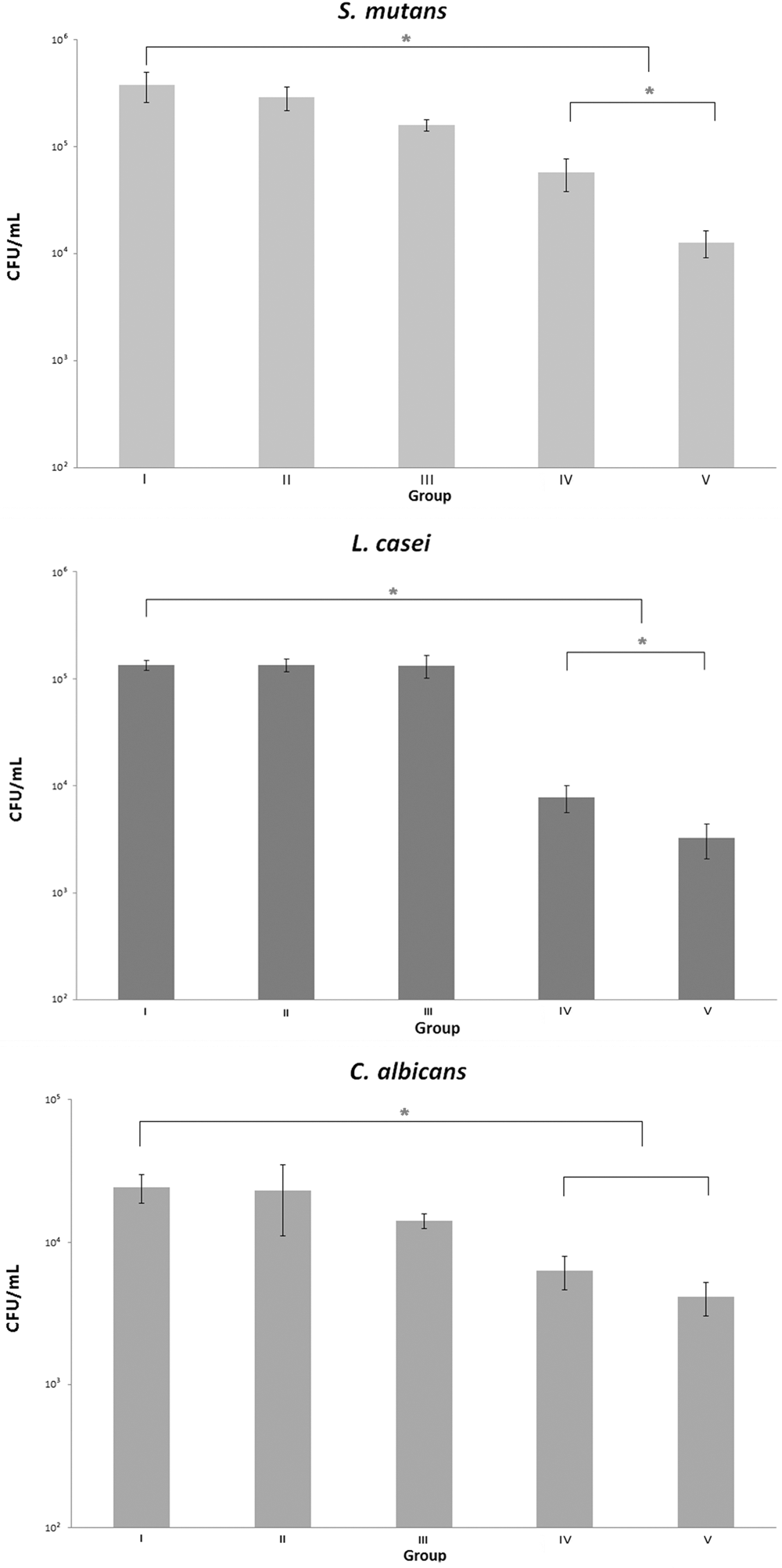
CFU counts of Streptococcus mutans (top), Lactobacillus casei (middle), and Candida albicans (bottom) after different photodynamic treatments. Group I: control group that was not subjected to irradiation; group II: erythrosine alone; group III: light irradiation; group IV: combined treatment with 40 μM erythrosine and light irradiation with blue LED light for 10 sec with a total of 18 J of energy; and group V: combined treatment with 40 μM erythrosine and light irradiation with blue LED light for 20 sec with a total of 36 J of energy. *Indicates significant difference (p < 0.05). CFU, colony-forming units; LED, light-emitting diode.
Effect of PDT on biofilm viability
The majority of the cells showed green fluorescence in the control group (group I), and the PDT-treated groups showed substantially enhanced red fluorescence, as shown in Fig. 2, which was stronger with increasing irradiation intensity.
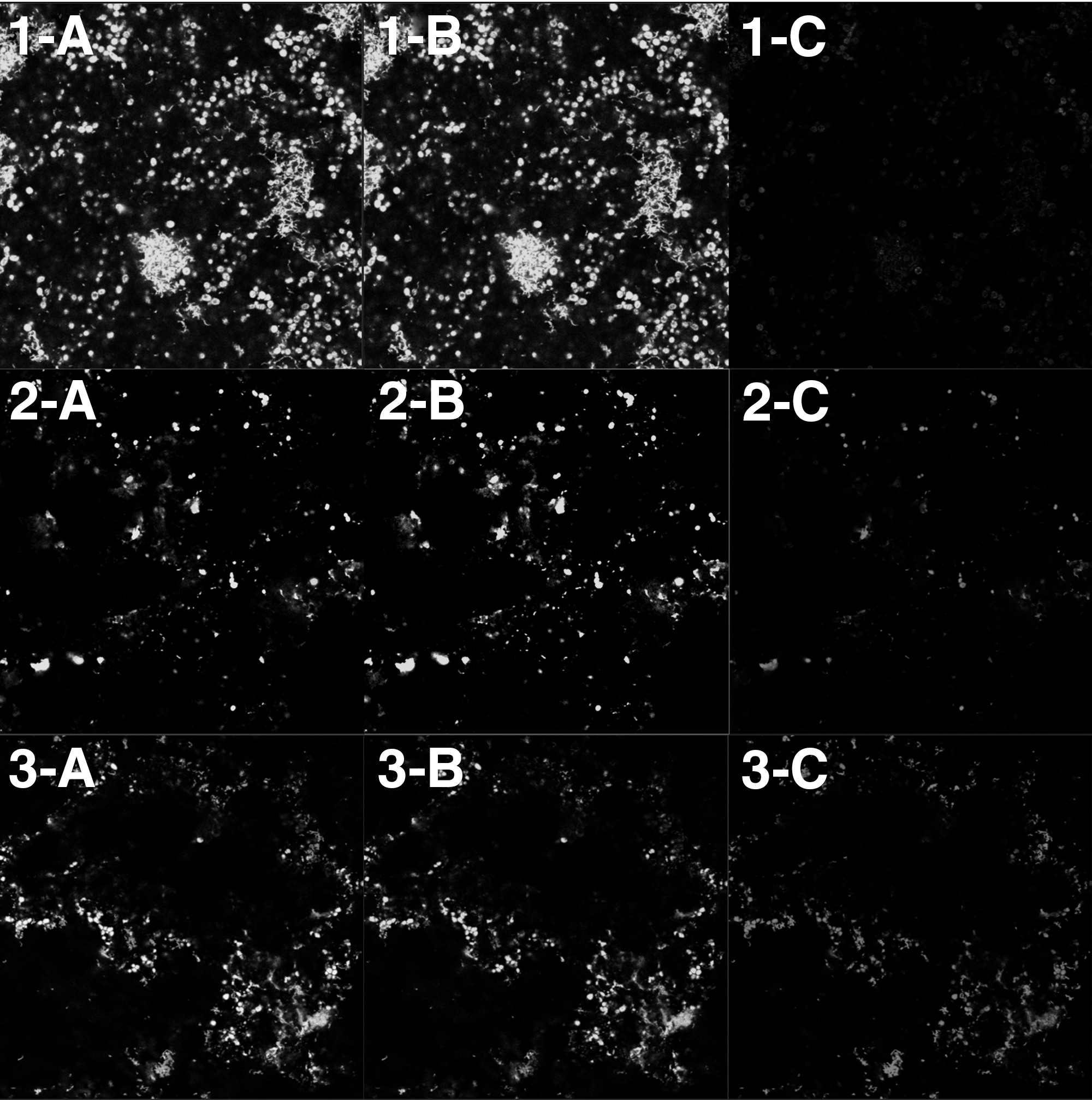
CLSM images of group I (Untreated control,
Discussion
In this study, we found that erythrosine-mediated PDT showed a significant microbial-killing effect on a multispecies cariogenic biofilm composed of S. mutans, L. casei, and C. albicans. This result is consistent with previous studies that evaluated the effect of erythrosine-mediated PDT on biofilms of S. mutans or C. albicans alone. 6,14 However, we further demonstrated that the three microorganisms in a cariogenic multispecies biofilm have synergistic interactions with each other. The average reduction of C. albicans in the multispecies biofilm assessed in this study was 83.33% (group V), which was greater than the average reduction (38.34%) in the biofilm containing only C. albicans, which was reported previously under the same experimental PDT condition. 14 This is inconsistent with the lower reduction in a multispecies biofilm of C. albicans, S. aureus, and S. mutans after PDT, compared with the single biofilm of each bacterium observed in another study. 15 This discrepancy may be related to the different types of growth media on which the biofilms were cultivated, the different components of the biofilm, and interactions among the species.
In the treatment of LED light irradiation for PDT, we set two different irradiation conditions to find optimum conditions for the inhibition of a multispecies biofilm. Thus, group IV was irradiated with 18 J, and group V was irradiated with 36 J, which was previously proved to inhibit a single S. mutans biofilm and single C. albicans biofilm, respectively. 6,14 Under both conditions, S. mutans and L. casei showed significant reductions, with a larger decrease of counts detected under an increased amount of irradiated energy. This proportional effect of PDT to the irradiation energy is consistent with a previous study using a halogen lamp as the light source of PDT for an S. mutans biofilm. 16
By contrast, there was no significant difference in C. albicans reduction concomitant with an increase of irradiation energy, which is in line with previous studies demonstrating that gram-positive bacteria are more sensitive to PDT when compared with fungi of the Candida species. 17 Nevertheless, a significant decrease in the C. albicans count was detected compared with the control under both 18 and 36 J irradiation. Therefore, inhibition of C. albicans can be achieved with less energy than previously reported conditions.
In this experiment, the erythrosine concentrations were set on the basis of studies conducted by Costa et al., 14 who reported significant inhibition of single S. mutans and C. albicans biofilms during PDT treatment with 40 μM erythrosine and Choi et al., 16 who suggested 20–40 μM erythrosine is optimum concentration for during PDT treatment on S. mutans biofilm. This concentration was much lower than 9–25 mM, which is used in clinical dental applications as plaque disclosing agents. The antimicrobial effect of PDT tends to be enhanced with increasing concentrations of erythrosine and treatment time. 16 In addition, high concentrations of erythrosine display antibacterial effects against some microorganisms even without light irradiation. 18 –20
In the present study, we developed a multispecies biofilm in vitro with three microorganisms that are closely related to the development and progression of ECC. Therefore, this study has significance in that it suggests the possibility of clinical use of PDT for ECC prevention. However, for substantial and clinical applications, in vitro studies determining optimal parameters and in vivo studies should be proceeded before. Consequently, to investigate the roles and interactions of the three microorganisms in the biofilm on the action of PDT, and to analyze the optimal conditions that can produce maximum antimicrobial effects, we suggest that further studies be designed with higher concentrations of erythrosine for both single and multispecies biofilms.
Conclusions
In the present study, we evaluated the effect of PDT on the multispecies biofilm in vitro with three microorganisms, S. mutans, L. casei, and C. albicans, using two different conditions of light irradiation. We confirmed positive effects of PDT with erythrosine and an LED light source for reduction of all three microorganisms in the multispecies cariogenic biofilm. In the case of C. albicans, we detected a greater reduction than previously reported for the single species biofilm. In addition, the inhibition effect was proportional to the amount of irradiated energy of PDT for S. mutans and L. casei.
Footnotes
Acknowledgments
This work was supported by the Scientific Research (SR1703) of Gangneung-Wonju National University Dental Hospital.
Author Disclosure Statement
No competing financial interests exist.