
Abstract
Objectives:
In this work we demonstrate the first laboratory study results of lens fragmentation with low-energy picosecond ultrashort laser pulses after artificial induction of cataract with microwave radiation on an ex vivo animal model.
Background:
This method will be evaluated with regard to the further development of lens fragmentation with novel ultrashort picosecond laser systems instead of ultrasonic phacoemulsification or the significantly more complex femtosecond laser fragmentation.
Methods:
As samples we used postmortem porcine eyes. The lenses were dissected and then irradiated in a microwave oven for artificial cataract induction. Subsequent computer-driven lens fragmentation was performed with a 12 ps, 1064 nm pulsed laser source with 100 µJ pulse energy, and 10 kHz pulse repetition rate.
Results:
Both the artificial cataract induction and the lens fragmentation were demonstrated. When inducing cataract, different degrees/stages of opaqueness and hardness could be achieved with different irradiation times and methods. The fragmentation with 12 ps pulses led to good results with regard to ablation depth and rate, especially for the softer lenses.
Conclusions:
As could be shown, low-energy picosecond ultrashort laser pulses are feasible for cataractous lens fragmentation on an ex vivo animal model with artificial cataract induction. Thus, this technique may influence future cataract surgeries by possibly being an alternative or extension to state-of-the-art methods. This will be evaluated with further tests and studies.
Introduction
Cataract is the most common removable visual impairment and cause of blindness worldwide, with a rising global clinical burden. 1,2 Therefore, cataract surgery is one of the most frequently executed surgeries in the world with about 20 million cases per year. 3 In this surgery, the opaque lens must be fragmented, removed from the eye, and replaced by an artificial lens (intraocular lens). For this lens fragmentation, ultrasound phacoemulsification is state-of-the-art. Here, the lens is irradiated with ultrasound, fragmented, and simultaneously aspirated from the eye. 4 However, the applied ultrasound has a non-negligible energy input possibly leading to additional clinical patterns and adverse effects—even though it is declared to be very safe. 5 In particular, ultrasound exposure can, for example, cause damage to corneal endothelial cells 5 –7 and may lead to retinal detachment. 7,8 Due to the adverse effects, other methods were investigated. Here, laser-based methods seemed to be auspicious. Two of these methods were Nd:YAG and Er:YAG laser phacoemulsification, in which high-energy nanosecond or microsecond pulses are applied to the cataractous lens. 9 Further, Yb,Er:glass lasers in burst mode were also investigated. 10 However, due to the comparatively long pulse width of the laser sources, the pulse energy has to be several tens or even several hundred mJ to reach the tissue threshold and achieve an appropriate ablation rate. Hence, effects on surrounding tissue are non-negligible. 10,11 In 2009, Nagy et al. 12 introduced a method using femtosecond ultrashort laser sources—likewise to laser-assisted in situ keratomileusis (LASIK) devices—whereby the lens is cut in a defined way with computer-driven cutting paths. It is used as an assistive adjunct in addition to the ultrasound fragmentation. This combination of methods was then tested and compared with the single ultrasound phacoemulsification in several studies. 13 –15 The studies show comparable results for both methods with regard to the ablation/cutting process. The energy, however, could be drastically reduced using femtosecond laser pulses, leading to, for example, drastically lowered damage on the corneal endothelial cells since none or only a small ultrasound exposure time is necessary for lens fragmentation. 5,6,16 The computer-controlled laser-based method also has the advantage that less training and experience are required to perform surgeries, while this is an important factor in manual ultrasound surgeries. 17,18 Further, with ultrashort pulses (USPs), it is possible to perform not only lens fragmentation but also corneal incision and capsulotomy. 12,19
However, femtosecond ultrashort laser systems have the disadvantage of very complex setups. Therefore, they are considerably more expensive than the simpler ultrasound phacoemulsification. The same applies to state-of-the-art picosecond ultrashort laser systems—which have also been tested and found to be suitable for different ophthalmical surgeries (including phacoemulsification). 20,21 This is where a new class of picosecond Q-switched microchip lasers come into play, combining the ablation capabilities of high-end USP laser systems with a simple and compact design. 22 –24 They have already been successfully tested for other ophthalmical applications such as laser iridotomy, (post)cataract treatment/capsulotomy, and selective laser trabeculoplasty and promise to become a universal tool for a whole range of different ophthalmological surgeries—not least because these picosecond USPs are easier to be coupled into a fiber than femtosecond pulses, which could lead to a small USP handheld device for direct application. 25
The first studies on the induction of artificial cataracts using microwaves were carried out as early as 1948. 19,26 Since then, this topic has been investigated further by several research groups, particularly for training purposes for ophthalmologists. 27 –29 It seems to be a simple and fast method for artificial cataract induction. However, according to Ruiss et al., it is quite difficult to prepare different cataract states, and the lenses are mostly completely white (cataractous). 29 Nevertheless, this method should be well suited for ablation investigations with picosecond laser pulses due to its simplicity.
This article discusses the first laboratory studies on picosecond laser lens ablation/fragmentation and emulsification after artificial cataract induction with microwaves. For the latter, a new approach is applied by only irradiating the lens.
Methods
The induction of artificial cataract in porcine lenses was achieved using a household 2450 MHz, 700 W microwave oven (MM720CA7-PM from Renkforce, Munich, Germany). The tissue samples are postmortem porcine eyes, which are processed only a few hours after slaughter (about 4 h) and constantly cooled until further processing. For cataract induction, we pursue a new approach by dissecting the lenses together with their lens capsule from the eyes and placing them directly on microscope slides, which are then placed into the microwave oven. Consequently, there is no surrounding tissue that could absorb microwave radiation, making the induction process more controllable. After a microwave irradiation of up to 3 sec at 700 W power, the cataractous samples were cooled down in a refrigerator at 7°C while being constantly moistened with water.
For lens fragmentation, a 12 ps USP laser source was used. This USP laser system consists of a passively mode-locked Nd:YVO4 oscillator and an Nd:YVO4 regenerative amplifier. The laser system emits a 1064 nm pulse train with Gaussian mode profile (TEM00) and an adjustable pulse repetition rate between 1 Hz and 300 kHz, and a pulse energy of up to 164 µJ. 30 For the results presented here, the laser parameters were set to 100 µJ pulse energy and 10 kHz repetition rate. An overview of all parameters is given in Table 1.
Parameters of the 12 ps USP Laser Source Used for the Lens Fragmentations
USP, ultrashort pulse.
The laser was focused on the cataractous lenses via a computer-controlled two-axis scanner head (SS-II-15 [Y] D2 from Raylase GmbH, Wessling, Germany) with an F-theta flat field objective (S4LFT3046/328 from Sill Optics GmbH & Co. KG, Wendelstein, Germany) with a focal length of 50 mm (resulting in a spot diameter of about 25 µm and a fluence of about 20 J/cm2 at 100 µJ pulse energy). The laser was moved with 20 mm/sec in a cross-hatched pattern over a circular area with a diameter of 6 mm and a line spacing of 0.33 mm (see Fig. 5b). This ablation pattern was chosen in accordance with state-of-the-art ablation pattern with femtosecond laser lens fragmentation. 31 Ablation was started at the lens surface and then gradually continued downward (i.e., anterior to posterior). The tests were executed at a constant room temperature of 22°C. The lenses were constantly moistened with distilled water during the process. For real-time observation and magnification, a stereo microscope (Stemi 508 trino from Carl Zeiss Microscopy GmbH, Oberkochen, Germany) was used. The results were then visualized and examined with high-resolution light microscopy. For this purpose, the incident light microscope Axio Lab.A1, the transmitted light microscope Primo Star, and the former mentioned Stemi 508 trino, all from Carl Zeiss AG, Oberkochen, Germany, were used.
In order to obtain information about the influences of the surrounding medium and to have a better comparison with the actual surgery procedure (i.e., a rough eye model), the same method was carried out but with a full water immersion in addition. Here, the lenses were placed in a 50 mL Petri dish during the microwave irradiation process and subsequently in a 7 mL cuvette during the laser process. Both containers were filled with distilled water at room temperature of 22°C. For cataract induction, the microwave irradiation time had to be increased to 15–20 sec since the additional water increased the amount of material being heated up.
Since natural cataract does not only lead to opaqueness but also an increase in hardness, the different lens states (nonopaque, partially opaque, and fully opaque) after artificial cataract induction via microwave radiation were tested for this parameter. Here, a pressing force of 2.3 N (i.e., weight with 235g) was applied to lenses with different opaqueness states, which were placed between two microscope slides.
Results
As already described in the literature (see the “Introduction” section), it is possible to induce an artificial cataract in porcine lenses using a microwave oven, but this usually leads to instantaneous opacity of the entire lens. Here, lens opacification on moistened lenses was achieved after only a few seconds (<5 sec) of irradiation. In Figure 1, different states of artificial cataract after microwave exposure are shown [bottom row (post)]. For comparison, the top row (pre) shows the lenses before microwave exposure. The tests show different cataract states: LM,1 has the lowest level, LM,2, LM,3, and LM,4 have a medium level, and LM,5 and LM,6 are completely opaque. Further, lens LM,5 bursts during heating. Since a natural cataract does usually not affect the entire lens before it is removed, state LM,1 seems to be the most comparable artificial cataract. All six lenses were irradiated with maximum power (700 W) with durations of 1 sec: LM,1 and LM,2; 2 sec: LM,3 and LM,4; and 3 sec: LM,5 and LM,6. Lower power adjustments would lead to comparable results since the microwave oven works always at 100% power but with temporal modulation to adjust the average power.

Six porcine lenses before (pre) and after (post) artificial cataract induction with a microwave oven. The lenses were moistened during the process. The pictures show different cataract states (arranged ascending LM,1 to LM,6). The lenses were irradiated with 700 W for 1 sec: LM,1 and LM,2; 2 sec: LM,3 and LM,4; and 3 sec: LM,5 and LM,6.
In comparison to the moistened lenses, the fully immersed lenses are shown in Figure 2. Lenses LW,1, LW,2, and LW,3 are irradiated for 15 sec, and lenses LW,4, LW,5 and LW,6 for 20 sec. As for the moistened lenses, different degrees of opaqueness were achieved, even though the opaqueness was less controllable than without the surrounding water. LW,4 and LW,6 burst during irradiation.

Six porcine lenses before (pre) and after (post) artificial cataract induction with a microwave oven. The lenses were immersed in water during the process. The lenses were irradiated with 700 W for 15 sec: LW,1, LW,2, and LW,3; and 20 sec: LW,4, LW,5, and LW,6.
Regarding the hardness of the lenses, exemplary results are given in Figure 3, where the lenses are shown before and after applying pressure. Here, the nonopaque (nonirradiated) lens was completely flattened, while the other states were only partially squashed. The fully opaque lens was squashed the least. Thus, the higher the opaqueness, the harder the lens.
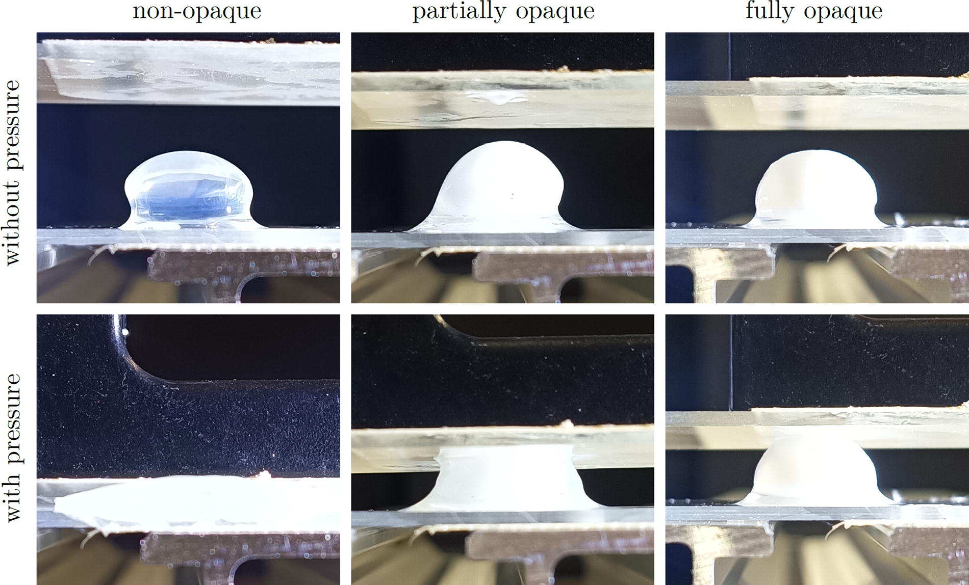
Pressure test for hardness estimation of different opaqueness states after artificial cataract induction with microwave radiation. The applied pressing force was 2.3 N.
After cataract induction, the lenses were irradiated with the 12 ps USP laser source. In Figure 4, a partially opaque porcine lens in a cuvette filled with distilled water is shown during the fragmentation process. Here, in the left image, the fragmentation laser can be seen. The turbidity of the water in both images is due to the ablated lens tissue during fragmentation and thus indicates a successful ablation process.
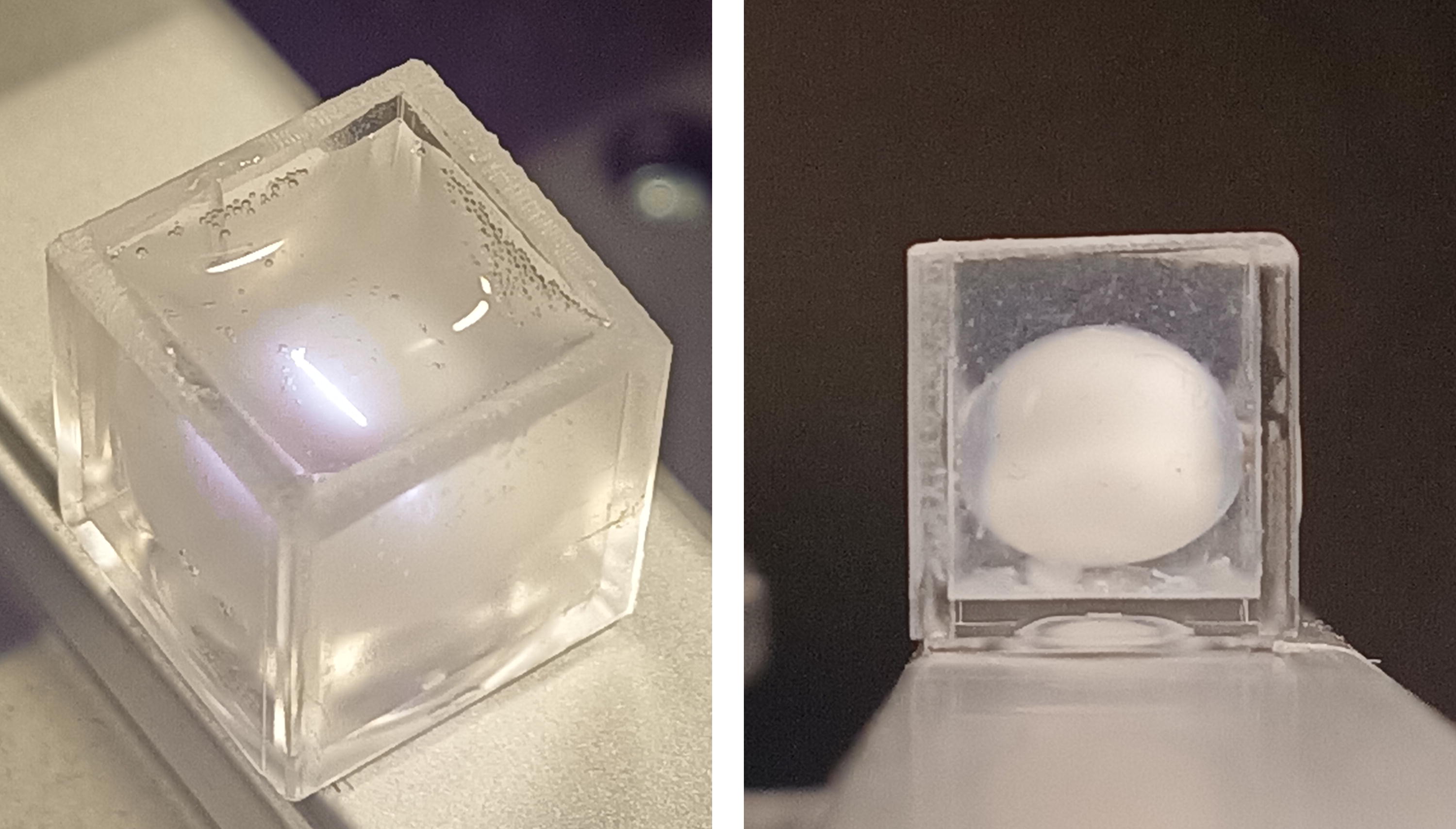
Laser fragmentation of a fully immersed porcine lens. The container is a 7 mL cuvette, which is filled with distilled water at constant room temperature of 22°C. The fragmentation laser is visible in the left image. The turbidity of the water is due to the ablated lens tissue during fragmentation.
As expected, lens fragmentation is possible with picosecond ultrashort laser pulses. Figure 5 shows a microwave processed (LM,1 from Fig. 1) and partially fragmented lens (partially because intentionally only the anterior part was fragmented). Here, (a) shows the entire lens, (b) the ablation pattern of the laser source, and (c) a magnified part of the lens. For size comparison, the ablation pattern is scaled to (a). The tissue was ablated with 12 ps pulses with a pulse energy of 100 µJ at a repetition rate of 10 kHz and a fluence of 20 J/cm2. The fragmentation process took 50 sec and resulted in an ablation depth of 1.5 mm. In images (a) and (c), the ablation pattern can be seen very clearly, both on the surface of the tissue and in the interior. Further, an emulsified accumulation of tissue bubbles is visible in the center of the lens. The ablation area has sharp and clear boundaries to surrounding nonirradiated tissue, which speaks for a very precise and well-controllable processing. Since this lens has both opaque and nonopaque parts, this result shows that picosecond pulses are feasible for both regions. Thus, they may be suitable for different states/opaqueness of cataract.
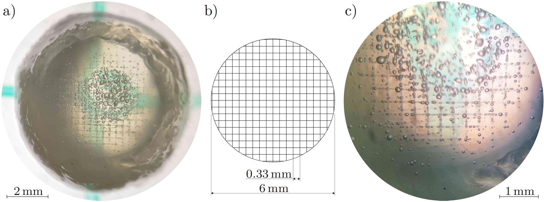
Lens fragmentation on a porcine lens (LM,1 from Fig. 1) with artificial cataract with a 12 ps ultrashort pulse laser source at 100 μJ pulse energy.
In addition to the moistened lenses, Figure 6 shows the cross-section of a sagittal bisected lens (Lens LW,1 from Fig. 2), which was irradiated in a microwave oven at 700 W while being fully immersed in distilled water. Here, image (a) shows the cross-section of the lens after partial laser fragmentation with 12 ps USPs with pulse energies of 100 µJ and 10 kHz pulse repetition rate. The fragmentation process took 120 sec and resulted in an ablation depth of 1.5 mm. The same ablation pattern as in Figure 5b was used. The subsequent sagittal bisection was done with a scalpel knife. The red ellipse marks the original shape of the lens, and the black dotted line marks the lower end of the fragmented area. The laser was irradiated from the top. Figure 6b shows the same lens with the fragmented part (above the dotted line) being removed afterward with a scalpel knife. The removal was necessary since this part was not sufficiently fragmented due to the increased hardness. The increased hardness of the lens also led to uneven edges during handling with forceps and to a longer fragmentation process.
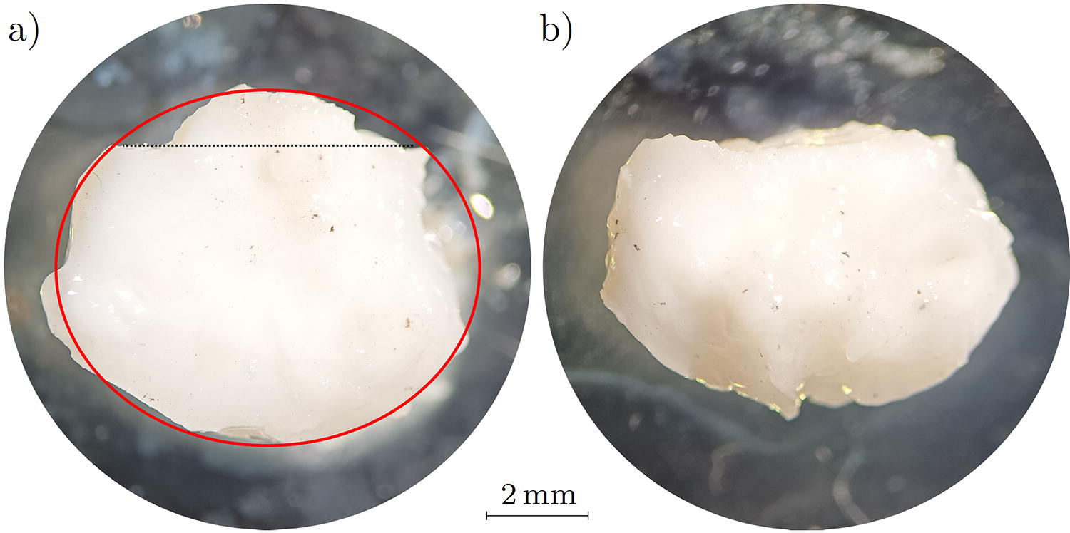
Cross-section (sagittal) of a USP laser fragmented porcine lens with artificial cataract induction (lens LW,1 from Fig. 2).
To evaluate the precision of the USP laser source, a noncataractous lens was irradiated with the ablation pattern mentioned at a spatial distance of 1 mm. The result is shown in a cross-section (sagittal) in Figure 7. Here, the distance is clearly visible and the patterns are neatly separated from each other locally. This indicates a high precision of the USP laser source during the fragmentation process, since only the target tissue was altered. Further, the bubbles appearing during the process indicate a phase transition from solid to gaseous during laser treatment.
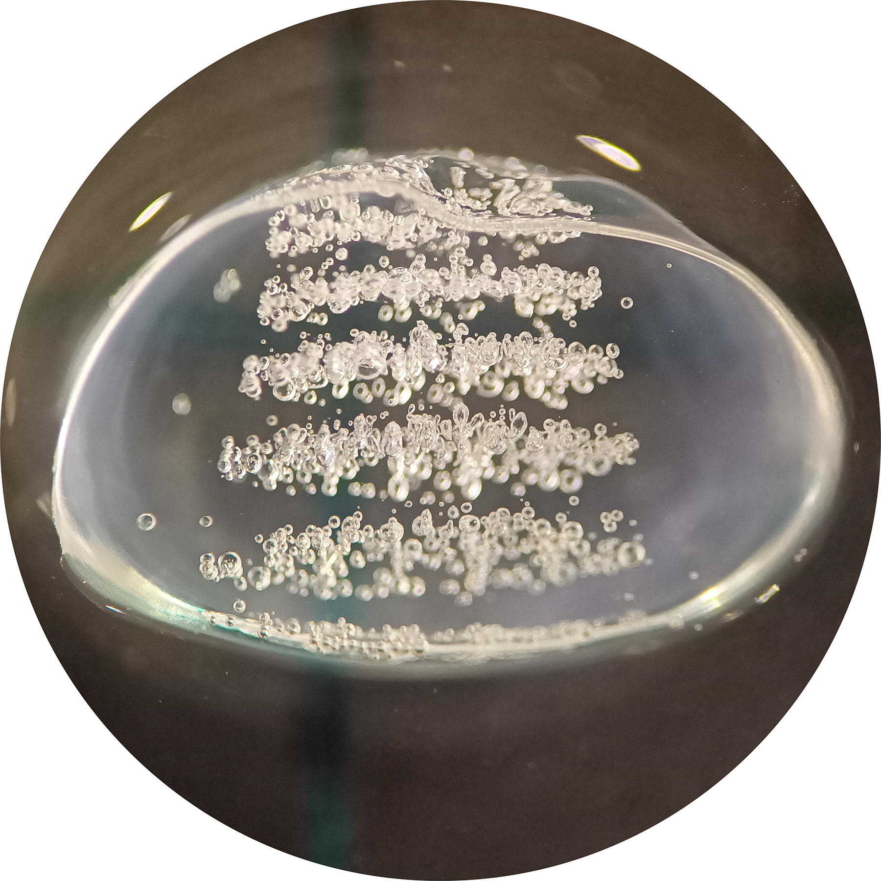
Photograph of a sagittal bisected porcine lens (cross-section) with six ablation patterns with a spatial distance of 1 mm. The bubbles indicate a phase transition from solid to gaseous during laser treatment.
Discussion
The results presented here demonstrate the fragmentation potential of picosecond ultrashort laser pulses on postmortem porcine eyes with artificially induced cataract. This method for lens fragmentation may be a good alternative or an assistive adjunct to ultrasound phacoemulsification or femtosecond- or nanosecond-laser lens fragmentation, which are the current state of the art. The results show very precise tissue ablation in opaque and nonopaque as well as soft and hard areas of cataractous lenses, whereby the ablation region can be precisely selected due to the comparatively low and spatially very defined energy input.
Compared to ultrasound methods, this is a major advantage since it reduces the possibility of adverse effects, especially in combination with the drastically reduced energy input. However, the harder the tissue (i.e., the higher the opaqueness of the lens), the lower the ablation rate and the harder it is to gain sufficient fragmentation. The latter could be counteracted by increasing the laser pulse energy, which will be tested and evaluated further in the aftermath.
It has been shown that it is possible to induce an artificial cataract with a household microwave oven. However, it is quite difficult to create different degrees/states of cataract this way. Here, our approach to irradiate the lenses only (after dissection) led to the expected results in this issue: We were able to induce different cataract states in the moistened lenses and thus simulate natural cataracts much better compared to fully cataractous lenses obtained with conventional microwave methods or the described water-inserted method. As the microwave oven works with temporal modulation to regulate the average power, it would be beneficial to use a power-adjustable microwave emitter to achieve more controllable results.
Conclusions
In conclusion, initial results in both artificial cataract induction and picosecond lens fragmentation were achieved. The artificial cataract could be induced in different opaqueness stages with microwave radiation of 700 W. Both methods (moistened lenses, water-inserted lenses) led to cataract induction. The moistened lenses got opaque after only 1–3 sec of microwave irradiation and showed different opaqueness stages. However, the hardness was only slightly increased. With the inserted lenses, it was difficult to induce different opaqueness stages, but the hardness increased drastically. The induction process took 15–20 sec.
The fragmentation/ablation was executed with 12 ps, 100 µJ laser pulses at a repetition rate of 10 kHz and a wavelength of 1064 nm. It resulted in appropriate ablation rates, with higher rates for softer lens tissue (soft: 1 mm/35 sec; hard: 1 mm/80 sec). The ablation rate could be increased with higher pulse energies or pulse repetition rates. Further, no emulsification was achieved for the hard tissue. However, picosecond ultrashort laser pulses seem to be feasible for (assisted) lens fragmentation in cataract surgeries since well-controllable and precise ablation was possible even at low energies.
Thus, these results provide a profound basis for upcoming extended animal models and clinical studies.
Footnotes
Authors Contributions
M.K.: Conceptualization (lead), formal analysis (lead), investigation (lead), methodology (lead), validation (lead), writing—original draft (lead). A.G.: Resources (equal) and writing—review and editing (equal). M.K.: Funding acquisition (equal), supervision (equal), and writing—review and editing (equal). F.L.: Investigation (supporting), methodology (supporting), and resources (equal). J.M.S.: Resources (equal) and supervision (equal). B.B.: Funding acquisition (equal), supervision (equal), and writing—review and editing (equal).
Author Disclosure Statement
All authors declare no conflicts of interest.
Funding Information
This project has received funding from the