




Abstract
Photoaging is the superposition of photodamage (ultraviolet [UV] radiation-induced) on the aging process. It is a major damaging consequence of free radical action. Curcumin (diferuloylmethane) is a phytochemical with diverse antioxidant and antiinflammatory properties. However, it shows a poor topical bioavailability. Therefore, we encapsulated curcumin in elastic vesicles (EVs) and investigated different doses (1, 3, 5 and 10 μmol) for its in vivo antiaging activity in mice. VICCO® turmeric served as the marketed control, and free curcumin dispersed in an ointment base was another control. The dorsal depilated skin surface was exposed to the whole UV range for 5 sec, five times a week for 6 weeks. Each exposure was followed by treatment with encapsulated curcumin (at different doses), free curcumin ointment, and VICCO® turmeric. The effectiveness was established in terms of macroscopic and histopathological evaluation of skin, pinch test, and redox homeostasis of skin homogenates. Skin evaluation demonstrated that 5- and 10-μmol doses of curcumin EVs and the marketed formulation were effective in preventing the formation of lesions and other changes. The pinch test showed that the reduction in recovery time with the 10-μmol dose of curcumin EVs was highly significant (p < 0.05). Histopathological studies further confirmed the protective role of curcumin EVs. The normal redox balance was restored with a 10-μmol dose, whereas a 5-μmol dose and the marketed formulation showed significant and equivalent activity. The free curcumin ointment group showed no improvement in redox levels. This study provides the first preclinical evidence for the use of topically delivered curcumin to attenuate photoaging.
Introduction
Studies by Dreher and Maibach 5 have indicated that regular application of skin care products containing antioxidants may be of the utmost benefit in efficiently preparing our skin against exogenous oxidative stressors occurring during daily life. A natural “antioxidant” composition offers added benefits in supporting skin texture, appearance, and tone. 6 The multifaceted effects include overall protective effects and antiinflammatory, antimicrobial, and immune system-supporting effects that are particularly relevant in the management of acne. However, antioxidant molecules are inherently unstable in nature, being especially susceptible to photodegradation and the presence of oxygen. Instability makes them difficult to be formulated in an acceptable, stable composition for therapeutic use. These observations have facilitated the entry of novel delivery systems in the development of antioxidants. These systems are developed to work in all areas of the delivery and thus can be applied to improve the solubility, permeability, and stability of antioxidants. 7
Curcumin (diferuloylmethane) is a food chemical present in turmeric (Curcuma longa linn.). Curcumin has been demonstrated to have potent antioxidant 8 –12 and antiinflammatory activity. 13 –19 Curcumin is reported to strongly repress matrix metalloproteinase-9 (MMP-9) gene expression as well as the activation of activator protein-1 (AP-1) induced by tumor promoters, c-Jun N-terminal kinase (JNK) activation induced by carcinogens, and radiation-induced lipid peroxidation. 20 AP-1 is implicated in the process of aging because it induces the formation of MMPs, which cause degradation of interstitial collagen fibers.
Kurien et al. have also reported on improving the solubility of curcumin by heating, but it is yet to be seen whether this can in any way help achieve an enhanced permeability or an improved therapeutics of curcumin. 21 In the current study, we investigated the in vivo antiaging activity of curcumin-encapsulated in elastic vesicles (EVs) using the mouse model, which was developed and reported by us earlier. 22 Free curcumin is, however, highly hydrophobic and cannot be administered topically due to poor bioavailability. Therefore, to enhance the bioavailability, curcumin was encapsulated into EVs and was dispersed in a suitable ointment base (development and characterization of EVs to be reported separately). This approach makes the agent amenable to topical dosing and circumvents the problem of poor topical availability that limits the utility of free curcumin. Kurien and Scofield 23 have reported the use of curcumin/turmeric solubilized in sodium hydroxide for inhibition of 4-hydroxy-2-nonenal (HNE)-protein modification, whereas another group 24 has reported the use of 10% curcumin dissolved in dimethylsulfoxide (DMSO) for wound healing. However, curcumin is reported to undergo hydrolytic degradation at a pH above neutral. 25 Therefore, the authors solubilized curcumin in alkali just before the assay, diluting it in phosphate-buffered saline and then using it rapidly. This method was possible for an in vitro assay, but it is unpredictable. It is also questionable whether a similar effect can be observed upon in vivo administration. Curcumin dissolved in DMSO also precipitates when diluted in aqueous buffer. The intention of preparing the EVs of curcumin in the present study was to present curcumin in a soluble or more appropriately bioavailable form and also to improve its permeability using EVs as a carrier system. Furthermore, EVs would act as a localized system targeted in the skin layers and would release the drug over an extended period of time.
The effectiveness of curcumin-loaded EVs was established in terms of the macroscopic evaluation of skin, pinch test, and the redox homeostasis of skin homogenates in terms of lipid peroxidation, catalase, and reduced glutathione levels.
Materials and Methods
Materials
Curcumin was a gift sample from Sanat Product Ltd. The sample constituted a mixture of three curcuminoids, namely curcumin (95%), demethoxycurcumin, and bisdemethoxycurcumin (later two constitute the remaining 5%). 5-5′-Dithiobis-2-nitrobenzoic acid, ethylene diamine tetraacetic acid, nitroblue tetrazolium, reduced glutathione, and thiobarbituric acid were obtained from Hi Media Laboratories Pvt. Ltd. (Mumbai, India). Disodium hydrogen orthophosphate, glacial acetic acid, potassium chloride, sodium chloride, and sulfosalicylic acid were obtained from S.D. Fine Chem. Ltd. (Mumbai, India). Hydrogen peroxide was obtained from Qualikems Fine Chemicals Pvt. Ltd. (New Delhi, India), hydroxylamine HCl from Merck Ltd. (Mumbai, India), pyridine from BDH Lab., Chemicals Division, and cholesterol from Sigma Aldrich Chemie GmbH. Sodium lauryl sulfate and chloroform were from LOBA Chem. Pvt. Ltd. (Mumbai, India). Phospholipid was a generous gift sample from Phospholipids GmbH, Germany. VICCO® turmeric skin cream (VICCO Laboratories, Goa) was used as the marketed control.
Histopathology studies
Histopathology of the skin samples was carried out at Medicos Centre (Chandigarh, India), under the supervision of by Dr. B.N. Datta (M.D., FAMS, FICP, ex-Professor of Pathology, Post Graduate Institute of Medical Education & Research, Chandigarh, India). All studies involving animals were approved by the Institutional Animal Ethics Committee of Panjab University, Chandigarh, India.
Preparation of curcumin elastic vesicles
EVs were prepared by the conventional rotary evaporation sonication method described by Cevc et al. 26 and El Maghraby et al. 27 Phospholipid, surfactant, and the drug were placed in a clean, dry, round-bottomed flask, and the lipid mixture was dissolved in chloroform. The organic solvent was removed by rotary evaporation under reduced pressure at 45–50°C. Final traces of solvents were removed under vacuum overnight. The deposited lipid film was hydrated with saline phosphate buffer (pH 6.4) by rotation at 100 rev/min for 15–20 min at 40–45°C. The resulting vesicles were left overnight to swell at room temperature and were subsequently incorporated into a hydrophilic ointment base. The ratio of phospholipid and surfactant was optimized on the basis of vesicle shape and type and number of vesicles per cubic millimeter. Ex vivo drug release with permeation was determined for the developed formulations.
Grouping of animals and UV irradiation
Female Laca mice (aged approximately 8 weeks) were obtained from the central animal house of Panjab University (Chandigarh, India). The animals were kept under a natural light/dark cycle and were given food and water ad libitum. Sixty mice were selected on similar body weight basis. They were divided into ten groups of six mice each according to Table 1. At the start of the experiment, the dorsal skin surface of the mice was shaved with a depilatory (Anne French®). Briefly, mice were anesthetized using ether (5 mL) and then depilatory was applied on the dorsal skin area (2 × 2 cm). After approximately 5 min, the depilatory was wiped off. Thereafter, the depilatory was applied as required (usually on alternate days).
U10 was evaluated only for any adverse histopathological changes, and the results are shown in Fig. 5.
UV exposure was 4.17 times the minimal erythemal dose, which was established earlier in the laboratory. 22 UV irradiation was carried out as described below using an in-house UV simulator. 22 Prior to irradiation, mice were anaesthetized using ether (5 mL) so that the movement of animals would be restricted during exposure and the irradiation would be homogeneous. Mice were irradiated using a UV bulb (Ultravitalux 300W Wotan®, Germany). The bulb gave the full spectrum of UV radiation, i.e., 260–400 nm, simulating the full solar spectrum. The distance from the lamp to the animals' back was kept constant at 35 cm. The animals were exposed five times a week for 6 weeks. Dose per exposure was 166.67 J/cm2, resulting into a cumulative dose of 5000.1 J/cm2.
Each UV exposure was followed by treatment with 100 mg of encapsulated curcumin (at different doses incorporated into a suitable hydrophilic ointment base), free curcumin ointment, and VICCO® turmeric. The ointments/creams were applied on the shaved dorsal area using a spatula and were spread evenly. The formulation/cream were rubbed thoroughly on the skin until no residue was observed. Caution was exercised to avoid applying outside the demarcated (shaved) area.
Macroscopic evaluation of dorsal skin
Skin was examined for photodamage every week for 6 weeks. The UV- exposed dorsal skin of each mouse was photographed while the mouse was under anesthesia. The grade of photodamage was determined using the evaluation criteria shown in Table 2, modified from Bissett et al. 4,28 The grading scale ranged from 0 for normal skin to 6 for severely photodamaged skin.
Evaluation of recovery from stretching (pinch test)
Pinch testing was carried out according to the method of Bryce and his co-workers. 29,30 The dorsal skin at the midline of mice was picked up with the fingers as much as possible (to a degree that does not lift the animal into the air), and the pinch was subsequently released (see Fig. 3A, below). The time(s) until the skin recovered to the original state was measured. The test was performed every week for 6 weeks.
Biochemical estimation
Preparation of skin homogenate
At the end of 6 weeks, the mice were sacrificed by cervical dislocation. The dorsal skin surface of the animals was excised and defattened using isopropanol. Excised skin was weighed and then finely cut into pieces. During this procedure and afterwards, skin was kept and maintained in ice-cold (0–4°C) 1.15% KCl. Depending on the weight, the finely cut pieces of the skin sample were mixed with 1.15% KCl in a relative proportion of 9 mL to 1 gram of wet skin sample (to obtain a 10% homogenate). The skin was then homogenized using a Teflon Potter glass (ground) homogenizer fitted into a Remi stirrer, keeping the temperature at below 4°C to prevent free radical generation or degradation of antioxidate enzyme catalase due to heat production. Homogenization was continued (it usually took 15–20 min) until a viscous turbid mixture with no solid particles was formed. The homogenate was then centrifuged at 4000 × g for 10 min, and supernatants were collected. After the protein estimation, the redox homeostasis status of the supernatants was determined using the following markers.
Protein estimation
The protein content of skin homogenates was measured using the Biuret method 31,32 and bovine serum albumin as the reference protein. In all, 0.1 mL of skin homogenate, 2.9 mL of sodium chloride, and 3 mL of working biuret reagent were mixed together. (Biuret reagent was prepared by mixing a solution of sodium potassium tartarate in 0.2 N NaOH with copper sulfate. Potassium iodide was added to this solution, and the total volume was made up to 100 mL with 0.2 N NaOH.) This mixture was kept at room temperature for 10 min, and its absorbance was measured at 540 nm.
Lipid peroxidation
The extent of lipid peroxidation in the mouse skin upon UV exposure was determined in terms of nanomoles of malondialdehyde/mg protein, as discussed by Ohkawa et al.
33
Malondialdehyde (MDA), an indirect index of lipid peroxidation, was assayed in the form of thiobarbituric acid reacting substances (TBARS).
33
To 0.2 mL of homogenate, 0.2 mL of 8.1% sodium lauryl sulfate, 1.5 mL of 20% acetic acid (pH 3.5), 1.5 mL of 0.8% thiobarbituric acid (freshly prepared), and 0.6 mL of distilled water were added. The mixture was heated for 1 h on a boiling water bath and then cooled under running tap water. Then 1 mL of distilled water and 5 mL of n-butanol:pyridine (15:1) mixture was added to the mixture. After vigorous shaking, the solutions were centrifuged and organic layer was separated. The absorbance of organic layer was measured at 532 nm. Calculations were done using the following formula:
where
Catalase activity
A number of procedures have been reported for measuring the catalase activity.
34
–38
We chose a method especially designed for the skin.
39
Because it was not possible to separate the epidermal and dermal skin layers in mice, an overall activity of the catalase in skin was measured. In this assay, to 0.05 mL of skin homogenate, 3 mL of H2O2 (30 mmol/L) in 50 mM phosphate buffer (freshly prepared) was added, and the change in absorbance was measured at 240 nm for 2 min at 30-sec intervals. Calculations were done using the following formula:
where, A1 and A2 are the initial and the final absorbance when the time interval Δt is 1 min.
Estimation of glutathione (reduced)
Glutathione (GSH) was estimated by the method of Ellman.
40
One milliliter of skin homogenate was mixed with 1 mL of sulfosalicylic acid (4%). The samples were incubated at 4°C for at least 1 h and then subjected to centrifugation at 1200 × g for 15 min at 4°C. The assay mixture contained 0.1 mL of supernatant, 2.7 mL of phosphate buffer (0.1 M), and 0.2 mL of 0.01M 5-5′-dithiobis-2-nitrobenzoic acid (4 mg/1 mL) to a total volume of 3.0 mL. The developed yellow color was read immediately at 412 nm on a spectrophotometer. The GSH concentration was calculated as nmol GSH/g tissue. For calculation, the following formula was used:
Histopathological studies
Hematoxylin & Eosin (H & E) as well as Alcian Blue stainings were performed after 6 weeks. Representative portions of skin were obtained from mice of each experimental group, fixed in 10% buffered formalin, embedded in paraffin, and sliced with a microtome. The sections were then stained accordingly. Finally, the stained specimens were observed under a high-power light microscope (Olympus) and were evaluated for their integrity and tested for stratum corneum, epidermis, and dermis thickness, and the extent of Alcian Blue staining.
Statistical analysis
Animal experiments were performed with 6 animals per treatment group. Quantitative data were expressed as the mean ± standard deviation (SD). Statistical significance was examined by the analysis of variance and the paired Student t-test. Differences were considered statistically significant if p < 0.05.
Results and Discussion
The developed EVs showed an average size of 11.39 μm. The maximum drug that could be loaded in the developed EVs was 20 mg and the per cent entrapment was 76.55%. The drug content of the EV dispersion was 2 mg/mL. The dispersion was then lyophilized and incorporated into a suitable hydrophilic ointment base. The developed ointment of curcumin EVs showed a flux of 2.631 ± 0.144 μg/h · cm2 and percent retention of 51.66 ± 2.259 in the skin. An enhancement of skin retention by a factor of 31.31 was achieved with respect to free curcumin when the above-mentioned ointment base was used.
Macroscopic effect of chronic UV irradiation on mice skin
Macroscopic effects of UV irradiation on mice skin are shown in Fig. 1. As compared to sham control mice (U2), the UV-irradiated mice group (U3) showed an onset of lesions starting from fifth week (2 out of 6 mice). At the end of the study, 5 out of 6 (83.33%) UV-treated mice showed extensive lesions (visual scoring 6). In the free curcumin ointment-treated group (U4), 3 out of 6 (50%) mice showed lesions (score 6) whereas the other 3 showed deep wrinkles (score 5) at the completion of the study period. No lesions were observed in EV-treated groups at doses above 1 μmol (1 μmol-treated animals showed lesions in 33.33% of animals). The marketed formulation treated group (U9) showed lesions in 16.67% of animals (1 in 6 mice). Results of macroscopic visual score evaluation are shown in Fig. 2.
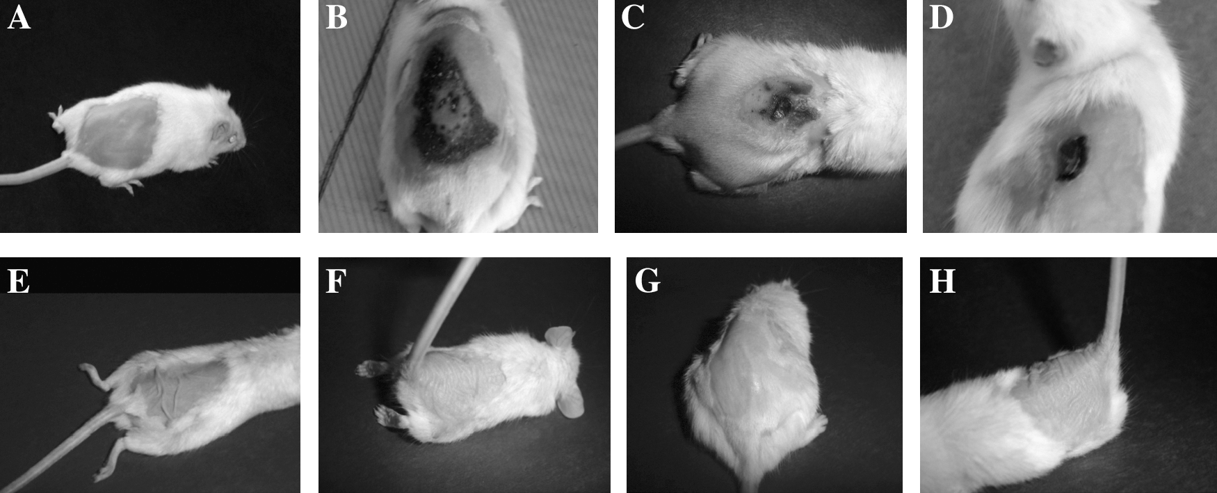
Pinch test in Macroscopic changes in the skin of mice upon various treatments at the end of experimental period of 6 weeks. (

Results of the visual score of different experimental groups during the 6-week study period.
The visual scores in the UV-irradiated group (U3) and free curcumin ointment- (containing 10 μmol of curcumin/application) treated group (U4) were significantly higher than the U7- (5 μmol EVs ointment), U8- (10 μmol EVs ointment), and U9- (marketed formulation) treated groups. However, 1- and 3-μmol doses (U5 and U6) did not show any significant improvement in scores, although the number of lesions formed was arithmetically less (only 33% in case of U5; the U6 group showed no lesions). There was no statistical significant difference between the results illustrated by 10 μmol (U8) and the marketed formulation (U9). These results indicate that curcumin EVs (10 μmol) and the marketed formulation effectively prevented the macroscopic changes and formation of lesions upon a UV exposure of 6 weeks.
Evaluation of recovery from stretching (pinch test)
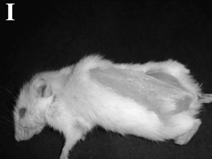
Figure 3 shows photographs of dorsal skins of various animal groups after being stretched for 1 sec. Figure 4 depicts the time in seconds taken by the pinched skin to return to normal for the different weeks of 6-week study period of UV exposure. Statistics were performed on the 6-week data for each group, and the results are documented below. All of the groups were significantly different from the sham control group (p < 0.05). The UV-irradiated group, free curcumin ointment-treated group, and 1 and 3 μmol dose-treated groups were significantly different (p < 0.05) from sham control, marketed formulation, 5- and 10-μmol dose groups. However, there was no significant difference (p < 0.05) within the four formerly stated groups. The 5-μmol dose-treated group did not show a statistically significant difference (p < 0.05) from the marketed formulation-treated group, whereas the 10-μmol dose-treated group showed a significantly better recovery (p < 0.05) from all other groups. There was no significant difference (p < 0.05) between the marketed formulation treated group and the 3- and 5-μmol dose-treated groups; all other groups were significantly different. The results show that although the reduction in recovery time with a 10-μmol dose of curcumin EVs was highly significant, it could not match the sham control values (there was significant difference from the sham control group). Similar findings have been reported by Tsukahara et al., 30 who showed decreased sagging of skin upon treatment with sunscreens.
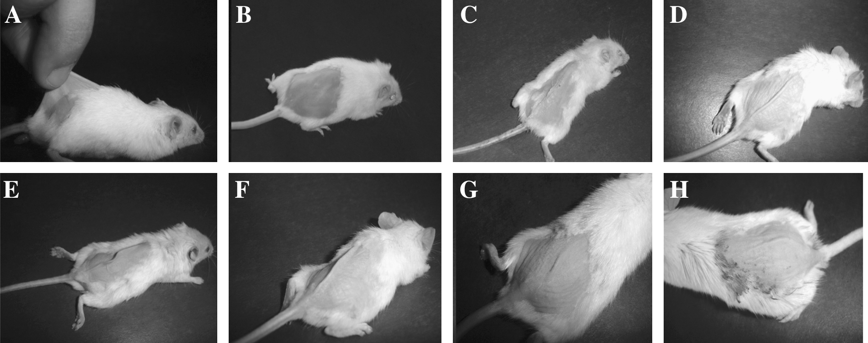
UV-irradiated groups with appropriate treatments. (

Results of the pinch test for different experimental groups during the 6-week study period.
Biochemical estimation
Lipid peroxidation
The MDA levels for the UV-irradiated group was approximately 4 times more than the naive/sham control group, indicating an increase in the MDA levels upon exposure to UV radiation (Table 3). Significant reduction (p < 0.05) in MDA levels was observed with curcumin EVs formulation-treated groups (U6, U7, U8) and marketed formulation- (U9) treated groups when compared to the UV-treated (U3) group. However, 1-μmol dose (U5) and free curcumin ointment- (U4) treated groups showed statistically insignificant changes when compared with the UV-irradiated control group (U3). The marketed formulation (U9) and 5-μ mol dose (U7) were statistically not significant from each other, showing that they achieved almost similar lowering of MDA levels, but they were statistically significantly different from the naïve/sham control group (U1 and U2). The 10-μmol dose-treated group (U8) of curcumin EVs showed a more significant reduction than the marketed formulation (U9), indicating it to be a very efficient free radical scavenger. The MDA values were reduced even beyond the control values (U1 and U2), although the difference was not significant, indicating that application of the 10-μmol dose could restore normal MDA levels in the UV-exposed skin of mice.
(*, **, #) No significant difference for similarly marked groups (p < 0.05).
UV, Ultraviolet; MDA, malondialdehyde; SD, standard deviation.
Catalase activity
An enzyme that scavenges H2O2 in the skin is catalase (CAT). Catalases are reported to be implicated in providing a buffer action against free radicals generated as a result of UV exposure. 41,42 Studies in the literature, report an increase in the catalase activity upon chronic UV exposure, whereas an acute exposure may show a fall in the enzyme levels. 37 Several workers have determined the activity, separately in the dermis and epidermis, and found the change in catalase activity to vary in these two layers. 37,38 In case of the mice model used by us, it was difficult to separate the two layers and an overall catalase activity of the skin was measured by us, which increased upon UV exposure, indicating that body's natural defense systems are triggered by an increased free radical production. Further, it has been observed that the quantitative increase in catalase activity in epidermis is more than the decrease in its activity in the dermis, 37,38 hence, on an average there should be an increase in the catalase activity; as observed by us (Table 4).
(#, *, **) No significant difference in activity for similarly marked groups (p < 0.05).
UV, Ultraviolet; H2O2, hydrogen peroxide; SD, standard deviation.
Chronic exposure of animals to UV irradiation showed approximately 3.5 times increase in the catalase activity as compared to naïve/sham control group. The 1-μmol dose of curcumin EVs and free curcumin ointment were found to be ineffective, as indicated by the insignificant difference in the catalase values for these groups and the UV-irradiated group. Doses at or above 3 μmol significantly reduced the increased catalase activity. Further, effects obtained with 5-μmol dose were not significantly different from those obtained with the marketed formulation, suggesting both to be equally efficient. They were however statistically different from the naïve/sham control group. Original catalase values were restored by the 10-μmol dose (Table 4).
Estimation of glutathione (reduced)
GSH (
This is also depicted in our results (Table 5). The GSH levels of the UV-irradiated group (U3) decreased to almost half of the naïve/sham control groups (U1 and U2). There was no significant difference between the free curcumin ointment-treated group (U4), 1-μmol dose (U5), and 3-μmol dose (U6) when compared to the UV-irradiated group (U3), showing these treatments to be ineffective against photoaging in terms of reduced GSH levels. Furthermore, there was no significant difference between the 10-μmol dose (U8) group and naïve/sham control groups (U1 and U2), indicating the restoration of normal GSH levels.
(#, *, **) No significant difference between groups marked similarly (p < 0.05).
(•, ♦) Significant difference between groups marked similarly (p < 0.05).
UV, Ultraviolet; SD, standard deviation.
The 5-μmol dose (U7) group and the marketed formulation (U9) showed no significant difference in their activity. The free curcumin ointment group (U4) was significantly different from 3-μmol dose (U6) group, but both of them were statistically insignificant from the UV-irradiated group (U3). Similarly, the 1-μmol dose and 3-μmol dose groups were statistically different from each other but insignificantly different from the UV-irradiated group.
H & E staining of mouse skin
The photoaged skin usually shows a variety of clinical manifestations, including coarseness, wrinkling, sallow discoloration, telangiectasia, irregular pigmentation, and a variety of benign, premalignant, and malignant neoplasms. 45 Old photo-protected skin may have increased laxity and fold accentuation, but it does not develop the leathery, sagging appearance of actinically damaged integument. The histologic hallmark of photoaging is dermal elastosis, which largely consists of thickened, tangled, and ultimately granular amorphous elastic structures. 46 –48 This elastotic material is postulated to result from direct UV-mediated damage to the dermal fibroblasts, which then produce abnormal elastin, or it may result from chronic low-grade enzymatic digestion of extracellular matrix by proteases elicited by inflammatory mediators. 49 The accumulation of elastotic material is accompanied by degeneration of the surrounding collagenous meshwork. 50 Thus, photoaged skin is characterized by epidermal hyperplasia, dermal elastosis, and matrix protein degradation 51,52 and by the presence of perivenular lymphohistocytic dermal infiltrates. 49
After 6 weeks of UV irradiation of mice, we detected the characteristic histologic features of epidermal and dermal thickening, associated with increased disorganization of elastic and collagen fibers in the dermis. Naïve control mice skin (U1) (Fig. 5A) and sham control mice skin (Fig. 5B) showed almost similar features. The epidermis had multiple layers of squamous cells, covered by thin layer of keratin and a small number of stratum granulosum cells. A basement membrane was seen beneath the basal layer, and the superficial dermis showed collagen fibers and elastic tissue fibers (Fig. 6A). Clusters of sebaceous glands were attached to the numerous hair follicles, and the sweat glands were mostly seen in the superficial dermis. Deeper dermis showed abundant fat with regularly distributed hair follicles; vascular channels were also seen in regular distribution. Beneath the dermal fat was a prominent layer of skeletal muscle fibers. This was followed by the subcutaneous fascia, which consisted of thin delicate fibrils of fibrocollagen. Sham control mice skin (U2) showed a normal epidermal layer; stratum corneum and stratum granulosum were also normal (Fig. 5B). The basement membrane was somewhat swollen and indistinctly fuzzy. There was diffuse edema of the skin separating the collagen and elastic fibers, as well as fat cells. Inflammation was not present (Fig. 6B).

Hematoxylin & Eosin (H & E) staining of: (
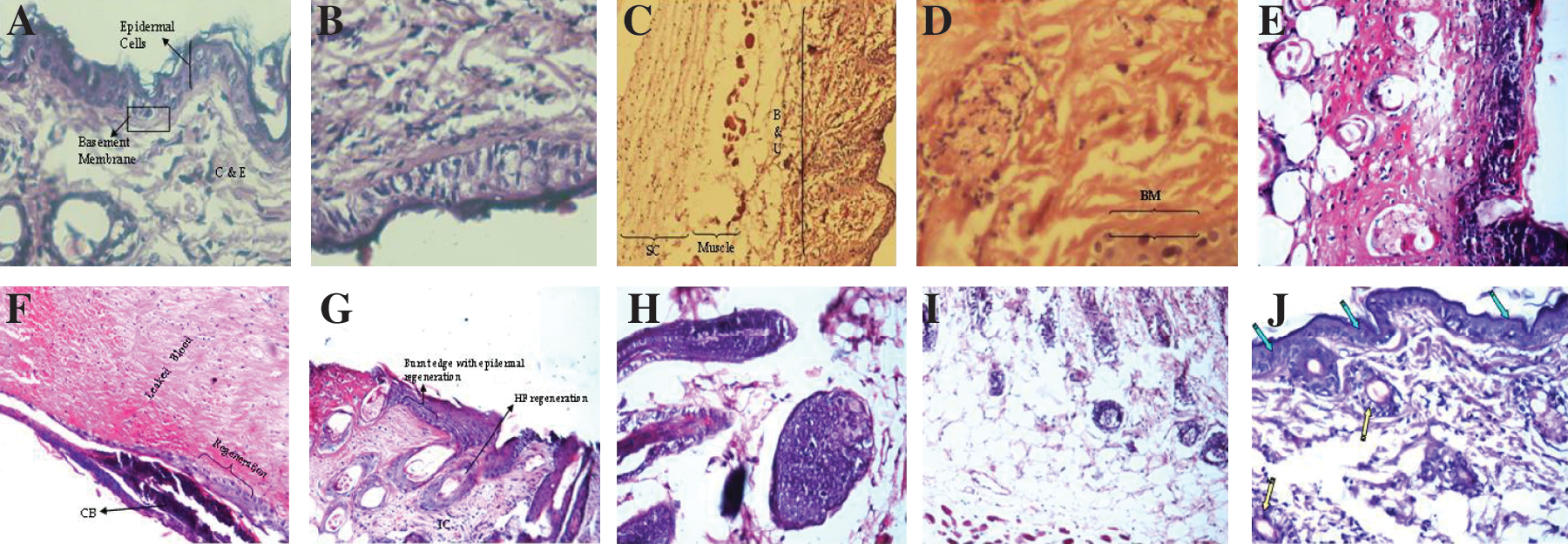
(
UV-irradiated mice skin (U3) (Fig. 5C) showed the following features. The epidermis and the subepidermis formed a coagulated eosinophilic mass. Cell outlines were not apparent, and irregular pyknotic nuclei and nuclear debris were scattered. The basement membrane was fuzzy and merged into the dermal collagen. In the central portion of the surface, there was ulcer formation and loss of elastic tissue distribution; the epidermis shed away exposing the dermis (Fig. 6C). Along the edges, there was invasion by leukocytes, including abscess formation. Superficial fat cells also underwent hyaline changes and collapsed. Hair follicles showed crowding and necrosis, with a thick eosinophilic outline and sebaceous glands disappeared. Inflammatory infiltration was seen in and underneath the fatty dermis. The muscle bundles showed disruption, vacoulation, and fragmentation, and the skin showed a coagulation necrosis of epidermis. Stratum corneum epidermis was atrophic, and individual cells lost distinction. Dermis showed edema and inflammation between fat cells and hair follicles, and capillaries were congested. The superficial dermis showed loss of elastic tissues. The deeper muscle layer was disrupted, showed edema that was myxoid, and contained excess Alcian Blue–positive material diffusely. On exposure to UV, the dermal fat was dissolved, and its loss was the main cause of reduced thickness of the skin. The hair follicles were crowded and, later on, these also disappeared. The fibrocollagen was increased in the superficial dermis, but the individual fibrils had lost their distinction to form a coagulated mass (Fig. 6D).
Empty EV-treated skin was normal (Fig. 5D). The epidermis was composed of three to four layers of squamous cells with a thin stratum corneum and distinct stratum granulosum; the basement membrane was also distinct. The dermal appendages, including sebaceous glands and sweat glands, as well as hair follicles, were normal. Vascular capillaries and fats were normal. The results confirmed absence of any adverse effects of EVs or the ingredient used therein on cutaneous tissue upon long-term use. The harmlessness of EVs application on skin is reported by us for the first time.
Free curcumin ointment-treated mice skin (U4) showed burns (UV induced) (Fig. 5E). The epidermal cells had coagulated into a red hyaline mass with loss of cell morphology. The collagen and elastic fibers had also coagulated into homogeneous eosinophilic material. The fat cells were preserved with eosinophilic change between the cells. The subcutaneous muscle fibers showed minimal change, and the subcutaneous fascia showed swelling. Furthermore, the tissue injury had invited an inflammatory reaction (Fig. 6E). Small clusters full of necrotic leukocytes formed in the surface beneath the keratin layer indicated such an inflammation reaction. The entire dermis and muscle and fat showed streaks of leukocytes, which included neutrophils as well as lymphocytes and plasma cells. Similar inflammation was also seen in the subcutaneous tissue. The histopathological markers were quiet similar to the UV-irradiated mouse skin.
The histopathology of the mousee skin treated with the 1-μmol dose (U5) of curcumin EVs ointment showed acute burns, but the onset of repair regeneration was also evident (Fig. 5F). The surface epidermis was replaced by an acute reaction, which also extended into the dermis. Healing in the form of fibroblastic proliferation had set in. The entire thickness of the skin was turned into an amorphous eosinophilic mass with bleeding; the epidermis was denuded, whereas the deeper layers showed extensive inflammation. Occasional plugging of hair follicles during acute phases, leading to accumulation of keratin to form cysts, was also evident. Some clusters of bacteria or microorganisms trapped in the superficial layers were also observed (Fig. 6F).
The UV-irradiated mouse skin treated with the 3-μmol dose (U6) showed hyperplasia of the epidermis with up to six cell layers, increased granulosa layer, and keratin (Fig. 5G). The hyperplasia involved the hair follicles also. Dermis appeared normal but fat layers were reduced. The process of regeneration could be seen along the edges. These include an increase in the number of epidermal squamous cells along the edges of the ulcerated area. Setting in of recovery was also visible as an increase in the thickness of epidermis along the edges of the ulcer, with an increase in the number of squamous cells. A thin unicellular layer of epidermis appeared to edge over the damaged area. The coagulated zone in the superficial dermis and the fatty layer were significantly reduced. Hair follicle recovery was irregular, so that the distribution of hair follicle was irregular. Similar to the epidermis, the follicular cells also showed hyperplasia forming multiple layers and some solid cell clusters. The muscle layer showed nuclear irregularities and inflammatory infiltration and fibroblastic proliferation was visible in the deeper layers. The epidermis showed wrinkling and the total skin thickness was reduced with loss of fat and hair follicles. It may be concluded that the skin showed significant regeneration (Fig. 6G).
UV-irradiated mice skin treated with a 5-μmol dose of curcumin EVs (U7) showed the regeneration process of hair follicles also. There was fragmentation in the muscles present in the deeper layers of the skin (Fig. 5H). Recovery of hair follicle was irregular, with hair follicles distributed irregularly (Fig. 6H).
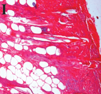
The skin of the marketed formulation-treated group (U9) showed extensive wrinkling of the epidermis with the persistence of inflammation (Fig. 5I). There were a decreased number of hair follicles with persistent muscle fragmentation (Fig. 6I). The process of regeneration of hair follicles had started which was indicated by an increased number of cellular layers.
The H & E staining of mice treated with the 10-μmol group (U8) showed a good smooth surface with no wrinkling; however, there was inflammation in the fatty layers (Fig. 5J). The upper portion of the skin showed a decreased number of hair follicles and slight inflammation. The collagen present was normal in nature and there was fragmentation of the muscle layer. The regenerating epidermis and hair follicles were also present (Fig. 6J).
A summary of the epidermal and dermal changes in all the animal groups are listed in Table 6 for better comprehension.
Alcian Blue staining of mouse skin
Chronic UV light exposure leads to various changes in the dermal connective tissue in humans, which are different from the cutaneous changes in chronological aging. 53 Histological studies on actinically damaged human skin showed an accumulation of elastotic materials and increased staining in glycosaminoglycans (GAGs). 54 GAGs are markers of photoaged skin. The changes in the dermal connective tissue components of hairless mice by chronic and repeated UV exposures have been investigated extensively as a model for photoaging because of their proposed similarities in dermal connective tissue of sun-damaged human skin. 53 The combination of UVA and UVB radiation may be more efficient to increase GAG synthesis than UVA or UVB alone. 55 It is now obvious that visible and physical skin changes induced by chronic UV exposures are the consequence of epidermal and dermal connective tissue alteration. Takahashi et al. 56 indicated the precise alterations of GAGs both in the total amount and in the composition, confirming the histochemical findings.
Alcian Blue staining of the mice skin samples was done to stain the GAGs blue in color. Our study demonstrated that there was a considerable increase in the amount of GAGs upon chronic exposure to UV radiation (Table 7). Naïve/sham control skin showed no or little Alcian Blue–positive material (Fig. 7A, B). UV-irradiated skin, the free curcumin ointment-treated group, and the 1-μmol dose-treated group, which were burned/inflamed in nature, showed an excess of GAGs, as the regeneration/healing and proliferation set in (Fig. 7C–E). The deposits were seen in the superficial collagen layers of dermis, around the hair follicles that showed hyperplasia as well as in the subcutaneous fibrous tissue. Doses of curcumin EVs at or above 3-μmol dose and the marketed formulation group showed less Alcian Blue–positive material (Figure 7F–I). Our findings are in agreement with the reports indicating an increase of skin GAGs in hairless mice exposed to UVA or UVB. 55, 57 –59

Alcian Blue staining of the mice skin samples. (
GAG, Glycosaminoglycan.
Conclusion
Aging proceeds by highly complicated biochemical processes, in which the involvement of the reactive oxygen species (ROS) and free radicals has been implicated. ROS are dramatically enhanced by exposure to the UV radiation. Free radical scavengers and antioxidants can thus provide a long-term protection against these changes. Curcumin (diferuloylmethane) is a component of the root of the plant Curcuma longa. Free curcumin is, however, highly hydrophobic and cannot be administered topically due to poor bioavailability. Encapsulation of curcumin into an EV makes this agent amenable to topical dosing and circumvents the problem of poor topical availability that limits the utility of free curcumin. The photoaged mice model showed promising results for curcumin-loaded elastic vesicles. The normal redox balance was restored with the 10-μmol dose of curcumin EVs, whereas the 5-μmol dose and marketed formulation showed significant and equivalent activity. Histopathological studies of the skin sample elaborately confirmed the protective role of curcumin EVs in photoaging. Bhagavathula et al. 24 reported on the wound-healing properties of a 10% curcumin (500 μL of 10% curcumin was used per application, which is equivalent to 50 mg; pretreatment before abrasions were induced). Use of the presently developed elastic vesicular system, though, in the photoaged mice model produced a 100% attenuation at a much smaller dose (3.6 mg, approximately equivalent to 10 μmol). Furthermore, we hereby establish the therapeutic rather than a protective/preventive role of curcumin. This validates its use as a drug rather than simply as a diet supplement. Present-day society needs preparations that can counter aging once it has set in. The performance evaluation of curcumin EVs against the mouse model of photoaging provided evidence that curcumin, when delivered through a suitable delivery system, has the potential to attenuate the UV-induced oxidative stress. This is the first time that the antiphotoaging effects of curcumin have been established in an animal model, although some in vitro studies indirectly implicate its antiaging effects.