
Abstract
Neonatal thymus grafts exert a rejuvenating action on various immunological and nonimmunological functions found altered in old mice. Commonly, half of a thymus is grafted under the kidney capsule. The invasiveness of the surgical procedure and the use of limited thymus tissue may explain why precedent survival kinetics remain unaffected. In this trial, we grafted two neonatal thymi into the axillary cavity of old mice, thus reducing the invasiveness of the intervention and increasing the amount of grafted neonatal tissue. Using a Piantanelli parametric model of survivorship, we found a significant change in mortality rate between the two groups (thymus graft and controls).
Introduction
However, investigations on the potential impact of this treatment on life extension were scarcely addressed, with few statements regarding any significantly change on mortality kinetics. 4 The reasons why the functional recovery does not result into an extension of mean or maximum lifespan were attributed mainly to the negative influence of physiological processes that cannot be corrected by the graft itself. In addition, the “benefits” of neonatal thymus grafts in old recipients may be limited to a transient period from 1 to 4 months. 8 In fact, factors extrinsic to the thymus necessary for its complete functional recovery decline with advancing aging. 12 Moreover, half- thymus grafts placed under the kidney capsule, as commonly performed, 13 might be a limit for a life-extension approach due to a likely invasiveness of the intervention in old mice.
Following this concept, in this work we grafted two neonatal thymuses directly into the axillary cavities, as suggested for the grafting of other tissues with an endocrine role. 14 The rejuvenating potential of neonatal thymic grafts was studied by comparing the survival curves of grafted old mice with those obtained from sham and normal controls using a sensitive mathematical model of survivorships able to fit data, even in the tail of survivorship curves, and accounting for the selection of the cohort at advanced ages. 4
Material and Methods
Animals
Experiments were performed on male BALB/c-nu mice from our own colony; this is same strain we previously used in our studies on aging. 7,8 The term “nu” refers to the recessive nude mutation introduced into inbred BALB/c mice by crossing them with athymic nude mutants. Introduction of the recessive nu mutation into the strain was of invaluable help in checking the influence of the thymus on functions that are altered in both athymic nude mice and old animals. 5 Animals (n = 128 mice) were bred as a close colony maintained under conventional conditions. They were randomly assigned to two experimental groups (n = 64 each group), housed 8 per cage with natural light/dark cycles, and fed with standard lab chow and tap water ad libitum. At 114 weeks, 118 mice were still alive (n = 59 to be used as recipients for thymus graft and n = 59 to be used as controls). The mean weight of the two experimental groups was not significantly different. At the first check following the transplant, n = 57 grafted mice and n = 51 controls were still alive. The use of mice was permitted by the Ethical Committee of INRCA following the DDL n. 641/S of 1974 from Italian Health Ministry.
Box 1. Outline of the Parametric Model
This model is based on two aspects of survival kinetics: the deterministic component of aging rate and the statistical distribution of vitality among subjects of a population. Vitality is used with the meaning of an index of comprehensive biological efficiency which allows the organism to survive.
Let Z(ν, t)dν be the probability that an individual of age t has vitality in the range ν − ν + dν:
where Z1 (ν, t) represent a normal distribution with mean μ(t) and standard deviation S(t). Z2 (ν, t) only differs from Z1 (ν, t) in having the mean shifted of an amount equal to 2S(t); in this way, the resulting distribution of the positive vitality values has the upper boundary ν = 1. Fc is a correcting factor which gives l(0) = 1: for t = 0.
If we assume unity as an upper bound for ν, that is, Z(ν, t) = 0 for all ν > 1, the probability l(t) that an individual survives to age t is given by
The function μ(t) is linked to S(t) as
μ(t) + S(t) = 1
and has the following structure:
Sometimes it can be more useful to represent the survival function reporting the absolute number of subjects L(t) alive at any age t instead of the survivorship probability l(t). Thus
Thus, the model here outlined contains only two parameters, ω and S0, whose values can be determined fitting specific survivorship curves. The parameters are related to deterministic and stochastic factors: ω, a deterministic component describing the environmental and genetic influence on physiological functions, also used as an index of the aging rate of the population; S0, a stochastic component representing the fluctuating interaction of the living organism and its environment. Roughly, the deterministic parameter ω is related to the maximal life span of the population studied, whereas the stochastic parameter S0 is linked to the shape of the curve.
Thymic grafting
Male mice 114 weeks old were anesthetized with ether. Thymic lobes removed from newborn pups were immediately grafted into the axillary cavities. The wound was closed with surgical glue, and the mouse was placed in a warm environment until it recovered. All mice were grafted with two thymic lobes, one for each axillary cavity.
Survival analysis
Survival data were analyzed using both Kaplan–Meier analysis (SPSS 14.0 package) and a parametric mathematical model of survivorship proposed by Piantanelli et al. 15,16 The method is based on two parameters as described in Box 1.
Results
The thymic graft into axillary cavity was preliminarily shown to be less invasive than the conventional graft performed under the kidney capsule by comparing the survival data of sham and controls (data not shown). Survival data of axillary cavity grafted mice versus controls starting from 114 weeks are reported in Fig. 1. According to Kaplan–Meier survival analysis, the two curves show no statistically significant differences (log rank Mantel–Cox statistic: chi-square = 2.618, p = 0.11), in spite of a visible difference in the tail of the curves. In fact, the maximal lifespan recorded was 182 weeks for grafted mice and 166 weeks for sham controls. Because nonparametric tests are safe but often lack power and do not give information about the possible interpretation of the differences eventually observed, 17 the same data were analyzed using the Piantanelli model (see Box 1). The resulting fitting curves are shown in Fig. 1. As expected, the two curves are very close to each other; the values of their estimated parameters together with their standard errors are given in the legend. The difference between the two values of the parameter S0 is not different, whereas the difference between ω values, although not dramatic, is significant (p = 0.0097) and may deserve some attention.
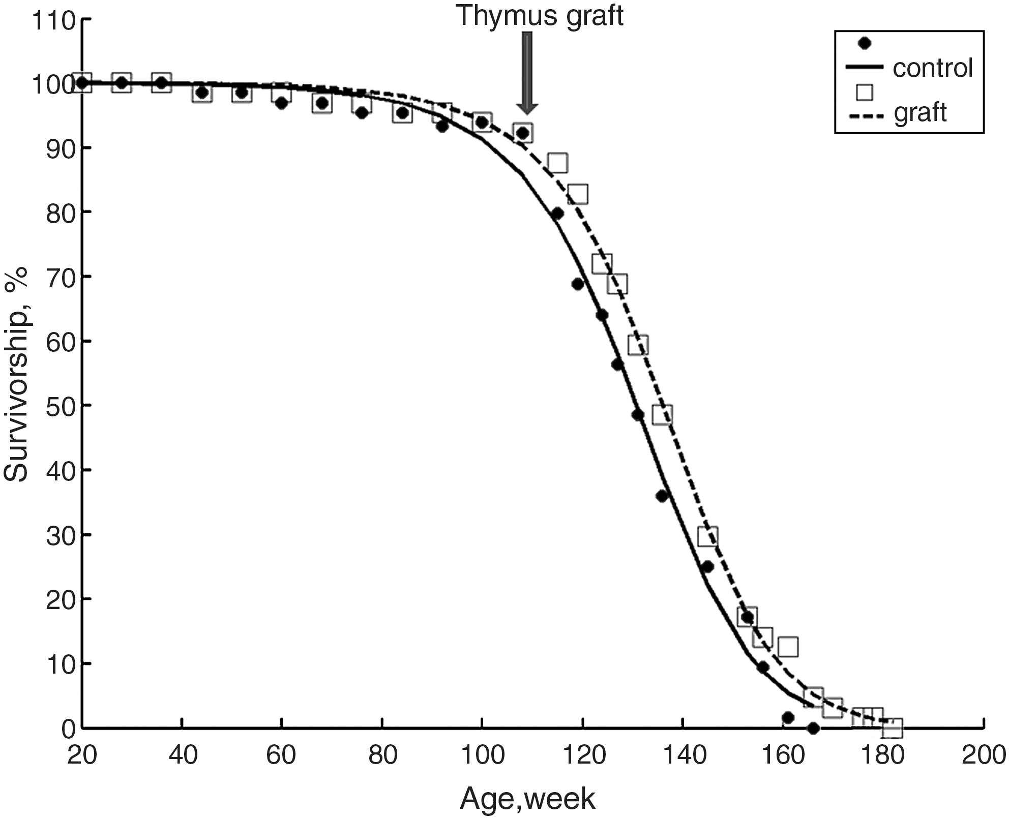
Survival data of old mice grafted with neonatal thymuses (filled circles) and control mice (open squares). Data from both groups are fitted using the mathematical model outlined in the Materials and Methods section and in Box 1. The values of the parameters ± standard error (SE) for the best fits are given as follows: grafted mice, ω =0.024114 ± 0.000105, S0 = 0.396286 ± 0.005731; controls, ω =0.024797 ± 0.000235, S0 = 0.412951 ± 0.011225. Grafted mice show a 3% decrease in ω value, which is statistically significant (p = 0.0097). The thymus graft was performed in old recipients at age 114 weeks, as shown by the arrow).
Discussion
The findings herein reported show for the first time that the longevity of experimental old mice can be slightly but significantly improved by transplanting two neonatal thymi in the axillary cavity with a minimal invasive intervention with respect to the thymus graft into kidney capsule. Scientific interest around studies examining longevity for these two types of transplants has been generally scarce, being the reverse of specific physiological processes that are the main target of most investigators. However, it has been reported that neonatal thymic grafts performed under the kidney capsule with a half-thymus does not significantly change the mortality kinetics when compared to sham or control mice. 4
These conclusions were drawn by comparing transplanted mice with controls without taking into account the impact of the invasive surgical intervention on the mortality kinetics of mice and without taking into account the limited amount of tissue (half-thymus) used for the transplant. 8 Thus, we decided to compare the survival of mice after grafting two neonatal thymi into the axillary cavities, as previously suggested for the transplant of other tissues with an endocrine role. 14 Also, in this case, conventional nonparametric tests did not show any significant change in survival between grafted old mice versus controls; however, survival analysis performed with a Piantanelli-sensitive parametric model 15,16 suggests that this kind of approach may be considered as a starting point for rejuvenating interventions. The very small difference between the two curves, which is not statistically significant according to the Kaplan–Meier analysis with log rank test, would have hardly an interest per se. The effects on survival are significant but still minimal, so that it is necessary to fit data with a sensitive parametric model to show the change.
When the parametric model herein described is used, the slight change induced in old grafted mice survival kinetics may reveal some interesting aspects. Although the difference in S0 values does not have much biological meaning (p > 0.05), the one observed in ω values deserves some attention despite being as small as 3%. Because ω is assumed to represent the deterministic component describing the environmental and genetic influence on physiological functions, the treatment is likely to influence few individuals with peculiar physiological capacity, thus resulting in an extension of maximal lifespan.
Previous data on thymic transplant suggest a general rejuvenating action on several physiological functions, 4 but it should be noted that these functions were tested in the short time following the transplant. Therefore, it can be argued that in most individuals the effect of the transplant is transitory, being limited by the old environment in which the neonatal thymus is implanted, whereas the conditions exist for a prolonged beneficial effect in a few individuals. On the other hand, it is worth noting that old thymus transplanted into young recipients can reverse to a juvenile condition, thus suggesting the relevance of environmental and important extrathymic factors involved in the endocrine functional maintenance of the thymus. 12,18,19
Therefore, it is reasonable that the effects of neonatal thymus grafts might be improved by adjuvant therapies using these putative extrathymic factors that are gradually lost in old age. These extrathymic factors might include hormones (melatonin, growth hormone, insulin-like growth factor-1), nutrients (zinc and arginine,) and cytokines (interleukin-7). 20 By combining interventions with these extrathymic factors and a neonatal thymus graft, a considerable improvement of the survival might be obtained.
In conclusion, we have shown for the first time that a neonatal thymus graft (into the axillary cavity) performed with a mild invasive intervention might extend the lifespan of old mice. In addition, we show the usefulness of a parametric model for analysis of survival data not only for the higher sensitivity, but also especially for the biological conclusions that could be drawn after parameter estimation.
Footnotes
Acknowledgments
This paper was supported by INRCA. The authors thank Mr. A. Antognini for technical support.