




Abstract
We assessed the effect of a sturgeon eggs–based nutraceutical (LD-1227) versus eicosapentaenoic acid (EPA)/docosahexaenoic acid (DHA) on the ultrastructure of spontaneously hypertensive rat (SHR) aortas. Sixty SHR were randomly divided into three groups that were fed (1) rat chow, (2) rat chow plus 10 mg of EPA/DHA, or (3) rat chow plus 10 mg of LD-1227, for 18 weeks. Afterward, aortas of these rats were used for blind measurements of the thickened intima area and examination by electron microscopy. Control SHR showed an expanded subendothelial space and leukocyte infiltration of the intima that were reduced in LD-1227–fed rats (p<0.05) and less in EPA/DHA group. Transmission electron microscopy showed endothelial alteration with severe subcellular injury and, unlike the EPA/DHA-group, LD-1227–treated rats displayed a significant reduction in endothelial alteration with severe subcellular injury (p<0.05). These data suggest that LD-1227 has stronger arterial protective properties and deserves further investigation in view of a preventive medicine strategy.
Introduction
I
Given the uniqueness of the composition of sturgeon eggs as compared to other marine species regarding polyunsaturated fatty acids (PUFAs) and phospholipid moieties, 8 and in consideration of our recent work proving the efficacy of a controlled caviar-derived homogenate, LD-1227 (Caviarlieri, LabDom, Switzerland), as an anti-oxidant/anti-inflammatory modulator with a wider efficacy properties as compared to docosahexaenoic acid (DHA), 9,10 we also tested this compound on the ultrastructure of the SHR aorta.
Materials and Methods
Sixty 2-month-old SHR with initial body weights varying from 140 to 160 grams were randomly divided into three groups of 25 rats each. The first group (control) was fed only rat chow; a second was fed with 10 mg eicosapentaenoic acid (EPA)/DHA added to the food, and a third was given 10 mg of LD-1227 added to the food for 18 weeks. During all treatment periods, the animals had free access to food and drinking water. All protocols were performed in accordance to the Guide for the Care and Use of Laboratory Animals (National Institutes of Health [NIH] Publication No. 85-23, revised 1996). Non-invasive measurement of tail-cuff pressure as an estimate of systolic arterial pressure was performed at baseline and 9 and 18 weeks afterward. Rats were warmed in a restraining chamber, and occluding cuffs and pneumatic pulse transducers were placed on the rat tail. A sphygmomanometer was inflated and deflated automatically, and the tail-cuff signals from the transducer were automatically collected using an IITC apparatus (IITC Inc., Woodland Hills, CA) connected to a computer. For each blood pressure measurement session, the mean of eight blood pressure readings was recorded for each rat. Blood lipid profiles and malondialdehyde (MDA) levels were checked as well.
Electron microscopy analysis
At the end of 4 months, the SHR were anesthetized by intra-peritoneal injections of Nembutal. The aorta of each SHR was dissected, making sure to clear it from surrounding tissues. Specimens from different parts of the aortic arch measuring about 4 mm long were obtained and soaked in cold 3% glutaraldehyde. All areas of the aortic sections were taken at the same site of the aortic arch of each rat, avoiding the branching sites. The same tissues were further cut to about 1 mm in size and fixed in the glutaraldehyde at pH 7.2 for 3 hr at 4°C. The aorta was cut at right angles to the longitudinal axis. Post-fixation of the specimens was performed in 1% solution of osmium tetroxide for 1 hr at 4°C. The specimens were then dehydrated in increasing concentrations of alcohol before being embedding in epoxy resin and polymerized in an oven at 60°C for 48 hr. Nine sections of 1 μm thickness from different parts of the aortic arch were obtained using an LKB I V ultramicrotome (LKP Produkter, Sweden), stained with Toluidine Blue, and observed under a light microscope.
During the electron microscopy session, the first encounter with two adjacent grid squares displaying a continuous arc of arterial intima was selected for study. For each section, two micrographs at approximately 12,000×final magnification were taken adjacent to grid bars at opposite ends of the two-grid-square span of intima. Moreover, every detection of abnormal structures in the intima, such as, mononuclear leukocytes, smooth muscle cells, or portions of cytoplasm, contained within the two-grid squares was photographed. The ultrathin sections obtained were collected on copper grids and stained with 3% uranyl acetate and Reynold lead citrate before being analyzed under an electron microscope with JEM 7A, and micrographs were taken. The adequacy of perfusion fixation was confirmed by the removal of corrugations from the elastic laminae in the thoracic aorta and the presence of an overall circumferential orientation outlook of medial smooth muscle cells. The blind examination was scored using a computer-assisted image analyzer.
Assessment of thickened intima area
Areas with the greatest focal thickenings were located and measured, the rest of the intima being relatively thin. The intima thickness was calculated and analyzed using the Pro-Plus 6.0 image system (Cybernetics, Inc., Bethesda, MD). All slices were investigated by an experienced pathologist who was blinded to the experimental design. The thickened areas of nine sections of the aortas of each rat were measured, averaged, and taken as the value for one aorta. A mean value of measurements was regarded as the representative results of each group. The data were then converted to its actual area by using a stage micrometer.
Malondialdehyde determination in plasma
MDA was assayed by spectrophotometric method, and the concentration of thiobarbituric acid (TBA) was calculated by the absorbance coefficient of the MDA–TBA complex 1.56×105 cm−1 M−1 and expressed in nmol/mL. The amount of phosphorus released by the enzymes was calculated as a quantitative measure of the activities of total adenosine triphosphatase (ATPase). 11 Tissue protein thiols were determined according to the methods described by Sedlack and Lindsay 12 by using Ellman reagent (5,5′-dithiobis 2-nitrobenzoic acid [DTNB]), which was reduced by thiol groups to form 1 mol 2-nitro 5-mercaptobenzoic acid/mol thiol and with maximal absorption at 412 nm.
Statistical analysis
Data are expressed as the mean±standard error of the mean (SEM) of multiple determinations from an experiment. Statistical significance was determined by the Student t-test when two data sets were analyzed with the statistical software StatView (Abacus Concepts, Calabasus, CA).
Results
Both groups of rats showed a comparable physiological weight increase and lipid profile without any significant difference among them (total cholesterol, μg/dL: 128±4.2 vs. 131±3.6 vs. 132±3.9, in the three groups as listed above, respectively; data not shown). As expected, the serum levels of MDA, as the index of oxidative stress, were significantly (p<0.05) increased in SHR as compared to a separate set of control healthy rats (MDA, nmol/100 mg: 1.2±0.3 vs. 2.1±0.6, p=0.05). Either EPA/DHA or LD-1227 supplementation did not significantly affect this parameter, although there was a trend in favor of LD-1227 [MDA, nmol/100 mg: 1.9±0.8 in EPA/DHA-treated SHR vs. 1.6±0.7 in LD-1227–treated SHR, not significant (NS)].
Systolic blood pressure in SHR was significantly higher than in normal Wistar rats and was not affected by any of the treatments during the observation period. In the aorta of untreated SHR, the most prominent change was an increase of the subendothelial space and penetration of mononuclear leukocytes into the intima as well as evident cellular elements and extracellular material in the subendothelial space of the thickened regions. Moreover, the thickened areas showed an apparent increase in the density of connective tissue elements, that is, the fibrils of collagen and those of some connective tissue cells together with fibroblasts, macrophages, and smooth muscle cells,
However, these findings were significantly reduced in LD-1227–fed rats and, to a lesser extent in the EPA/DHA group (p<0.05 vs. control and EPA/DHA group) (Fig. 1A–C). The areas of thickened intima were significantly reduced in EPA/DHA-treated rats (5530 μm2 vs. 7141μm2, p=0.05) and this appeared to be further reduced in LD-1227–treated SHR rats (4110 μm2 vs. 5530 μm2, p=0.05). Proliferation of smooth muscle cells and their migration from the medial into the intimal layer is also part of the pathology of hypertension. However, the extent of the lesions was significantly less relevant in the LD-1227–treated animals (p<0.05). SHR showed an increase in the density and frequency of occurrences of cytoplasmic vacuoles and cytoplasmic processes of endothelial cells, with longer and more slender cytoplasmic processes. Moreover, transmission electron microscopy (TEM) analysis also showed structural alteration of endothelial cells with severe subcellular injury (atypical mitochondria of smooth muscle cells, such as swollen and vacuolated mitochondria, and several vacuoles and electron-lucent chromatin). Unlike the EPA/DHA-group, LD-1227–treated rats displayed a significant reduction of such changes (p<0.05) (Fig. 1D–F). No changes were apparent in the adventitia of any of the three groups of SHR.
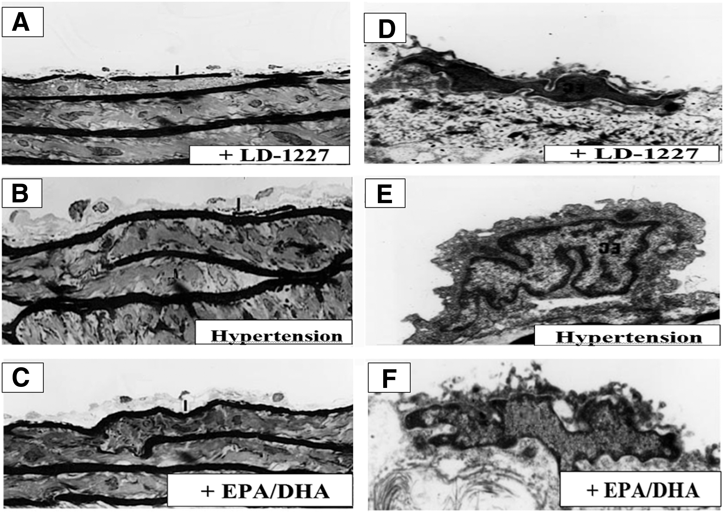
Light microscopy and electron microscopy analysis of arterial structure in spontaneously hypertensive rats (SHR): Effect of LD-1227.
Discussion
Hypertension represents a main risk factor of cardiovascular diseases linked with endothelial dysfunction. Structural abnormalities of endothelium in vessels observed in hypertensive animals 13 –15 indicated their likely contribution to the development or maintenance of the high blood pressure. SHRs were used in this study in an attempt to unveil the anti-atherogenic effect specific marine nutraceuticals may have on the process of atherosclerosis. Hypertensive rats and other animals, such as rabbits, have been shown to develop atherosclerotic lesions when their plasma lipoprotein levels are increased by dietary cholesterol feeding; however, rats are more resistant to the occurrence of atherosclerosis, thus representing a more feasible human model. Hypertension is associated with structural changes in blood vessels known as “vascular remodeling” that include dynamic structural changes involving cell growth, migration, phenotypic changes, and extracellular matrix synthesis and degradation. 16 Effects of LD-1227 on expression of angiotensin-converting enzyme 2, mitogen-activated protein kinase (MAPK) signaling, and peroxisome proliferator-activated receptor-γ in aortas of SHR are underway, and preliminary in-house data show a significant beneficial modulation exerted by this marine nutraceutical. Although no effect was noted on systolic blood pressure in our study, this may suggest further properties related to a protective modulation against vascular hypertrophy. Although further detailed biochemical research is needed, following a recent research direction in this field, 17 these preliminary data suggest that LD-1227 has stronger arterial protective properties than EPA/DHA and it is worth further longer-term studies in view of a preventive cardiovascular protective medical strategy.
Author Disclosure Statement
No competing financial interests exist.