




Abstract
Aberrant crypt foci (ACF) and mucin-depleted foci (MDF) are pre-neoplastic lesions identified in the colon of carcinogen-treated rodents and in humans at high risk for colon cancer. The present study was carried out to divulge the protective potential of the probiotic Dahi containing Lactobacillus acidophilus LaVK2 and Bifidobacterium bifidum BbVK3 alone or in combination with piroxicam (PXC) on the development of early biomarkers of colorectal carcinogenesis in male Wistar rats administered 1,2-dimethylhydrazine (DMH). DMH was injected subcutaneously at the rate of 40 mg/kg body weight per animal twice a week for 2 weeks. A total of 120 male Wistar rats were randomly allocated to five groups, each group having 24 animals. The rats were fed with buffalo milk or probiotic supplement (20 grams) alone or as an adjunct with PXC in addition to a basal diet ad libitum for 32 weeks. Group I was offered buffalo milk (BM) and served as the control group. Group II was administered DMH along with BM and served as the DMH-control group; group III was administered BM-DMH-PXC, in which besides administering BM-DMH, PXC was also offered. Group IV was offered probiotic LaBb Dahi and DMH, and group V was offered both probiotic LaBb Dahi and PXC along with DMH. The rats were euthanized at the 8th, 16th, and 32nd week of the experiment and examined for development of ACF, aberrant crypts per ACF (AC/ACF), mucin-depleted foci (MDF), large MDF, and proliferating cell nuclear antigen (PCNA) labeling index. Administration of DMH in rats induced pre-neoplastic lesions (ACF and MDF) and increased the PCNA index in colorectal tissue. A significant (p<0.05) reduction in the number of ACF, AC/ACF, MDF, large MDF, and PCNA labeling index were observed in the probiotic LaBb Dahi group compared with the DMH control group. Feeding rats with LaBb Dahi or treatment with PXC diminished the initiation and progression of DMH-induced pre-neoplastic lesions and the PCNA index, and treatment with LaBb Dahi and PXC combined was significantly more effective. The dietary intervention of probiotics and PXC significantly protects against the development of CRC in the rat-DMH model. These observations suggest that probiotic LaBb Dahi alone or as an adjunct with PXC may have anti-neoplastic and anti-proliferative activities. Moreover, probiotic LaBb Dahi possesses the medicinal properties to prevent colorectal carcinogenesis.
Introduction
C
Aberrant crypt foci (ACF), easily visualized in unsectioned colon tissue, are reliable intermediate biomarkers for colon carcinogenesis and considered to be surrogate pre-neoplastic lesions because they are found in the colon of carcinogen-treated rodents 10 –12 and in patients at high risk for colon cancer development. 13 Biochemical, genetic, and morphological studies have shown that ACF and colon tumors share similar alterations, further supporting the hypothesis that ACF are precursors of CRC. 13 Mucin-depleted foci (MDF) have been recently identified as dysplastic lesions or crypts devoid of mucins that occur in the colon of carcinogen-treated rats and also in humans at high risk of colon cancer. 14,15 MDF harbor mutations in genes affecting colon carcinogenesis and, like colonic tumors, show Wnt signaling activation, features suggesting that these lesions are pre-cancerous. 16,17 Also, the expression of MUC2, the most abundant mucin in the colon, is dramatically reduced in MDF, leading to the speculation that the focal loss of the protective mucous layer might activate local inflammation, because MDF would be more exposed to noxious agents present in the colonic lumen. This local inflammation might drive carcinogenesis to more advanced stages. 18 The number of MDF per colon increases in rats treated with promoters of colon carcinogenesis, such as cholic acid, whereas it is decreased by chemopreventive agents. 18 MDF are induced dose dependently by dimethylhydrazine (DMH), and progressively increase in size after carcinogen administration. 16 Recently, it has been shown that MDF carry alterations in the Wnt signaling pathway and mutations in the β-catenin gene, 16,19 characteristic phenomena in colon tumorigenesis. Therefore, although MDF have been used so far in a limited number of studies, 14 –20 they are a promising biomarker for study of the effect of chemopreventive agents in colon carcinogenesis. Aberration in epithelial colonial crypt cell proliferation leads to hyperplasia with higher-risk colon cancers both in humans and experimental models. 21 Assessment of PCNA expression as an indicator of colonic crypt cell proliferation is suggested as a putative intermediate marker of colon cancer risk. 22
Probiotics may inhibit cancer development by limiting exposure to carcinogens (e.g., inhibitors of carcinogen formation and blocking agents) and acting as a inhibitors of tumor initiation acting by different mechanisms, including reactive oxygen species (ROS) scavenging, changes in phase I and II enzymes of carcinogen metabolism, and further DNA damage. 23 As humans are exposed to hydrazines, heterocyclic amines, and other mutagens/carcinogens through the diet, 24 possible strategies for cancer prevention could be achieved using potential alternate biotherapeutic agents, such as probiotics, to reduce or eliminate the deleterious effects of human exposure to potential carcinogens. Dahi is a component of daily Indian diet throughout the country, hence it could be a very effective medium for the delivery of probiotics to the Indian population. Probiotics can be easily and economically introduced into Dahi. Our laboratory has developed technology for incorporation of probiotic L. acidophilus and B. bifidum strains into Indian Dahi. Several studies from our laboratory have demonstrated the potential of probiotic Dahi, prepared by co-culturing selected strains of L. acidophilus and B. bifidum and Dahi culture, to reduce tumor incidence in the gastrointestinal tract of rats treated with DMH. 25 This probiotic Dahi also upregulates glutathione-S-transferase (a carcinogen detoxifying activity) in liver, and downregulates β-glucuronidase (a carcinogen-activating enzyme) in the intestinal lumen, attenuates hypercholesterolemia, 26 and stimulates immune functions 27 and anti-oxidant status in rodents. 28 Being a fermented milk product, Dahi can be an excellent medium for delivery of probiotic strains that can provide protection against CRC.
In this context, the present study was carried for the first time to evaluate the chemopreventive effects of probiotic LaBb Dahi alone as well in combination with PXC on the development of pre-neoplastic biomarkers such such as ACF and MDF and cell proliferation markers such as the proliferating cell nuclear antigen (PCNA) labeling index in male Wistar rats.
Materials and Methods
Bacterial strains
Lactococcus lactis spp. cremoris NCDC-86 and L. lactis spp. lactis biovar diacetylactis NCDC-60 were obtained from National Collection of Dairy Cultures, National Dairy Research Institute (NDRI), Karnal, India. Lactobacillus acidophilus LaVK2 and Bifidobacterium bifidum BbVK3 are our laboratory isolates with probiotic attributes tested through a battery of tests as per Food and Agriculture Organization/World Health Organization (FAO/WHO) guidelines. These isolates were identified by molecular-typing methods and studied extensively for their functional and probiotic attributes, viz., acid and bile salt tolerance, cell-surface hydrophobicity, autoaggregation, and Caco-2 cell-binding, as well as anti-bacterial and anti-oxidative activities (unpublished data). Lactobacilli and lactococci were propagated and maintained in MRS broth and M17 broth (Himedia Laboratories Pvt. Ltd., Mumbai, India) at 37°C and 30°C, respectively, and were stored at 4–8°C between transfers. B. bifidum BbVK3 was cultured and propagated in anaerobic conditions at 37°C for 24 hr.
Preparation of Dahi and probiotic Dahi (LaBb Dahi)
Bacterial cultures were revitalized three times in reconstituted and autoclaved skim milk prior to use for preparation of fermented milk. Buffalo milk (BM) obtained from the cattle yard of the institute and standardized to 3.0% fat was heated to 90°C for 15 min and then cooled to 37°C. Dahi was prepared by culturing standardized BM with Dahi starter culture (L. lactis ssp. cremoris and L. lactis ssp. lactis biovar diacetylactis, 1% each) at 30°C for 8 hr. Probiotic LaBb Dahi was prepared by culturing BM with L. acidophilus LaVK2, B. bifidum BbVK3 and Dahi culture (1.0%). The final product contained 2×10⁹ cfu/gram of lactococci, B. bifidum, and L. acidophilus each.
Chemicals
1,2-Dimethylhydrazine dihydrochloride (DMH), PXC, Alcian Blue stain, Harris' Hematoxylin, monoclonal anti-PCNA antibody, and 3,3-diaminobenzidine were purchased from Sigma-Aldrich Chemical Co. (St. Louis, MO, USA). The source of mouse ABC immunostaining system was Santa Cruz Biotechnology (Santa Cruz, CA, USA), N,N′-dimethyl-m-phenylene diamine and N,N′-dimethyl-p-phenylene diamine were procured from National Chemicals Ltd., Bangalore, India. All other chemicals were obtained from s.d. Fine Chemicals Ltd., Mumbai, India, or Hi-Media Lab. Ltd., Mumbai, India.
Animals and diet
Male Wistar rats (3 weeks of age) were obtained from Small Animal House of National Dairy Research Institute, Karnal, India. The animals were housed in stainless steel cages (three animals per cage) throughout the study, and room temperature was maintained at 25±2°C, 55±5% humidity, and at a 12-hr light/12-hr dark cycle. The animals were used and cared for in accordance with the principles and guidelines for humane use, and the protocol was approved by the Institutional Ethics Committee. The composition of basal diet was starch (63%), casein (20%), soybean oil (5.5%), cellulose (5%), mineral mixture (5%), vitamin mixture (1%),
Experimental design
Animals were randomly distributed into five groups, with 24 in each group, and fed with the BM or probiotic supplements (20 grams) in addition to basal diet for 32 weeks. Rats of group I were fed with BM and served as control group. Rats of group II (BM-DMH) were administered DMH and served as the DMH control; DMH-induced rats of group III (BM-DMH-PXC) were administered BM and PXC. Rats of group IV (LaBb Dahi-DMH) were fed probiotic Dahi and DMH, and DMH-induced rats of group V (LaBb Dahi-DMH-PXC) were offered both probiotic Dahi and PXC. Following 28 days of feeding, DMH (40 mg/kg body weight) was administered subcutaneously (s.c.) to the rats of the corresponding groups twice a week for 2 weeks. 22 PXC (4 mg/rat or 200 mg/kg of supplements) was given daily orally along with the supplements (BM/probiotic Dahi), and its feeding started 1 week after the last dose of DMH and continued until termination of the experiment. 6 Rats were sacrificed by cervical dislocation at the 8th, 16th, and 32nd weeks of experimental time, and colorectal tissues were examined for ACF, MDF, and PCNA labeling index.
Identification of ACF
Colorectal segments were removed, washed with saline, cut open longitudinally, fixed flat in 10% buffered formalin, and stained with Methylene Blue. 30 The ACF were characterized by their enlarged crypts, elevated crypts from the surrounding epithelium, thickened epithelial layer, increased pericryptal space, and their round or elongated luminal openings.
Identification of MDF
After ACF determination, the colorectal segments were processed for high iron diamine Alcian Blue staining. 31 The MDF were characterized by having or very little mucin and fulfilled the criteria according to Caderni et al. 14 The colons were coded and scored independently by two observers. The numbers of MDF per colon and crypts per MDF were recorded.
Immunostaining for PCNA
Following MDF determination, the colorectal tissues were embedded in paraffin so that the crypts could be sectioned longitudinally. The proliferative activity in the mucosa was evaluated by determining PCNA immunoreactivity.
31
Sections were cut from paraffin-embedded tissue and mounted on poly-
Finally, after rinsing 3× with PBS, the slides were treated for 5 min with 3,3′-diaminobenzidine, washed with deoinized water for 5 min, and counterstained with Harris' Hematoxylin. Proliferative activity was evaluated by counting PCNA-positive nuclei in at least 10 full longitudinal crypt sections (from the base to the bottom of the crypt) under the light microscope. 24 The dark brown–stained nuclei were considered PCNA positive. The PCNA-positive index was calculated as the proportion of positively stained nuclei expressed as a percentage. The microscopic slides were coded and read independently by two observers.
Statistical analysis
The results were expressed as the means±standard error (SE) for each group (n=8) and analyzed by one-way analysis of variance (ANOVA) followed by the Tukey post hoc test (SYSTAT v. 6.0.1, SPSS Inc., Chicago, IL, USA). Differences were considered significant at p<0.05.
Results
Effect on feed intake and body weight
A significant decline in average feed intake as well as in body weight gain was observed in rats treated with DMH (Table 1). Treatment of DMH-induced rats with PXC helped to increase growth significantly (p<0.05), but the gains in body weight were still lower than in untreated control group. Treatment of DMH-induced rats with either LaBb Dahi or PXC alone or LaBb Dahi combined restored feed intake to normal levels and increased the body weight significantly (p<0.05). The DMH-induced animals treated with LaBb Dahi or PXC and LaBb Dahi combined grew at rates even faster than the control rats.
Values are mean±standard error (SE) for n=8.
Mean values within a column with different superscript letters are significantly different (p<0.05).
DMH, 1,2-dimethylhydrazine dihydrochloride; PXC, piroxicam.
Effect on aberrant crypts and ACF formation
The ACFs in DMH-treated rats were present throughout the length of colorectal segment, with the incidence higher in mid and distal regions; these regions accounted for approximately 90% of the total ACF. Furthermore, the incidence of ACF increased almost linearly with time in animals treated with DMH. The DMH-induced rats fed withLaBb Dahi or PXC or PXC and LaBb combined showed significantly lower (p<0.05) numbers of ACF and aberrant crypts (ACs) than the rats from the DMH control group. The development of DMH-induced ACF was decreased significantly (p<0.05) by treatment with PXC and LaBb Dahi.
Figure 1a shows the data on total number of ACF in the colorectum at different time intervals of 8, 16, and 32 weeks that clearly indicate the protective potential of probiotic LaBb Dahi alone or in combination with PXC against CRC. The total number of ACF at 8 weeks was decreased by 49.5%, 55.4%, and 81.6% in rats fed with PXC, LaBb Dahi, and PXC combined, respectively, relative to the BM-DMH group. Although the total number of ACF at 16 weeks increased in each group, the proportion relative to the BM-DMH group remained almost the same in rats fed PXC or LaBb Dahi or LaBb Dahi and PXC combined (i.e., 46.8%, 59.8%, and 81.0%, respectively). Whereas the total number of ACFs continued to rise beyond 16 weeks in the BM-DMH group, it almost plateaued in rats fed with PXC or LaBb Dahi or PXC and LaBb Dahi combined. The proportion of ACF having one crypt was 53.0%–76.0% in animals fed PXC or LaBb Dahi or PXC combined, compared to 22.0% in the DMH-treated control (Fig. 1b). The proportion of ACFs having more than three crypts was reduced to 20.4%, 29.3%, and 16.6% in rats fed with PXC, LaBb Dahi, and PXC combined, respectively, compared to 37.1% in BM-DMH group (Fig. 1c, d). When expressed as a percentage, the total number of ACFs at 32 weeks decreased by 59.2%, 71.9%, and 84.9% in rats fed PXC, LaBb Dahi, and LaBb Dahi and PXC combined, respectively, relative to the BM-DMH group. Furthermore, the proportion of ACFs having more than three crypts at 32 weeks was 48.4% in BM-DMH group, and it was reduced to 14.9%, 27.9%, and 10.1% in animals fed PXC, LaBb Dahi, and PXC combined, respectively (Fig. 1d). The LaBb Dahi treatment was more efficacious than PXC in reducing the number of ACFs in the colorectum. A further reduction in the number of ACFs in the colorectum was observed when animals were treated with probiotic LaBb Dahi and PXC combined; hence, the effect of PXC and LaBb Dahi in reducing the number of ACF was additive.
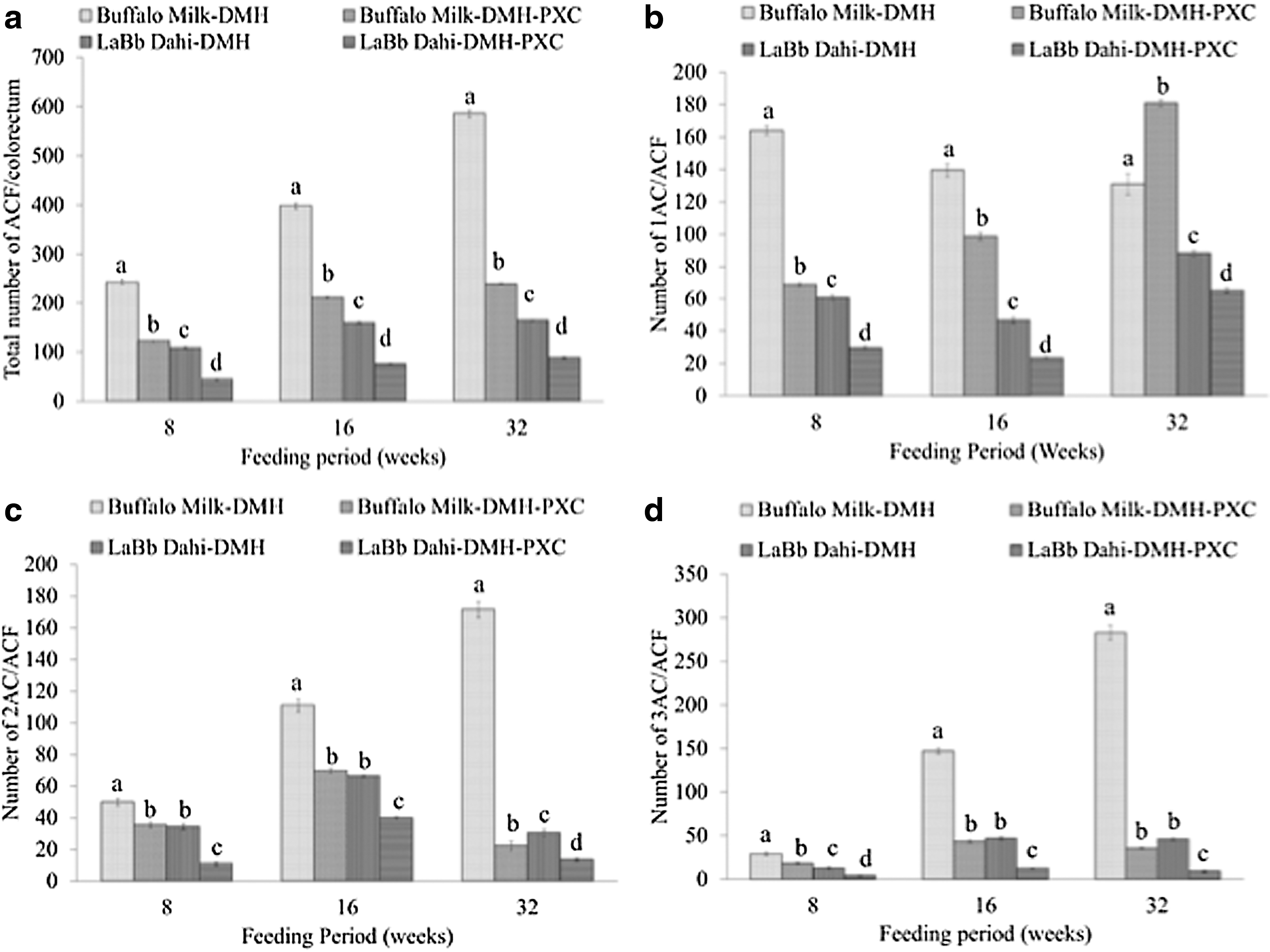
Number of aberrant crypt foci (ACF)/colorectum and aberrant crypts (AC)/ACF in colorectal tissue of dimethylhydrazine (DMH)-treated rats at 8, 16, and 32 weeks. (
Effect on MDF formation
The administration of DMH in rats resulted in development of MDF in the colorectum, which decreased in number after 16 week (Fig. 2a). The number of DMH-induced MDF decreased significantly (p<0.05) by treatment with PXC or LaBb Dahi. The extent of reduction in the number of MDF was almost similar in animals treated with PXC and those treated with LaBb Dahi. When DMH-injected animals were treated with a combination of PXC and LaBb Dahi, the number of MDF in the colorectum was significantly (p<0.05) lower than in groups treated with either PXC or LaBb Dahi, suggesting that LaBb Dahi improves the efficacy of cancer treatment with PXC. The number of large MDF (containing more than 4 AC) in the BM-DMH group increased with time throughout the experimental period (Fig. 2b). The treatment with PXC or probiotic Dahi significantly decreased the number of total as well as large MDF in the colorectal segment. Unlike the BM-DMH group, there was no increase in number of large MDF beyond 16 weeks in groups treated with PXC or probiotic Dahi or probiotic Dahi and PXC combined. The probiotic Dahi was more effective than PXC in reducing the progression in number as well as in size of MDF. Furthermore, in animals treated with a combination of PXC and probiotic Dahi, the number as well as the size of MDF in colorectal segments were significantly lower than in groups treated with PXC or probiotic Dahi alone, suggesting that probiotic Dahi improves the efficacy of cancer treatment with PXC.
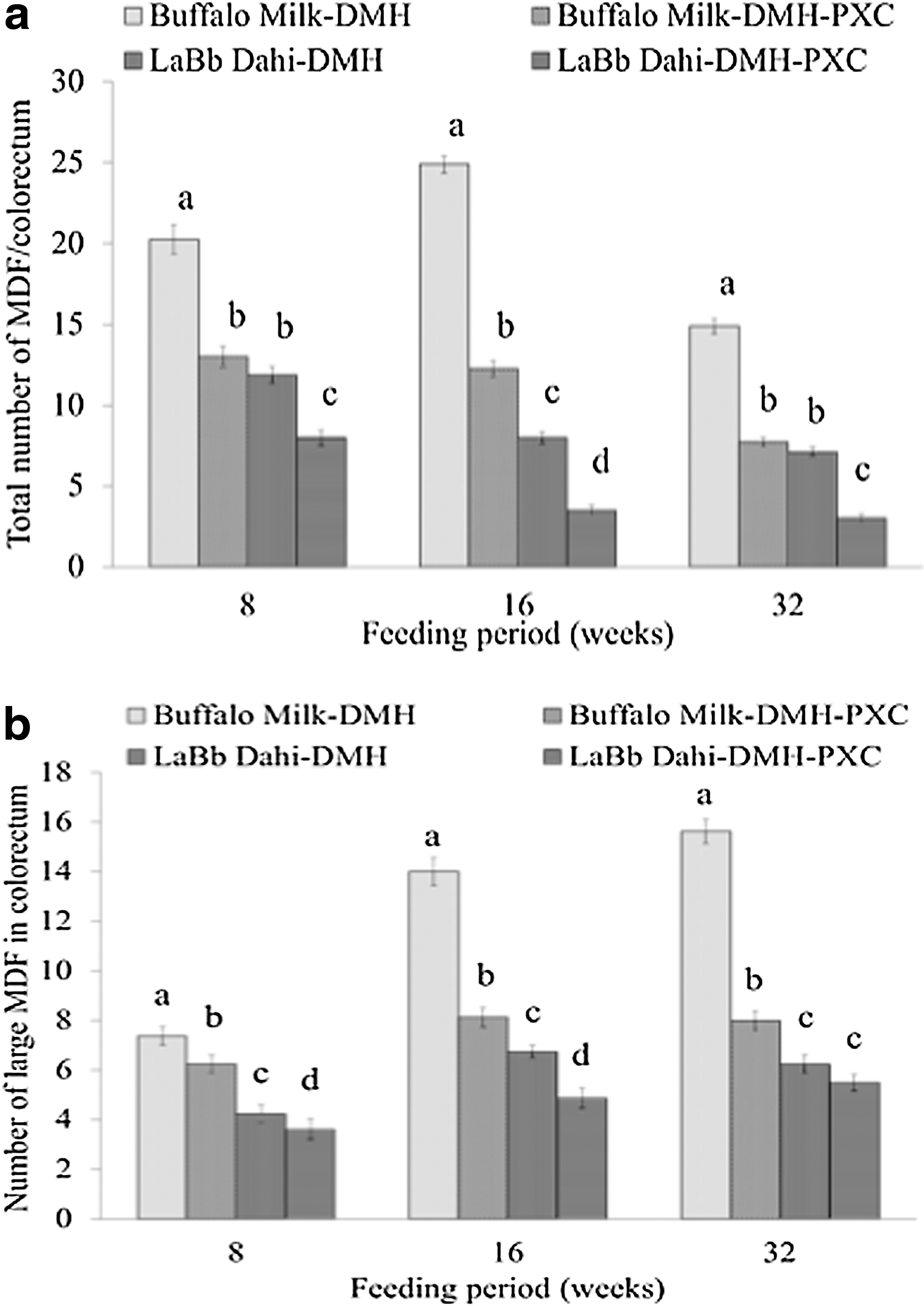
Number of mucin-depleted foci (MDF)/colorectum and larger MDF/colorectum in colorectal tissue of dimethylhydrazine (DMH)-treated rats at 8, 16, and 32 weeks. (
At 16 weeks, the number of MDF was reduced by 50.6%, 67.9%, and 86.0% in animals treated with PXC, LaBb Dahi, and PXC combined, respectively, and the corresponding diminutions in large MDF were 42.1%, 51.4%, and 65.0%, respectively. At 32 weeks, the total number of DMH-induced MDF decreased by 47.6%, 52.3%, and 79.9% in rats fed PXC, LaBb Dahi, and PXC combined, respectively, relative to the BM-DMH group. The corresponding decrease in large MDF (containing >4 AC) was 48.7%, 59.6%, and 64.7%, respectively. These results show that LaBb Dahi was more effective than PXC in reducing the total number of MDF in the colorectum. Furthermore, LaBb Dahi was more effective than PXC in reducing the progression in number as well as in size of MDF. The diminutions in the number of large MDF were more pronounced in animals treated with a combination of LaBb Dahi and PXC compared to those treated with any of these two individually, suggesting that probiotic Dahi improves the efficacy of cancer treatment with PXC.
Effect on PCNA labeling index
PCNA is a non-histone nuclear protein essential for DNA replication and repair. The content of PCNA is increased during carcinogenesis, is used as prognostic marker in development of cancer. The data represented in Fig. 3 show that the content of PCNA in cells is increased in normal animals and reaches 48% above the 0-day level at 32 weeks. When animals were injected with DMH, the accumulation of PCNA protein was substantially enhanced, reaching six-fold that of the 0-day level at 32 weeks. The treatment with PXC or LaBb Dahi decreased DMH-induced accumulation of PCNA in epithelial cells of the colorectum. PXC and LaBb Dahi were almost equally effective in reducing DMH-induced accumulation of PCNA in epithelial cells of the colorectum. The reductions in DMH-induced accumulation of PCNA in epithelial cells of the colorectum were more pronounced in animals treated with the combination of PXC and LaBb Dahi. The content of PCNA in epithelial cells of the colorectum at 32 weeks of the experimental period reached a level similar to that in untreated normal animals, suggesting this combination of treatment is most effective in preventing initiation and progression of carcinogenesis.
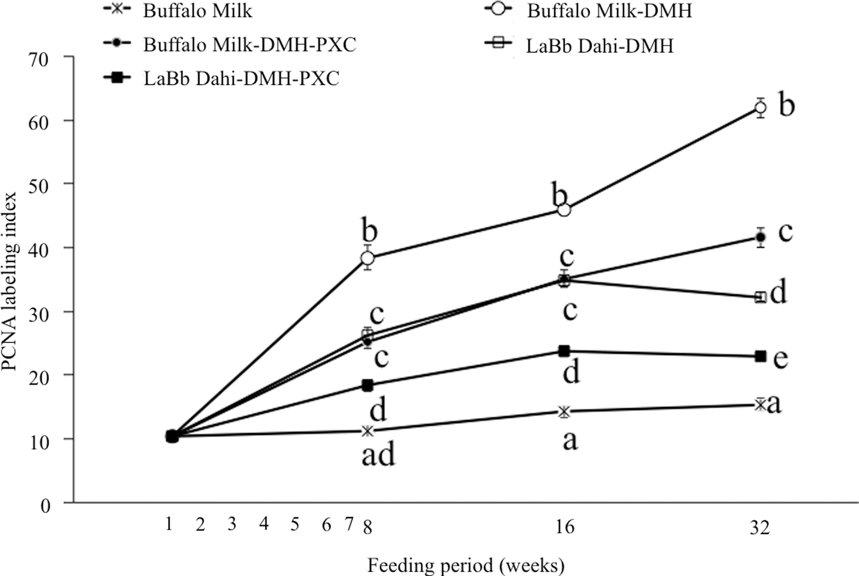
Effect of feeding probiotic Dahi and piroxicam (PXC) treatment on proliferating cell nuclear antigen (PCNA) labeling index in colorectal tissue of dimethylhydrazine (DMH)-treated rats. PCNA labeling index was determined as proportion of PCNA-positive cells in a crypt. Values are mean±standard error (SE) for n=8. a,b,c,d,e,fValues with different superscripts letters are significantly different (p<0.05).
Discussion
To the best of our knowledge, this is the first study reporting the intervention of probiotic LaBb Dahi and PXC, a well-known non-selective NSAID, can effectively reduce pre-neoplastic biomarkers such as ACF, MDF, and the PCNA proliferation index in a rat model. These results showed that LaBb Dahi alone or in combination with PXC can suppress the formation of both pre-neoplastic lesions and proliferation at initiation, promotion, and progression stages in the colorectal tissues of rats.
ACF appear predominantly in the distal colon and are the precursors of colon cancer. 32 In the present study, ACF were mainly distributed in the middle and distal colons in all of the DMH-induced rats; these ACF accounted for approximately 90% of the total ACF, and a significant reduction (p<0.05) in the number of ACF was observed. The rats from all of the LaBb Dahi, PXC, or groups with their combinations treated had significantly lower (p<0.05) total numbers of ACF and ACs than the rats from DMH control group. ACF growth is reported to occur through the mechanism of “crypt fission,” 33 and, therefore, ACF with more crypts indicate a more advanced cancer stage. In the present study, the rats in the PXC and LaBb Dahi groups showed a significant reduction (p<0.05) in the number and proportion of ACF having more than three crypts, which suggested that combination of LaBb Dahi and PXC may be effective against colon carcinogenesis at an early stage of ACF development.
In the present study, the development of DMH-induced ACF was decreased significantly (p<0.05) by treatment with PXC and LaBb Dahi. The LaBb Dahi was more efficacious than PXC in reducing the number of ACF in the colorectum. A further reduction in number of ACF in the colorectum was observed when animals were treated with LaBb Dahi along with PXC; hence, the effect of PXC and LaBb Dahi in reducing the number of ACF was additive. In this study, incidence and multiplicity of ACF were increased almost linearly with time in animals treated with DMH, similar to an observation made by Kristiansen et al. 34
Because the dietary treatment of LaBb Dahi preparation was started 4 weeks before carcinogen administration, the present results suggest that probiotics are active during the initiation and early promotional phases of the carcinogenic process and effectively prevent the early biomarkers of colorectal carcinogenesis. There is also substantial evidence that probiotic, prebiotic, or synbiotic intervention may decrease exposure of the colonic epithelial cells to cytotoxic and genotoxic agents or may modulate the balance of colonic cell proliferation and apoptosis, and/or enhance the production of butyrate/acetate, thereby improving mucosal structure. 35 –38 Recently, the use of fatty acids as adjunctive chemotherapeutic agents for CRC treatment has been explored. 39,40 Fatty acids are basic, bioactive, aliphatic monocarboxylic acids categorized by the number of carbon atoms in the aliphatic chain, demonstrating a number of important anti-neoplastic properties relevant for use in adjunctive chemotherapy for CRC. Short-chain fatty acids (SCFAs, i.e., butyrate) (<C8:0) are the by-products of anaerobic fermentation of dietary fiber in the large bowel. 41,42 Medium-chain fatty acids (MCFAs) (C8-14:0) and long-chain fatty acids (LCFAs) (>C16: ω3-9) are more complex in their chemistry due to the number and position of double bonds and are obtained mainly through dietary intake. 43,44 The fatty acids investigated to date have differing mechanisms of action, including cell cycle arrest, epigenetic modification, intracellular redox modulation, modification of Wnt/β-catenin expression, lipid peroxidation, induction of tumor necrosis factor (TNF)-related apoptosis-inducing ligand (TRAIL)-mediated apoptosis, inhibition of telomerase activity, down-regulation of Ras/MAP kinases, and cell cycle proteins and upregulation of p53 and p21, all promoting the induction of apoptosis. 45 –47 Thus, because intestinal bacteria are capable of activating or de-activating proximal carcinogens, behaving as promoters, and producing fatty acids and some anti-promoters in colon carcinogenesis, 37 the probiotic intake could act by decreasing de-conjugation or de-activation of DMH metabolites.
Recent studies suggest that MDF are considered to be a promising biomarker for studying the effects of chemopreventive agents on colon carcinogenesis. 14,37,38 The treatment with DMH in the present investigation resulted in formation of MDF in the colorectum that increased up to 16 weeks followed by a decrease at 32 weeks. Femia et al. 16 also showed that MDF appear in the colon of DMH-treated rats 5 weeks after first dose of DMH (150 mg/kg×2 times) and increased in number up to 13 weeks; at 28 weeks, when macroscopic tumors appear, their number decreases. The regression during carcinogenesis of pre-neoplastic ACF lesions has been reported in colon. 48 In our study, reduction in the number of MDF at 32 weeks might be due to their transformation into tumors. The number of large MDF in the DMH-treated control group continued to rise throughout the experimental period, and the results are in agreement with those of Femia et al. 16 As in our study, Femia et al. 18 also showed that treatment with PXC for 15 weeks significantly reduced the total number of MDF as well as large MDF in the colon of F344 rats. The treatment with LaBb Dahi significantly decreased the number of large MDF in the colorectum. Unlike the DMH-induced control group, there was no increase in the number of large MDF beyond 8 weeks of the experimental period in rats treated with PXC or LaBb Dahi. The diminutions in the number of large MDF were more pronounced in animals treated with combined LaBb Dahi and PXC than in those treated with these two individually, suggesting that LaBb Dahi improves the efficacy of cancer treatment with PXC. A combination of LaBb Dahi and PXC was most effective in reducing the number of total MDF as well as large MDF (containing more than 4 AC) in the colorectum of DMH-induced rats.
Recently, studies from our laboratory have shown that LaBb Dahi also down-regulates carcinogen-activating cytochrome P450 enzymes in the liver and up-regulates carcinogen-detoxifying activities in the liver and colon tissue in rats. 49 Moreover, the potential of this LaBb Dahi in reversing the age-related decline in macrophage and lymphocyte functions 50 and anti-oxidative status 51 has been demonstrated in rodents. In the present investigation, a mechanism for prevention of carcinogen-induced colonic ACF, MDF, and proliferative activity by LaBb Dahi could be due to down-regulation of carcinogen-activating cytochrome P450 enzymes in liver, 49 up-regulation of carcinogen-detoxifying activities in liver and colon tissue, 49 decreased activation of pro-carcinogens to active carcinogenesis by way of reduction in β-glucuronidase activity in the intestine, decreased accumulation of lipid peroxidation products, thiobarbituric acid–reactive substances in liver and colorectal tissues, decreased progression from adenoma to carcinoma in the gastrointestinal tract, increased expression of the pro-apoptotic bax gene in colorectal tissue, and decreased expression of the anti-apoptotic bcl-2 and c-myc genes in colorectal tissue (unpublished data).
PCNA is an auxiliary protein of DNA polymerase delta that reaches an expression peak during the S phase of the cell cycle and plays an important role in cell proliferation, 52 and it has been used as an intermediate biomarker in chemoprevention of CRC. 53 The fraction of PCNA-expressing cells is used as an indicator of DNA synthesis and cellular proliferation. 52 The PCNA index increased significantly (p<0.05) in epithelial cells of rats injected with DMH, and the treatment with PXC or LaBb Dahi decreased this rise in the PCNA index. The reductions in DMH-induced accumulation of PCNA in epithelial cells of colorectal mucosa were more pronounced in animals treated with the combination of PXC and LaBb Dahi. This suggests that the colorectal tissues were in a more active state of proliferation in DMH-injected control rats, which is consistent with the results from other DMH-induced colorectal carcinogenesis in Wistar rats. 31 It was concluded that suppression of cellular proliferation may represent one of the mechanisms through which probiotic Dahi alone or in combination of PXC exerts a protective potential against DMH-induced CRC in the initiation and progresssion of pre-neoplastic lesions to invasive tumors in the post-initiation phase in this rat model. Therefore, the additive/synergistic protective effect of the combined treatment with LaBb Dahi and PXC on biotransformation of DMH metabolites could influence the amount of DNA injury to the colonic mucosa, as confirmed by lower cell proliferation (PCNA labeling index) and reduced pre-neoplastic biomarkers. Moreover, LaBb Dahi was more effective than PXC in reducing pre-neoplastic lesions and the PCNA labeling index in colorectal tissues of DMH-treated rats. Furthermore, the combination of PXC and probiotic Dahi treatment decreased DMH-induced initiation and progression of neoplastic lesions more effectively.
Therefore, LaBb Dahi can be used as a potential nutraceutical intervention in prophylaxis and treatment of CRC. These results suggest that probiotic Dahi is active during the initiation and early promotional phases of the carcinogenic process. However, future research may be needed to explore the molecular mechanism underlying the anti-carcinogenic potential of probiotic Dahi alone or in combination with PXC to reduce the carcinogenic effects of DMH. In conclusion, the present study demonstrated that probiotic LaBb Dahi administered alone or in combination with PXC has a chemopreventive effect during the initiation and progression phases of DMH-induced colorectal carcinogenesis in the rat. This study also provided evidence that probiotic Dahi significantly suppressed the progression of pre-neoplastic lesions and cell proliferation, and this might make probiotic Dahi a promising agent to combat the progression of pre-neoplastic lesions into malignant metastatic tumors and to manage CRC. These findings suggest that probiotics could have a therapeutic protective potential to decrease the risk of CRC and to be used as neutraceutical intervention in the prophylaxis and treatment of CRC.
Footnotes
Acknowledgments
The authors wish to acknowledge the research fellowships and necessary facilities provided by University Grant Commission (UGC), Department of Biotechnology (DBT), and Indian Council of Agricultural Research (ICAR), New Delhi, and NDRI, Karnal, India.
Author Disclosure Statement
No conflicting financial interests exist.