




Abstract
Terminalia chebula (TC), a kind of Combretaceae, is a widely used herb in India and East Asia to treat cerebrovascular diseases. However, the potential mechanism of the neuroprotective effects of TC at the metabonomics level is still not clear. The present study focused on the effects of TC on metabonomics in a stroke model. Rats were divided randomly into sham, model, and TC groups. Rats in the TC group were intragastrically administered with TC for 7 days after a middle cerebral artery occlusion (MCAO) operation. The sham and the model groups received vehicle for the same length of time. Subsequently, the neuroprotective effects of TC were examined by evaluation of neurological defects, assessment of infarct volume, and identification of biochemical indicators for antioxidant and anti-inflammatory activities. Further, metabonomics technology was employed to evaluate the endogenous metabolites profiling systematically. Consist with the results of biochemical and histopathological assays, pattern recognition analysis showed a clear separation of the model group and the sham group, indicating the recovery impact of TC on the MCAO rats. Moreover, 12 potential biomarkers were identified in the MCAO model group, involving energy (lactic acid, succinic acid, and fumarate), amino acids (leucine, alanine, and phenylalanine), and glycerophospholipid (PC [16:0/20:4], PC [20:4/20:4], LysoPC [18:0], and LysoPC [16:0]) metabolism, as well as other types of metabolism (arachidonic acid and palmitoylcarnitine). Notably, it was found that metabolite levels of TC group were partially reversed to normal. In conclusion, TC could ameliorate MCAO in rats by affecting energy metabolism (glycolysis and the TCA cycle), amino acid metabolism, glycerophospholipid metabolism, and other types of metabolism.
Introduction
I
Herbal medicines have historically been used for stroke, and their related natural compounds are recognized as essential sources for drug discovery. 5 Terminalia chebula (TC), the dried ripe fruit of TC Retz, is one of the oldest traditional medicines in India and East Asia. TC has been used for the treatment of cardiovascular and cerebrovascular diseases. 6,7 TC extracts are rich in tannins, including chebulagic acid, chebulinic acid, and corilagin. 8,9 Tannins are water-soluble polyphenols that are found in many plant foods and have been shown to have several functions, 10,11 including their involvement in energy metabolism and their protein digestion properties in experimental models. Notably, previous reports on experimental models of cerebral ischemia have demonstrated that TC extracts protect against ischemia–reperfusion (I/R) injury, partly via the inhibition of oxidative stress. 12 However, the fundamental mechanism of the neuroprotective effects of TC are unclear, particularly at the metabonomics level.
In present study, a metabonomics approach based on ultra performance liquid chromatography/quadrupole time-of-flight mass spectrometry (UPLC-Q/TOF-MS) was used to explore the biochemical changes in a middle cerebral artery occlusion (MCAO) model and the neuroprotective potential of TC. Furthermore, potential biomarkers relating to the perturbed metabolic pathways were identified, which should provide insight for exploring the mechanisms of action of TC.
Materials and Methods
Extraction of TC
TC was purchased from Xi'an Jinlv Bioengineering Co. Ltd. (Xi'an, China; batch no. 160520). TC (100 g) was grinded, and the powder was then extracted using methanol (1000 mL), deionized water (1000 mL), and 95% ethanol (1000 mL) with a reflux condenser (Sigma–Aldrich, St. Louis, MO) three times. The mixture of extracts was then evaporated and lyophilized until completely dry, with a yield value of 38.1%. Quality control analysis of TC was conducted using a high-performance liquid chromatography (HPLC) fingerprinting method established previously. 13 Briefly, an Agilent 1200 series HPLC with an Agilent TC-C18 column (150 × 4.6 mm, 5 μm; Agilent Technologies, Santa Clara, CA) was utilized, and the total number of peaks was determined for the quality aspects of the extract.
Animals
This experiment was approved by the Ethics Committee of Animal Experimentation of the Fourth Military Medical University and according to the principles of the Guide for the Care and Use of Laboratory Animals (National Institutes of Health). All rats were kept in a specific pathogen-free colony of the laboratory at 23°C ± 2°C, humidity of 55% ± 4%, and a 12-h light/dark cycle, with free access to tap water and standard rat chow.
MCAO model and drug administration
Male Sprague–Dawley rats (12 weeks old), weighing 250 ± 20 g, were provided by Fourth Military Medical University (the qualified production no. was SCXK-[Shan]-2014-002). In this study, a total of 100 rats were assigned to one of three sets, with three groups in each set. Rats were allowed to acclimatize for 7 days before the experiments were conducted. In all sets, the rats were randomly assigned to one of three groups: sham-operated group, model group, and TC-treated group.
Before surgery, the rats were fasted for 12 hours but were allowed access to water. Then, the rats were anesthetized with 2.0%–3.0% isoflurane and maintained using 1.0%–1.5% isoflurane (both in 70% N2O/30% O2). The operating procedures for MCAO were performed as described previously. 14,15 At 2 hours after the induction of ischemia, the filament was slowly withdrawn, and the neck incision was closed. The rats were allowed to recover and then survived for 7 days.
After surgery, the sham and model groups received an intragastrically administered physiological solution of sodium chloride (1 mL), and the TC group received intragastrically administered TC (500 mg/kg) for 7 days. The dose selection for TC (500 mg/kg) was based on preliminary experiments, where a dose–response (100, 300, and 500 mg/kg) study was conducted on TC (shown in Supplementary Fig. S1; Supplementary Data are available online at
Sample collection
After 7 days, rats were anesthetized with 10% chloral hydrate. Blood samples were obtained using the carotid artery cannulation technique. The rats were then sacrificed, and the brains were collected immediately. Subsequently, the blood was centrifuged for 10 minutes. Serum supernatant was collected and stored at −80°C. Brain tissues were washed with saline buffer for further analysis.
Neurological defects evaluation
The neurological function of rats was assessed after 7 days. The neurological scores were recorded based on five-point scale: 0, no deficits; 1, difficulty in fully extending the contralateral forelimb; 2, unable to extend the contralateral forelimb; 3, mild circling to the contralateral side; and 4, severe circling. 17 Immediately after reperfusion, dying rats were rejected, and other rats were recruited. Scoring for each rat was performed within 1 minute and was repeated three times.
Measurement of cerebral infarct volume and edema
Cerebral infarct volumes were evaluated using the 2,3,5-triphenyltetrazolium chloride (TTC) staining method. The rats were sacrificed, as indicated above, 1 hour after TC or vehicle treatment of the three rat study groups. Each brain was sectioned into six 2 mm-thick coronal slices, stained with 2% TTC at 37°C for 30 minutes in the dark, and then fixed with 4% paraformaldehyde solution at 4°C overnight. After staining with TTC, the normal tissue displayed a rose-red color, while the infarct tissue remained white. The images of the stained slices were photographed and recorded. Both the adjusted infarct volume and the hemisphere areas of each slice were determined by Image J software (National Institutes of Health, Bethesda, MD). The total infarct volume for each brain was calculated by adding the lesion areas of all brain sections (infarct volume × thickness [2 mm]) from the same hemisphere. Infarct volume (%) = (right ischemic pale area/2 × left nonischemic area) × 100. Edema (%) = [(right brain area − left brain area)/left nonischemic area] × 100.
Histopathology assessment
Eight rats from the three groups were used for histopathology assessment. After 7 days, rats were deeply anesthetized with an overdose of 10% chloral hydrate and transcardially perfused with 0.9% sodium chloride solution followed by 4% paraformaldehyde. The brain of each rat was removed. A portion of brain tissue was immersed in 10% neutral-buffered formaldehyde solution. The tissues were dehydrated, embedded in paraffin, and cut at 4 μm thicknesses. Four-micrometer sections of paraffin-embedded brain tissue were subjected to hematoxylin and eosin (H&E) staining using commercial kits (Sigma–Aldrich, St. Louis, MO) according to the manufacturer's protocol. The observed brain regions were the ischemic penumbra selected within the cortical area and, in groups, the contralateral cortex used as control tissue (bregma 0.7 mm as reference).
Terminal deoxynucleotidyl transferase dUTP nick end labeling (TUNEL) staining of the paraffin-embedded sections was conducted with an In Situ Cell Death Detection Kit (Roche, Mannheim, Germany) to assess DNA damage. 18 Staining was executed based on the manufacturer's directions. A color change was used to identify TUNEL-positive cells: the nuclei of the normal brain cell fluoresced blue, and the apoptotic cells fluoresced green. The stained cells were visualized at a magnification of 40 × on a light microscope (Olympus, Tokyo, Japan) by two experienced pathologists blinded to the experimental setup.
Assessment of oxidative stress and inflammation
Rats were sacrificed with 10% chloral hydrate 7 days after TC or vehicle treatment of the three study groups. Blood samples were obtained by carotid artery cannula. The cortex of the ischemic hemisphere was separated, homogenized with ice-cold saline, and centrifuged at 3000 g for 10 minutes at 4°C. Then, 1 mL of the supernatant was collected for assay using a microplate spectrophotometer. Antioxidant properties of TC treatment were evaluated by analyzing superoxide dismutase (SOD) and glutathione peroxidase (GSH) activities, and malondialdehyde (MDA) contents in the cortex tissue was determined using a commercial analysis kit (Jiancheng Institute of Biotechnology, Nanjing, China) according to the manufacturer's protocol.
The levels of serum tumor necrosis factor-α (TNF-α) and interleukin-6 (IL-6) were assayed using enzyme-linked immunosorbent assay (ELISA) kits (Jiancheng Biological Engineering Institute, Nanjing, China). Absorbance was measured at a wavelength of 450 nm. Protein concentration was determined by interpolation onto absorbance curves, generated by recombinant TNF-α or IL-6 protein standards with an ELISA reader (Model ELX800; BioTek, Winooski, VT). The specific experimental manipulations were carried out according to the manufacturer's instructions. 19
Preparation of metabonomic samples
Before the analysis, the serum samples were thawed on ice at 4°C for 30–60 minutes. Serum (100 μL) was aliquoted into a labeled 1.5-mL microcentrifuge tube, and 160 μL of internal standard solution 1 and then 140 μL of methanol was added. This was thoroughly mixed on a vortex mixer for 15 seconds, and the protein precipitate was pelleted in a centrifuge operating at 4°C and at 12,000 g for 10 minutes. The supernatant (100 μL) was then transferred to a 200 μL vial insert for analysis.
UPLC-Q/TOF-MS analysis
The UPLC analysis was conducted by a Waters Xevo G2-XS Q/TOF mass spectrometer instrument. An ACQUITY UPLC CSH C18 Column (2.1 × 100 mm, 1.7 μm; Waters, Elstree, United Kingdom) was used for chromatographic separation. Two phases of aqueous and organic solvents were established for separation. Mobile phase A was acetonitrile/water (60:40), and mobile phase B was isopropanol/acetonitrile (10:90). Both phases A and B contained 0.1% formic acid and 10 mmol/L of ammonium acetate. The optimized HPLC gradient conditions were: 0–1.0 minutes, 20% B; 1.0–16.0 minutes 20.0%–100.0% B; 16.0–18.0 minutes, 100.0% B; 18.0–20.1 minutes, 100%–20% B; and 20.1–22.0 minutes, 20% B. The flow rate was maintained at 300 μL/minute, and the injection volume was 1 μL for each run.
MS was performed using a Waters Xevo G2-XS Q/TOF mass spectrometer equipped with a HESI-II probe. The parameter settings for the MS were as follows: the positive (pos) and negative (neg) HESI-II spray voltages were 3.7 and 2.2 kV, respectively; the heated capillary temperature was 320°C; the sheath gas pressure was 30 psi; the auxiliary gas setting was 10 psi; and the heated vaporizer temperature was 300°C. Both the sheath gas and the auxiliary gas were nitrogen. The collision gas pressure was 1.5 mTorr. The full mass scan parameters were set as follows: 70,000 resolution; auto gain control target <1 × 10⁶; maximum isolation time 50 ms; and m/z range 150–1500. The dd-MS2 scan parameters were set as follows: 17,500 resolution; auto gain control target <1 × 10⁵; maximum isolation time 50 ms; a loop count of top 10 peaks; isolation window m/z 2; normalized collision energy 30 V; and intensity threshold <1 × 10⁵. All acquisition and data analysis were controlled by Masslynx v4.1 (Waters).
Data analysis
The mass data acquired were imported into Progenesis QI data analysis software (Nonlinear Dynamics, Newcastle, United Kingdom) for peak alignment, picking, and normalization to produce peak intensities for retention time (t R) and m/z data pairs. The resultant data matrixes were imported into the EZinfo v3.0 software for principal component analysis (PCA), partial least squares-discriminate analysis (PLS-DA), and orthogonal PLS-DA (OPLS-DA). They are powerful statistical modeling tools that provide insight into separations between experimental groups. The variable importance in projection (VIP) values and a weighted sum of squares were used to select biomarkers. Variables with a VIP value >1 demonstrated a higher influence on classification. Mean squared error analysis was used to assign metabolite peaks or interpreted with available biochemical databases, such as HMDB, ChemSpider, Lipidmaps, and KEGG.
Other statistical analyses performed were a one-way analysis of variance (ANOVA), followed by Fisher's post hoc test, using PASW Statistics for Windows v18.0 (SPSS, Inc., Chicago, IL). Neurological deficit (ND) data were analyzed using the Kruskal–Wallis test followed by Mann–Whitney's test and Bonferroni post hoc correction. The ND scores were expressed as median and ranges. Differences were considered statistically significant when p < 0.05.
Results
MCAO-induced neuronal injury assessment
Results of neurobehavioral abnormality score, infarct volume, and degree of brain edema are shown in Figure 1. Cerebral infarct volume, extent of brain edema, and ND score in MCAO group, which were attenuated by TC treatments, were all statistically significant. At 7 days, the mortality rate in the vehicle-treated rats in the MCAO group was 30%; in the TC-treated rats, it was 15%.

The results of cerebral infarct volume
Histopathological assessment
TUNEL staining and H&E staining of the ischemic cortex demonstrated a protective effect of TC against cerebral ischemic injury (Fig. 2). In the sham group, the cells displayed an orderly arrangement, with a clear outline and compact structure. In the model group, the cells were arranged irregularly, mostly shrunken with a triangulated pycnotic nucleus. In contrast, the damage was substantially reduced in the TC group. DNA fragmentation was detected in situ by performing a TUNEL assay in the brain sections, and it was found that TC reduced ischemia-induced DNA damage, as revealed by less fragmentation staining. TC group samples were closer to the samples in the sham group compared to the model group.

Representative images of H&E staining and TUNEL staining performed on sections from ischemic cortex. Scale bar = 20 μm. H&E, hematoxylin and eosin; TUNEL, terminal deoxynucleotidyl transferase dUTP nick end labeling.
Assessment of oxidative stress and inflammation
Levels of biochemical indicator in the model group were significantly altered compared to the TC group (p < 0.05), with the indicators returning to near sham-treated levels in the TC-treated group. TC notably decreased the concentration of MDA, TNF-α, and IL-6 and increased the activity of SOD and GSH (p < 0.05), as shown in Table 1. These results indicate that TC can attenuate neuronal injury by exhibiting antioxidant and anti-inflammatory effects.
Biochemical Indicator Levels in the Cortex at 7 Days After Middle Cerebral Artery Occlusion in Each Group
Data are expressed as means ± standard deviation (n = 8).
p < 0.05 model group versus sham group; ** p < 0.01 model group versus sham group; # p < 0.05 TC group versus model group.
IL-6, interleukin-6; MDA, malondialdehyde; SOD, superoxide dismutase; TC, Terminalia chebula; TNF-α, tumor necrosis factor-α.
Method development and validation
The precision and repeatability of the UPLC-Q/TOF-MS method were validated via reduplication of the analysis by quality control and parallel samples using the identical assay method. The relative standard deviations of retention time and peak area are <0.85% and <4.2%, demonstrating that the precision and repeatability of the method were satisfactory.
Metabonomic profiling and multivariate data analysis
The UPLC-Q/TOF-MS system was used to obtain metabolic profiling of the serum samples in the positive and negative ion mode. The typical base peak intensity chromatograms of serum samples from the sham group, model group, and TC group are presented in Figure 3.

The characteristic chromatographic peaks of different groups were obtained by analyzing the samples of different groups in both positive and negative mode.
To determine whether TC would likely alter the metabolic pattern of MCAO rats and to determine whether the metabolite concentration would change significantly (i.e., potential biomarkers), multivariate data analysis techniques were used, including PCA, PLS-DA, and OPLS-DA, based on the UPLC-Q/TOF-MS data. In this study, the score plot of the PCA showed that the sham, model, and TC groups were clearly distinct, with the TC group being much closer to the sham group than the model group (Fig. 4). This observation demonstrates that their metabolic profiles had changed and that TC could restore the pathological process of MCAO rats.

The score plot of PCA in rat serum among the sham, model, and TC groups. PCA, principal component analysis.
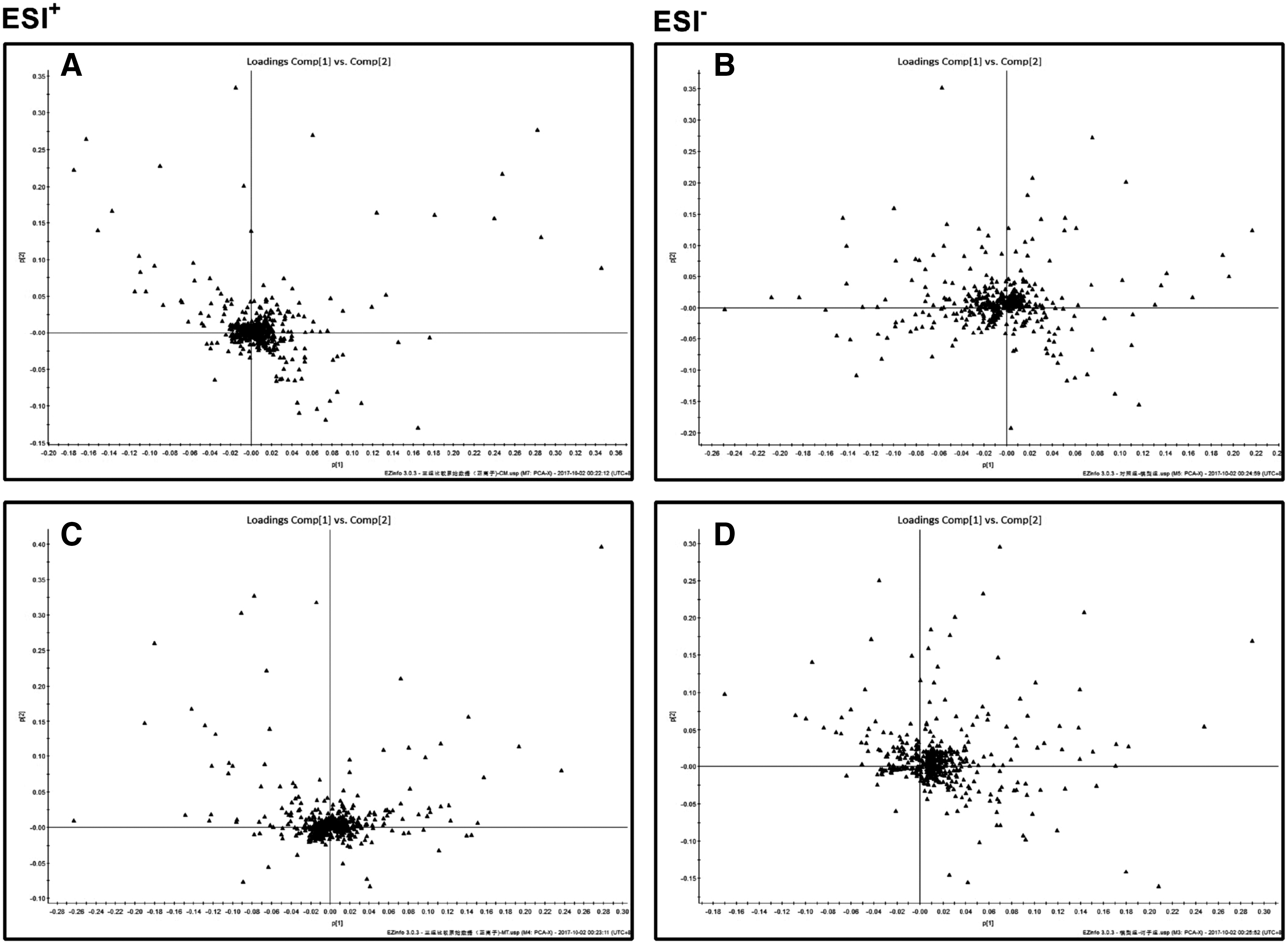
Additionally, PLS-DA and OPLS-DA were further used to maximize differences between groups (Figs. 5 and 6). The results suggested the generation of the MCAO model was successful and that TC has a protecting effect on the model. The OPLS-DA was built to clarify distinctions and potential biomarkers among groups.

Differential profiles in rat serum among the sham, model, and TC groups.
Biomarker elucidation
In the score plot, scattered points of various samples segregated into three groups, which suggested that the proposed OPLS-DA model could reveal proper MCAO-related patterns and that the serum metabolic pattern significantly changed after TC treatment. The loading plot displayed 12 potential MCAO-related metabolites according to their VIP values, which was set to be >1.0, and significance test, with p < 0.05. To identify these metabolites, first candidates were searched for in the HMDB (
In this study, 12 compounds, including phospholipids and amino acids, were tentatively identified by MSE fragmentation data and i-FIT values, as shown in Table 2. Many of these identified metabolites have also been reported in other MCAO studies, such as lactic acid and LysoPC (16:0), while in the current study, the metabonomics approach was employed, and more MCAO-related metabolites were discovered.
Metabolites as Biomarkers Characterized in Serum Profile and Their Change Trends (n = 8 Per Group)
The levels of potential biomarkers are labeled as ↓ (downregulated) and ↑ (upregulated; * p < 0.05).
Change trend compared to the sham group.
Change trend compared to the model group.
LysoPC, lysophosphatidylcholine; VIP, variable importance in projection.
Metabolic pathway and function analysis
A systematic pathway analysis of the metabonomics was carried out to explore the biomarkers. MCAO-associated metabolic perturbation was evaluated using pathway analysis. Based on an impact value >0.1, four perturbed metabolic pathways—valine, leucine, and isoleucine biosynthesis; arachidonic acid metabolism; glycerophospholipid metabolism; and alanine, aspartate arid glutamate metabolism—were revealed (Supplementary Fig. S2A). These pathways may be the potential targets of TC for cerebral ischemic injury. Without TC treatment, the data showed profound derangements of energy metabolism (glycolysis and TCA cycle), amino acid metabolism, glycerophospholipid metabolism, and other types of metabolism (Supplementary Fig. S2B).
Discussion
This study investigated the potential protective effects of TC against MCAO injury in a rat model. TC-treated MCAO rats (TC group) had a smaller cerebral infarct size compared to the untreated MCAO group. Biochemical indicators and metabolic profile analysis (PCA, PLS-DA, and OPLS-DA score plots) also supported the protective effects of TC treatment.
Oxidative damage induces lipid peroxidation, protein degradation, and DNA lesions, leading to cell death.
20,21
The elevated levels of amino acids (
Energy metabolism
After MCAO surgery, glycogen is rapidly depleted and produces a significant amount of lactic acid, resulting from anaerobic glycolysis, and is a marker in ischemic tissue. 3,24 It has been demonstrated that this lactate shift is a compensatory mechanism for generating much less energy from glucose under a local hypoxic condition in stroke. 25 This study found that the levels of lactate in serum were significantly increased in the MCAO group compared to the sham group (Table 2), a finding that is in keeping with the fact that the brain energy supply pattern changed from aerobic glycolysis to anaerobic glycolysis. On the other hand, the level of lactate in the TC group significantly decreased, which could be related to the improvement in energy supply pattern corresponding to the operation of the TCA cycle.
Accordingly, the levels of succinic acid and fumarate, which are in TCA cycle intermediates, decreased in the MCAO group (Table 2), indicating that the rat brain injury could downregulate the TCA cycle. When glycometabolisms reduced, glycolysis occurred rapidly, which might have resulted in increased lactic acid production, observed with the MCAO serum assays. These findings are in agreement with published reports 26,27 in which it was demonstrated that the levels of succinic acid and fumarate in the TC group showed an upward trend, while the level of lactic acid showed a downward trend—an indication of recovery TCA cycle operation and overall energy metabolism. Thus, the increased succinic acid and fumarate levels suggest brain protection against ischemic injury of MCAO rats via anti-inflammatory and antioxidant activities.
Further, it is concluded that anaerobic glycolysis (lactic acid) and TCA cycle (fumarate and succinic acid) indicators were affected, which are directly related to energy metabolism. These results indicate that the therapeutic effects of TC on cerebral ischemia are partially due to its ability to repair energy metabolism.
Amino acid metabolism
Leucine and alanine, which have protective functions for cerebral ischemia, are intermediates in the TCA cycle. 28 In the model group, the level of leucine and alanine showed a downward trend. Leucine and alanine have close links to metabolic pathways such as glycolysis, gluconeogenesis, and the TCA cycle. In this study, the levels of leucine and alanine tended to be normal in the TC group, indicating that TC offers protection against brain damage.
Phenylalanine, one of the essential aromatic amino acids, can be used to produce various proteins in cells of body tissue under normal circumstances. 29 What is more, it can produce fumarate, which is involved in the TCA cyle. 30 The results of the current study show that the content of phenylalanine became higher in the MCAO group. This may be due to brain damage affecting the phenylalanine metabolic pathway. After TC treatment, the level of phenylalanine significantly reduced.
Glycerophospholipid metabolism
Lipids are the primary components of biological membranes. Alterations in the synthesis and degradation of many lipids are considered to be closely related to impairment and repair mechanisms of neurocytes during cerebral ischemia-associated risk of stroke and contribute to the progression of brain disease. 31,32 It is a known fact that the relational protein of LysoPC is involved in several motility-related processes such as angiogenesis and neurite outgrowth. 33,34 In the current experiments, many lipids decreased in the model group compared to the sham group, consistent with a previous study. 29 Specifically, levels of lipids, including phosphorylcholine (PC) and LysoPC, all distinctly reduced in the model group compared to the sham group. However, after TC treatment, the levels of PC (16:0/20:4), PC (20:4/20:4), LysoPC (18:0), and LysoPC (16:0) showed a trend toward normalcy. These results indicate that the therapeutic effects of TC on cerebral ischemia are partially due to repair of glycerophospholipid metabolism. Furthermore, PC has a marked fluidity effect on cellular membranes, and decreased cell-membrane fluidity has been associated with neurological diseases and cell death, thus confirming TC's ability to restore the aberrance membrane fluidity that might have occurred in the model group.
Other metabolism
Arachidonic acid is a metabolite of fatty acids, which could induce IL-6 production in the endothelial and smooth-muscle cells, and contributes to vascular inflammation in the brain in neurological disorders. 35 This type of fat-related brain metabolism disorder not only increased adenosine triphosphate (ATP) consumption while decreasing ATP synthesis, but also increased oxygen consumption in the brain, which speeds up the process of brain dysfunction. Thus, the altered levels of arachidonic acid were overcome by the protective role that TC plays in the ischemic brain.
Palmitoylcarnitine (PLC), a type of long-chain acylcarnitine, was detected at higher levels in the model group compared to the sham group. PLC is an essential intermediate in the degradation of acyl lipid in the mitochondria. It has been proven that long-chain acylcarnitines are involved in the synthesis of complex lipids, interact with neural membranes, and modulate proteins. 27 In additional, carnitine and its derivatives, as endogenous metabolites, were reported to protect brains subjected to oxidative stress. 36 In this study, the TC-treated group displayed a decreased level of PLC compared to the model group, and this might account for the protective effects of the brain subjected to ischemia or oxidative stress.
UPLC-Q/TOF-MS-based serum metabonomic strategy results indicate that the therapeutic effects of TC on MCAO injury are partially due to interference with impaired energy (lactic acid, succinic acid, and fumarate), amino acids (leucine, alanine, and phenylalanine), glycerophospholipid (PC [16:0/20:4], PC [20:4/20:4], LysoPC [18:0], and LysoPC [16:0]) metabolism, and other types (arachidonic acid and palmitoyl carnitine) of metabolism (Fig. 6B), which were consistent with results of the current study and a previous review, and demonstrate the anti-inflammatory and anti-oxidative effects of TC.
Conclusion
Combining pharmacodynamics with UPLC-Q/TOF-MS analysis, the metabolomics approach was used first to evaluate the protective effects of TC on ischemic stroke. The identified metabolites were related to a variety of metabolic pathways, including energy metabolism, amino acid metabolism, and lipid metabolism. However, this metabonomics study did not determine which type of cell in brain or the signal pathway was involved in the neuroprotective effects of TC. The exact underlying mechanism by which TC alleviates cerebral ischemic injury should be clarified in the future. The findings suggest that TC could ameliorate MCAO rats by intervening in energy metabolism (glycolysis and the TCA cycle), amino acid metabolism, glycerophospholipid metabolism, and other metabolisms.
Footnotes
Acknowledgments
The research is funded by the National Natural Science Foundation of China (no. 81603385; no. 81673631; no. 81501003; no. 81601149), the Science Foundation of Shaanxi Province (no. 2017JM8006), and the China Postdoctoral Science Foundation (2015 M580465).
Author Disclosure Statement
No competing financial interests exist.
References
Supplementary Material
Please find the following supplemental material available below.
For Open Access articles published under a Creative Commons License, all supplemental material carries the same license as the article it is associated with.
For non-Open Access articles published, all supplemental material carries a non-exclusive license, and permission requests for re-use of supplemental material or any part of supplemental material shall be sent directly to the copyright owner as specified in the copyright notice associated with the article.
