




Abstract
Crude ethanolic extracts from Phlomis viscosa Poiret leaves from the Judea region (Israel) are renowned for their remarkable geroprotective properties: anti-inflammatory, anti-diabetic, and anti-cancer. A phytochemical investigation carried out in this study revealed that the tested plant might belong to a particular distinct chemotype because its phytochemicals are different from compounds that were mentioned in the literature. Among the compounds identified by us was diosmin, the synthetic derivatives of which were further obtained and investigated. In particular, activities of the isolated compounds and synthesized diosmin derivatives were assessed. Our results revealed that the following compounds significantly lessened secretion of some pro-inflammatory cytokines: diosmin, himachala-2-diene, and 5,7-dihydroxy-2-(3-hydroxy-4-methoxyphenyl) chromen-4-one. In addition, diosmin, synthesized diosmin derivatives, and some identified terpenes were found to have anti-diabetic activities. A significant anti-cancer effect of the whole extract on U-87 (human glioblastoma carcinoma cells line) and MCF7 (human breast carcinoma cell line) was also demonstrated, and it was better than that of DOX (doxorubicin). Collectively, the results obtained in the in vitro models suggest a wide spectrum of beneficial bioactivities of the extract and its active compounds.
Introduction
Around 173 million people suffered from type 2 diabetes mellitus (noninsulin-dependent diabetes mellitus [NDDM]) in 2000, and the global number of people with this disease is estimated to increase to 370 million in 2030. 1 This disease is characterized by a strong inflammatory component 2 and is age related. Thus, there is an ever-increasing need for novel and improved anti-inflammatory and anti-diabetic agents. Current therapeutic strategies for NDDM are limited and are centered on insulin and four main classes of oral anti-diabetic agents that stimulate pancreatic insulin secretion, reduce hepatic glucose production, delay digestion and absorption of intestinal carbohydrate, or improve insulin action. 3 Each of these drugs has generally inadequate efficacy and a number of serious adverse effects. Characteristic symptoms of NDDM include hyperglycemia, chronic inflammation, and insulin resistance. In addition, some studies demonstrated that excess lipid accumulation (an acknowledged risk factor for diabetes) may result in impaired insulin signaling through the induction of inflammation and the subsequent production of inflammatory cytokines by macrophages, which, in turn, impairs insulin action. 4
The incidence and morbidity of another age-related disease cancer was 14.1 and 8.2 million, respectively, in 2012 worldwide. 5 Doxorubicin (DOX) is widely used in modern anti-cancer therapy because this drug is able to inhibit tumors but its low drug utilization and strong toxicity to organs limit its application. 6
Thus, no option should be neglected in searching for new effective anti-cancer, anti-inflammatory, and anti-diabetic agents to improve human well-being and fight aging. From this perspective, application of herbal extracts and their phytochemicals could be an excellent source of these new effective agents. The flora of some regions is known to be exposed to highly stressful conditions that cause local herbs to accumulate relatively high levels of bioactive compounds. The Judea region in Israel is one of such stressful phytogeographic zones because it is situated between the Judean Hills, with an elevation of up to 1000 m above sea level, a rainy Mediterranean climate, and the Dead Sea, which is the lowest place on earth (more than 400 m below sea level), with constantly warm and dry conditions. As a result of this permanent stress, on a narrow strip of land of only 30 km in width, both endemic and widely distributed Mediterranean plants growing in the area have acquired unique chemotypes with various medicinal uses. 7 –11
In this study, we have isolated and identified some active compounds from Phlomis viscosa Poiret from the Judea region (Israel). It is an ever-green shrub that grows only in Israel, Turkey, Lebanon, and Syria. 12 Although pro-wound healing properties of P. viscosa from the Judea region (Israel) were reported, 10 its phytochemicals have not yet been identified.
Screening of the various plant extracts from the Judea region for the anti-diabetic, anti-cancer, and anti-inflammatory activities revealed the most potent effect of P. viscosa and prompted this study. 10 Here, we present the results of our phytochemical investigation. All the determined phytochemicals have been a subject of intensive research aimed at examining their anti-inflammatory and anti-diabetic properties. In addition, synthetic diosmin derivatives were obtained and investigated.
Materials and Methods
Compounds
Diosmin, 1-octen-3-ol, himachala-2,4-diene, and etoposide were purchased from S.l. Moran (Israel). High-performance liquid chromatography (HPLC) solvents were purchased from Merck. The following compounds were purchased from Tzamal D-Chem Laboratories (Israel): germacrene D, β-Caryophyllene, alloaromadendrene, humulene, diosmin, 1-octen-3-ol, himachala-2,4-diene, Isovaleraldehyde, 2,4-hexadienal, and 2-hexenal.
Preparation of plant material
The tested plants were collected from the Hebron Hills region near Moshav Carmel (Israel). Leaves, flowers, and stems of P. viscosa were dried by lyophilization and ground for gas chromatography/mass spectrometry (GC/MS) analysis. Ethanol extract was prepared from leaves, flowers, and stems of P. viscosa. Plant extract preparation and its fractionation was performed as previously described. 13
High-performance liquid chromatography
Gradient elution was performed with solution A, composed of water–acetic acid (97:3 V/V) and solution B, composed of methanol, as previously described. 12 A UV detector at 360 nm with a reverse-phase column (Betasil C-18, 5 μm, 250 × 0.46 mm; Thermo-Hypersil, United Kingdom) was used.
Liquid chromatography-mass spectrometry instrumentation and conditions
LC-MS Agilent 1100LC series (Waldbronn, Germany) and Bruker Esquire 3000plus MS (Bremen, Germany) instrument consisting of a C18 column (Betasil C18, 5 μm, 250 × 4.6 mm; Thermo- Hypersil) and methanol–water as the mobile phase (see section “High-performance liquid chromatography” in the method above) were used. The UV detector was set at 360 nm, the flow rate at 1 mL/min, and the injection volume at 10 μL. The MS conditions were optimized as follows: atmospheric pressure ionization electron spray interface, negative mode polarity, a drying gas flow of 10 L/min, a nebulizer gas pressure of 60 psi, a drying gas temperature of 335°C, a fragmentor voltage of 0.4 V, a capillary voltage of 4451 V, and a scan range of m/z 25–1000, at 1.15 s/scan.
GC/MS instrumentation and conditions
Identification of volatile compounds was performed according to the GC principles. 14 Specifically, GC/MS, a Varian CP 3800 GC-MS analytical system, was used. Headspace injection mode was used to perform qualitative analysis without extracting active compounds. Five grams of the given sample was placed in a headspace vial and sealed by a magnetic cap with septum (Chromofor).
The sample heating and shaking were performed at a temperature of 95°C, and equilibrium time was 1 hour. The headspace content was injected into the chromatograph; the injector temperature was fixed at 250°C. The GC was equipped with the ZB-5 column operating with helium as carrier gas at a constant flow of 2.0 mL/min.
A modified (data collected and updated for 5 years on the basis of the current experience) analytical library (National Institute of Standards and Technology [NIST] standard reference database) was applied. Probability of compound identification was measured by comparing its spectrum with those found in the NIST library.
Chemical synthesis
Diosmetin [5,7-dihydroxy-2-(3-hydroxy-4-methoxyphenyl)Chromen-4-one] was obtained by acid hydrolysis of diosmin; 3′,5-dihydroxyethyl-diosmine was obtained by alkylation of diosmin by bromethanole in the presence of cesium carbonate in methyl ethyl ketone medium.
Cell cultures
Cell culture reagents: Dulbecco's modified Eagle's medium (DMEM), fetal calf serum (FCS), l-Glutamine, and antibiotics were obtained from Biological Industries (Beit Haemek, Israel).
Human pulmonary fibroblasts (HPFs), U-87 (human glioblastoma carcinoma cells line), MCF7 (human breast carcinoma cell line), HEK293 (primary human embryonal kidney cell line), NIH/3T3 (Mus musculus, mouse embryo fibroblasts), and 3T3-LI adipocytes were grown in DMEM supplemented with 10% FCS, 1% l-glutamine, and 1% penicillin/streptomycin/nystatin and incubated at 37°C in humidified air containing 5% CO2. After 3T3-LI cells approached confluency, they were grown in differentiation media containing 100 nM insulin, 500 nM dexamethasone, and 0.5 mM isobutylmethylxanthine. Then, 3T3 cells were plated at a concentration of 105 cells × 1 mL on 96-well plates and differentiated. After 7–10 days, mature adipocytes were identified based on their morphology and used for study.
Cytotoxicity examination
Nontoxic concentrations of the herbal extracts, their fractions and compounds were determined by the (2,3-Bis-[2-Methoxy-4-Nitro-5-Sulfophenyl]-2H-Tetrazolium-5-Carboxanilide) assay as previously described. 15 Briefly, after the 3-day incubation period, 25 μL of 1 mg/mL XTT solution containing 0.2 mM phenazine methosulfate was added, and the cells were incubated for an additional 1 hour at 37°C. The OD values were measured by using SpectraMax Paradigm multi-mode detection platform (Molecular Devices, Sunnyvale, CA) at 450 nm with a reference wavelength of 650 nm.
According to the obtained results, the CC50 (a concentration that causes 50% toxicity) of the tested compounds was determined.
Treatments
Old or young HPFs were treated with specified concentrations of different plant extracts, their fractions or compounds for 3 days. After that, the medium was collected and the concentrations of interleukin-6 (IL-6) and IL-8 were measured by using ELISA kits according to the protocols of R&D Systems. Standard curves were generated for each plate to determine sample concentration.
Absorbance was determined by using SpectraMax Paradigm multi-mode detection platform (Molecular Devices), and data were analyzed by using GraphPad Prism software (version 6; GraphPad Software, La Jolla, CA).
DOX (doxorubicin) served as a positive control in experiments with U-87 (human glioblastoma carcinoma cells line), MCF7 (human breast carcinoma cell line), and HEK293 (primary human embryonal kidney cell line). The effect of extracts on cell viability was examined compared with the control NIH/3T3 cell line (noncancerous cells). The viability assay was performed by using the CellTiter Fluor Cell Viability Assay Kit® (Promega), according to the manufacturer's instructions. This assay measures protease activity within live cells and, therefore, serves as a marker of cell viability.
The 3T3-LI fibroblasts were grown in DMEM containing 10% FCS. After cells approached confluency, they were grown in differentiation media containing 100 nM insulin, 500 nM dexamethasone, and 0.5 mM isobutylmethylxanthine. Then, 3T3 cells were plated at a concentration of 105 cells per mL on 96-well plates and differentiated. After 7–10 days, mature adipocytes were identified based on morphology and used for study. Differentiated adipocytes were transferred to low-glucose serum-free media overnight before treatment with extracts or _insulin for 4 h. After incubation, 200 μM of the fluorescent glucose analog 2-(N-(7-nitrobenz-2-oxa-1,3-diazol-4-yl)amino)-2-deoxyglucose (Invitrogen, Carlsbad) was added for 1 h. Cells were subsequently rinsed in cold phosphate-buffered saline, and fluorescence was measured with a fluorescence microplate reader (BMG Polarstar, Ortenberg, Germany).
Statistical analyses
All data were analyzed by using Statistica for Windows software (StatSoft, Inc., Tulsa, OK). p < 0.05 was chosen as the minimal acceptable level of significance.
Results and Discussion
Therapeutic features and phytochemicals of many species from the genus Phlomis are well known, 15 but those of P. viscosa were not investigated in depth. β-Caryophyllene, germacrene D, alloaromadendrene, and humulene were identified in P. viscosa from Turkey. 10 Our first hypothesis was that the compounds mentioned earlier might be also found in the ethanolic extract of the Israeli chemotype, and possibly that they stand behind its anti-diabetic and anti-inflammatory properties. The second hypothesis was that the tested plant might contain other unique active phytochemicals. To test the first hypothesis, a comparison with commercially available standards of β-Caryophyllene, germacrene D, alloaromadendrene, and humulene was performed; however, it did not confirm their presence in the tested plant. Identification of diosmin in the extract was performed by using liquid chromatography-mass spectrometry as confirmed by comparing it with commercially available standard compounds (Table 1). Purchased diosmin standard with retention times of 23.45 was compared with a pick of flavonoid fraction that had a similar retention time; the analytical spike test confirmed the presence of diosmin. Volatile compounds were identified by GC/MS. Table 1 summarizes the results of the analytical part of the research.
Identification of Major Compounds of Phlomis viscosa
HPLC, high-performance liquid chromatography; LC-MS, liquid chromatography-mass spectrometry; GC/MS, gas chromatography-mass spectrometry.
The identified phytochemicals clearly showed that our second hypothesis about the unique chemotype of the tested plant was true. Although the pro-wound healing activity of P. viscosa has already been reported, 10 many other important beneficial properties of the Israeli chemotype and its associated compounds have not been investigated so far.
An important indicator of aging and age-related diseases is activation of pathways leading to chronic inflammation. 17 Pro-inflammatory cytokine interleukins, IL-6 and IL-8 are among the key mediators of inflammation, during which their levels increase. 16 Although some medicinal plants from the Judea region were screened for potential geroprotective activities, 10 many plants from this region have not yet been investigated.
In the framework of this study, we considered the impact of some plant extracts such as Helichrysum angustifolium, Calotropis procera, Ephedra aphylla, P. viscosa (leaves, and flowers), and other (data not shown) on the secretion of IL-6 and IL-8 by etoposide-treated versus control fibroblasts. It is known that etoposide is able to turn young cells into senescent ones and this process is accompanied by elevated proinflammatory cytokines such as IL-1, IL-6, IL-8, and tumor necrosis factor alpha (TNF-α). 18 Figure 1 demonstrates that P. viscosa extracts significantly decreased secretion of IL-6 and IL-8 (p < 0.001).
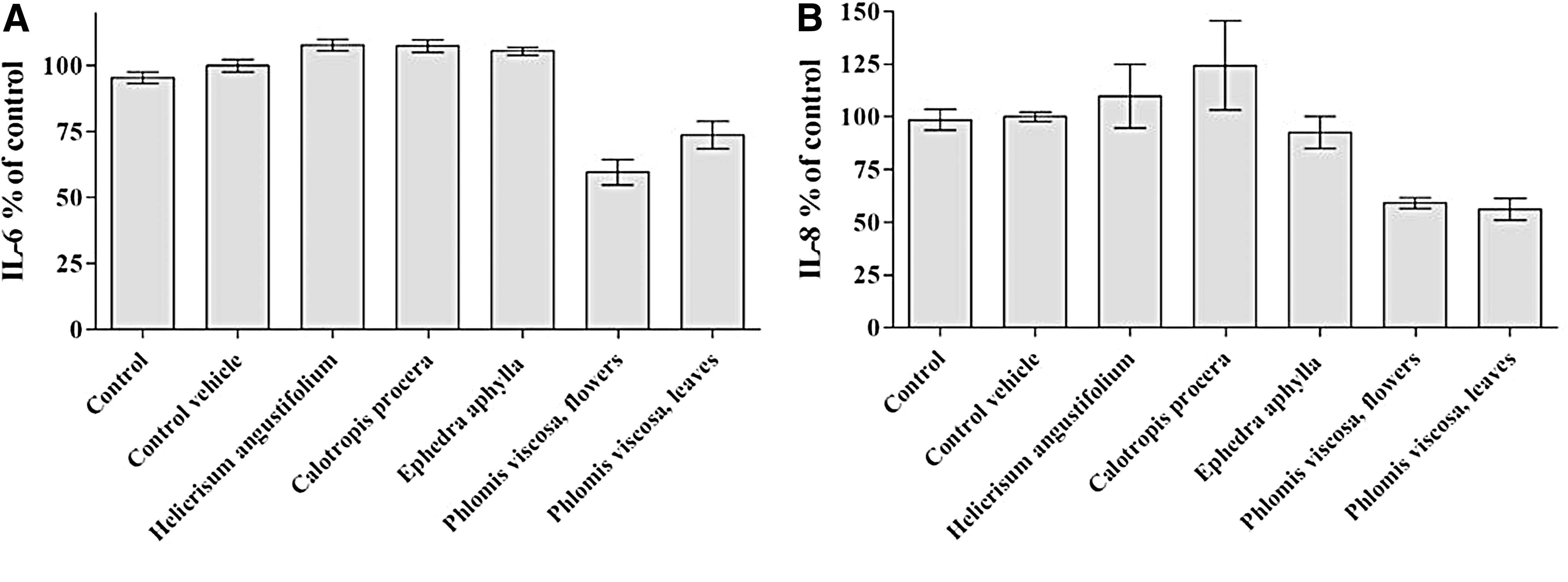
Effect of plants extracts on the secretion of pro-inflammatory cytokines, IL-6
The identified phytochemicals of P. viscosa (Table 1) and synthetic diosmin derivatives mentioned earlier were investigated from the perspective of chronic inflammation. The following compounds were tested: number 1 (diosmin), number 2 (1-octen-3-ol), number 3 (3′,5-dihydroxyethyl-diosmine), number 4 (himachala-2,4-diene), number 5 (Isovaleraldehyde), number 6 (2,4-hexadienal), and number 7 (5,7-dihydroxy-2-(3-hydroxy-4-methoxyphenyl)chromen-4-one) (Fig. 2). Only diosmin, himachala-2,4-diene, and 5,7-dihydroxy-2-(3-hydroxy-4-methoxyphenyl)chromen-4-one significantly (p < 0.001) decreased secretion of pro-inflammatory cytokines (Fig. 2). The last synthetic compound was more effective than diosmin in reducing secretion of pro-inflammatory cytokines, in particular in the case of IL-8 (Fig. 2B). Anti-inflammatory properties of diosmin are in agreement with the fact that in case of ethanol-induced hepatic injury diosmin managed to modulate inflammation by alleviating ethanol-induced nuclear factor-kappaB (NF-κB) activation, and enhancing expression of TNF-α, cyclooxygenase-2 and inhibitor, and nitric oxide synthase. 19 Although 5,7-dihydroxy-2-(3-hydroxy-4-methoxyphenyl)chromen-4-one (Diosmetin) was identified in some plants, its concentrations were below the detectable levels by ultra-high performance liquid chromate graphy-quadrupole time of flight mass spectrometry, 20 so the market price of 5,7-dihydroxy-2-(3-hydroxy-4-methoxyphenyl)chromen-4-one is very expensive, and, thus, we fulfilled the synthesis of this compound.
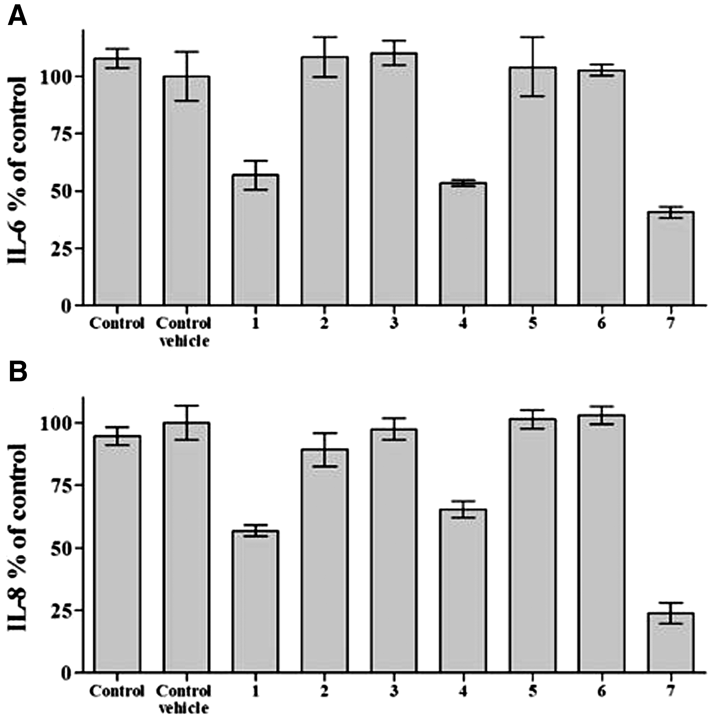
Effect of herbal and synthesized compounds on the secretion of pro-inflammatory cytokines, IL-6
We expected to identify the influence of diosmin on glucose uptake of the 3T3 adipocytes since it was reported that diosmin modulated the NF-κB signal transduction pathways and downregulation of some oxidative stress markers in alloxan-induced diabetic nephropathy. 21 Our results confirmed this notion. Adipocytes were treated with various ethanolic extracts of P. viscosa, identified and synthesized compounds. Figure 3 demonstrates that diosmin and its synthetic derivatives were more effective than crude extracts in stimulating glucose update. Altogether, all tested extracts and compounds significantly (p < 0.001) increased glucose uptake of the 3T3 adipocytes.

Anti-diabetic activity of plant extracts and several compounds. The glucose uptake of the 3T3 adipocytes was determined. Negative control consisted of untreated adipocytes (1); insulin (2) was used as a positive control. Adipocytes were treated with diosmin (1), 5,7-dihydroxy-2-(3-hydroxy-4-methoxyphenyl)chromen-4-one (2), 3′,5-dihydroxyethyl-diosmine (3), 1-octen-3-ol (4), himachala-2,4-diene (5), leaf ethanol extracts of P. viscosa (6), flower ethanol extracts of P. viscosa (7), and stem ethanol extracts of P. viscosa (8). Data from three independent experiments are shown (mean ± SD).
With regard to its anti-cancer properties, Figure 4 demonstrates that P. viscosa extracts significantly decreased the viability of cancer cell lines at concentrations of 300 and 200 μm (p < 0.001) in comparison to NIH/3T3 (noncancerous cells). We have found that P. viscosa actually causes the death of cancer cells, which are known to have high resistance to chemotherapy. We have also examined the effect of P. viscosa extract on additional cancer cell lines. We found that the P. viscosa extract causes death of the brain cancer cells (U87) even more efficiently than the DOX chemotherapeutic agent. A possibility to obtain more effective and less toxic drugs than the DOX ensued. Thus, the investigation of phytochemicals derived from P. viscosa extract warrants further study.
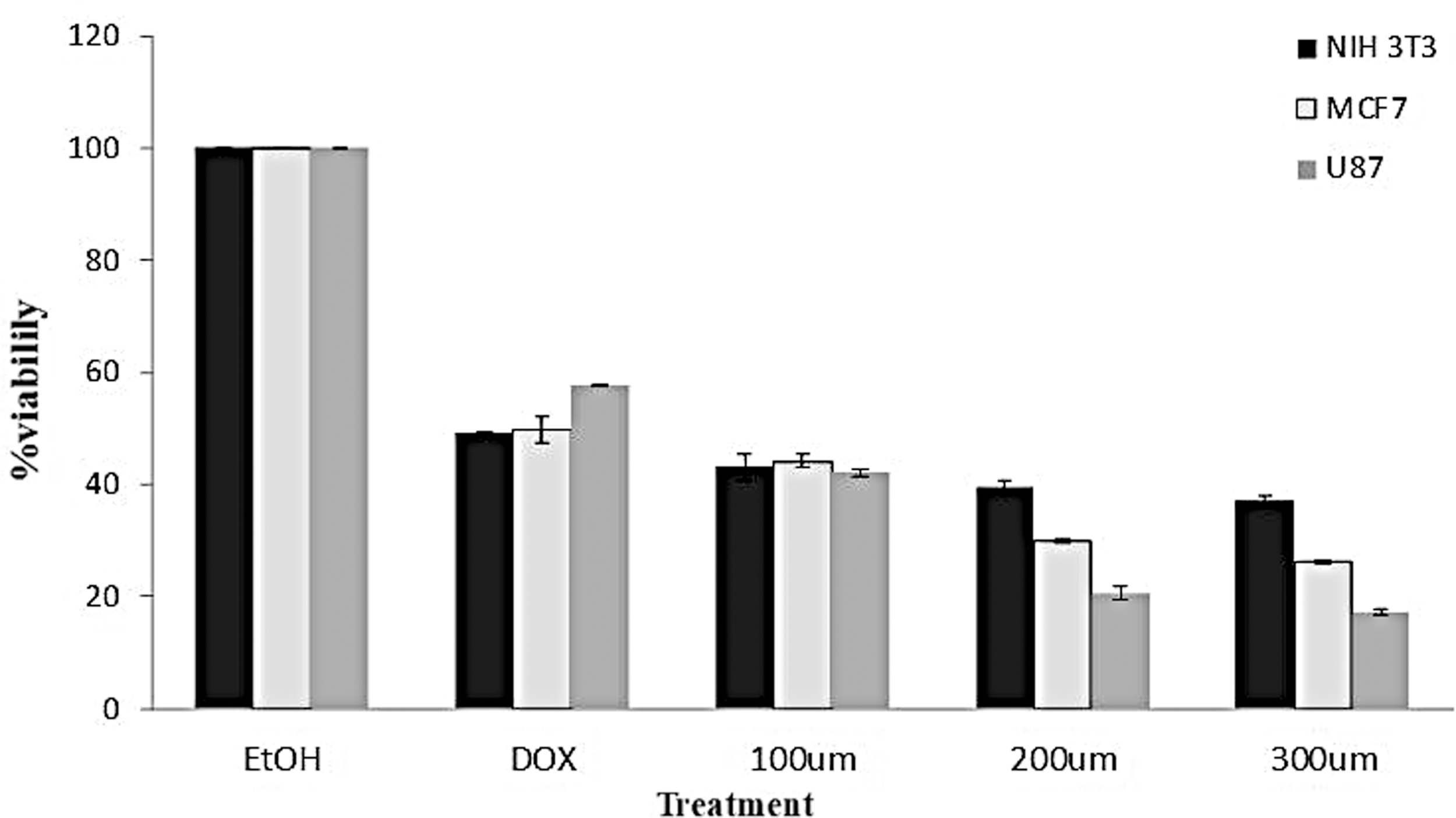
Anti-cancer activity of P. viscosa (leaves) extracts. The effect of the extract at concentrations of 300, 200, and 100 μm on MCF7, U87 (cancer cell lines) viability was examined as compared with NIH/3T3 (noncancerous cells). Doxorubicin at a concentration of 20 μm served as a positive control. Data from three independent experiments are shown (mean ± SD).
Conclusions
In conclusion, various bioactivities of the tested extract and compounds demonstrate their geroprotective potential. Nevertheless, the molecular mechanisms involved in the action of the tested compounds remain to be investigated and should be the focus of future studies. These results obtained on the in vitro models suggest a mechanistic basis for the potential use of plant phytochemicals and diosmin synthetic derivatives in the treatment of many chronic age-related diseases.
Footnotes
Acknowledgments
This study was supported by the Israeli Ministry of Science, Technology, and Space, Grant Number 3/13584. The authors are grateful to Dr. Yulia Solomonov for her assistance in data analysis and preparation of the figures.
Author Disclosure Statement
No competing financial interests exist.