
Abstract
Hydroxysafflor yellow A (HSYA), an active component from Chinese medicinal herb, has been applied to the prevention and treatment of cerebral ischemia/reperfusion injury (CIRI). To clarify the comprehensive mechanisms HSYA for stroke, we used label-free quantitative proteomic analysis to investigate the modulated proteins of rats subjected to CIRI and their alteration by HSYA. Neurological examination, infarct assessment, and biochemical assay were performed to validate the effects of HSYA, and the results indicated that HSYA played a significant role in brain protection. A total of 13 proteins were identified as overlapped proteins by label-free quantitative proteomic analysis. Gene Ontology and pathway analysis showed that these differentially expressed proteins were mainly enriched in the hypoxia-inducible factor 1 (HIF-1) signaling pathway. Furthermore, networks were constructed with respect to protein function interactions. The results suggested that seven proteins were identified as hub proteins between model and sham groups, while 25 proteins were identified as hub proteins between HSYA and model groups. In addition, the expressions of three overlapping proteins were validated by Western blot, and their levels were consistent with the results of label-free analysis. In conclusion, Eftud2, mTOR, Rab11, Ppp2r5e, and HIF-1 signaling pathways have been detected as key hub proteins and pathways in HSYA against CIRI through proteomic analysis. Our research has provided convincing explanations for the mechanism of HSYA against CIRI and the identified key proteins and pathways might provide novel therapeutics for CIRI.
Introduction
Ischemic stroke is a devastating neurological disorder with high morbidity and severe mortality. 1 It accounts for ∼80% of stroke deaths in the world. Ischemia is caused by sudden interdiction of blood flow to the lesioned area, which initiates a train of metabolic events and eventually leads to ischemic injury. 2 In addition, the reperfusion after transient cerebral ischemia could aggravate brain injury. Since the pathophysiological processes in cerebral ischemia/reperfusion injury (CIRI) are complex and extensive, it is a huge challenge for researchers and doctors to find effective treatment for it. The contemporary thrombolytic therapy for ischemic stroke or CIRI in clinical practice is not satisfactory due to its extremely narrow therapeutic time window following the infarct. 3 Therefore, it is essential to find a potential compound that can effectively relieve CIRI and elucidate its therapeutic mechanism.
Hydroxysafflor yellow A (HSYA, molecular formula, C27H32O16; molecular weight, 612.53 g/mol) is one of the active ingredients of Carthamus tinctorius L., which has been predominantly used for activating blood and dissolving stasis. 4 HSYA has been widely used in clinical practice in China for the prevention and treatment of ischemic injury. The value of safflower yellow injection for the treatment of acute cerebral infarction has been investigated by a randomized controlled trial. 5 HSYA can resist inflammation, oxidation, and apoptosis, improve neurological function, alleviate infarct volume, inhibit thrombosis formation and platelet aggregation, reduce brain edema, and protect blood vessel. 6 –9 Furthermore, the mechanism of HSYA on cardiovascular disease has been analyzed on the basis of metabolomics and genomics. 10,11 Currently, neuroproteomic studies have offered new insight into the molecular mechanism underlying CIRI. 12 With the rapid development of proteomics and its wide application to medical research, it is feasible for us to discover the distinctive features of HSYA against CIRI and investigate its mechanism through proteomic analysis.
In recent years, many researches have investigated the mechanism of some traditional Chinese medicinal herbs against cerebral ischemia through proteomic analysis. For example, curcumin showed the function of regulating the expression of various proteins in cerebral ischemia based on two-dimensional gel electrophoresis and mass spectrometry. 13 Gastrodia elata displayed obvious effects on brain tissues in rats by using isobaric tags of relative and absolute quantification technique. 14 The findings have demonstrated that proteomic analysis could reveal the mechanism of the differences between normal and injured brains in rats through the treatment of such traditional Chinese medicinal herbs as curcumin and Gastrodia elata.
In the present study, we aim at analyzing the differentially expressed proteins (DEPs) of HSYA against CIRI by label-free quantitation and bioinformatic techniques to determine the multieffects of safflower constituents on stroke with blood stasis. First, a rat model of middle cerebral artery occlusion and reperfusion (MCAO/R) was established to simulate CIRI. Second, the pharmacological effects of HSYA on rats were evaluated. Third, label-free quantitation techniques were used to identify the DEPs of HSYA against CIRI. Finally, bioinformatic analysis was conducted to reveal their effects and mechanism.
Materials and Methods
Animals
Adult male Sprague-Dawley rats (280 ± 20 g) were obtained from the Experimental Animal Center of the Air Force Medical University (Xi'an, China). All the experimental rats have been approved by the Ethics Committee for Animal Experimentation of the Air Force Medical University and in compliance with the National Institutes of Health (NIH) Guide for the Care and Use of Laboratory Animals. They were housed in air-conditioned animal quarters in a constant 12-hour light/dark cycle at 25°C ± 2°C temperature and 50% ± 10% air humidity, with free access to pellet food and water.
Establishment of CIRI model
After 7 days of acclimatization, the rats were randomly divided into three groups: sham operation group (Sham), MCAO/R model group (Model), and HSYA treatment group (HSYA). The rats in Model and HSYA groups were operated to establish the MCAO/R model as previously described. 15 In brief, the rats were anesthetized with 5% isoflurane (diluted in 30% oxygen and 70% nitrous oxide). The right side of common carotid artery and external carotid artery (ECA) was exposed and then the right internal carotid artery (ICA) was isolated. A 4–0 monofilament nylon suture (Doccol, MA) was used to occlude the right middle cerebral artery from ECA to ICA. The nylon was slowly withdrawn 2 hours after the occlusion to induce reperfusion injury. The rats in the Sham group were operated to establish MCAO with the same procedure except the monofilament insertion.
HSYA administration
HSYA (purity >95%; Mansite Bio-technology, China) was dissolved in 0.9% sodium chloride and stored at 4°C. The rats in the HSYA group were administered intraperitoneally with 6 mg/kg HSYA at 12 hours and at 30 minutes before MCAO operation. The dosage for HSYA was chosen based on our previous study on HSYA. 16 Then, they were treated with the same dosage of HSYA every 12 hours immediately after the onset of reperfusion. The rats in both Sham and Model groups were injected with equal doses of vehicle, respectively.
Neurological examination and infarct assessment
Neurological deficits of the rats were blindly evaluated at 72 hours after reperfusion according to Zea Longa's 5-point scale system. 17,18 The neurological deficiency scores are defined as follows: 0 means no neurological deficit, 1 means being unable to extend left forelimb fully, 2 means circling to the contralateral side, 3 means falling to the affected side, and 4 means being with no spontaneous walk or in a comatose state. Soon after the neurological deficiency examination, the rats were deeply anesthetized and their brains were quickly removed for infarct volume assessment. The brains were cut into coronal sections at 2 mm, immersed in 1% 2,3,5-triphenyltetrazolium chloride (TTC; Sigma-Aldrich, St. Louis, MO) at 37°C for 30 minutes, and then fixed with 4% paraformaldehyde. The infarct volume was measured with Image Pro Plus 7.0 by a blinded investigator. The percentage of infarct volume was calculated by the following equation: ([total contralateral hemispheric volume] − [total ipsilateral hemispheric stained volume])/(total contralateral hemispheric volume) × 100%.
Biochemical assay and ELISA
The rats were sacrificed and their brains were removed rapidly after 72 hours of reperfusion. The cortex samples of the cerebral ischemic hemispheres were weighed. Homogenates were centrifuged at 2000 g for 15 minutes, and the supernatant obtained was used for measurement according to the following procedures. The levels of such oxidative stress factors as superoxide dismutase (SOD), malondialdehyde (MDA), and glutathione peroxidase (GSH-PX) were measured by commercially available assay kits (Jiancheng, Nanjing, China). 19 Inflammatory factors such as tumor necrosis factor-α (TNF-α), interleukin-1β (IL-1β), and interleukin-6 (IL-6) were detected by commercial ELISA kits (Boster, Wuhan, China). The experimental procedures were followed in accordance with the instructions designated by the manufacturer.
Label-free quantitation analysis
Label-free quantitation techniques were used to screen the dysregulated proteins of HSYA against CIRI through the Q-Exactive proteomics platform (Thermo Fisher Scientific, Waltham, MA). The proteome was estimated using peak area method according to the manufacturer's instructions. 20 First, the peptide mixtures from the brain proteins were separated by HPLC system and then analyzed by data-dependent tandem mass spectrometry (MS/MS) acquisition with a mass range of 300 to 1500 m/z. Second, precursor ion areas were extracted at 10 ppm mass precision with m/z and retention times recorded for precursor area quantification through Proteome Discoverer 1.4 (Thermo Fisher Scientific). Finally, the area of each protein was normalized by a number of potential peptides of given proteins. Three biological replicates with a total of 12 MS runs in each group were performed for biological reliability.
Bioinformatic analysis
Bioinformatic analysis was performed to explore the functions of the obtained DEPs. All the proteins were uploaded to GENECODIS (
Western blot analysis
Brain tissues were harvested as soon as the rats were sacrificed after 72 hours of reperfusion. Total proteins were extracted from the brains, and protein concentration was determined by a bicinchoninic acid protein assay kit (Beyotime, Shanghai, China). Protein samples (60 μg) were separated by 10% sodium dodecyl sulfate/polyacrylamide gel electrophoresis and then transferred to poly vinylidene fluoride membranes. The membranes were incubated with such primary antibodies as anti-mTOR (1:500; Abcam), anti-Rab11 (1:1000; CST), and anti-Ppp2r5e (1:1000, GeneTex) at 4°C overnight after they were blocked with 5% nonfat milk at room temperature for 1 hour. Then, the blots were incubated with a peroxidase-conjugated secondary antibody at room temperature for 90 minutes and visualized with ECL-Plus reagent. The relative protein levels were determined by Image Pro Plus software (IPP 6.0; Media Cybernetics, MD).
Statistical analysis
Statistical analysis was carried out with SPSS version 19.0 (SPSS, Inc., Chicago, IL). Data are presented as mean ± standard deviation, except for the neurobehavioral score. Statistical differences among different groups were analyzed through analysis of variance followed by the Student-Newman-Keuls test. Neurological deficit scores were presented as the median (interquartile range) and were assessed by the Mann–Whitney U test. A p < 0.05 was regarded statistically significant.
Results
HSYA improved neurological deficit and reduced infarct volume
The neurological deficit scales in the Model group were significantly increased compared with Sham group. Administration of HSYA remarkably improved the neurological deficit of the MCAO rats (Fig. 1A). Furthermore, TTC staining results showed that no injured area was found in the Sham-operated group, while an extensive lesion was found in the Model group. However, compared with the Model group, the area of cerebral infarction in HSYA group was obviously decreased (Fig. 1B).
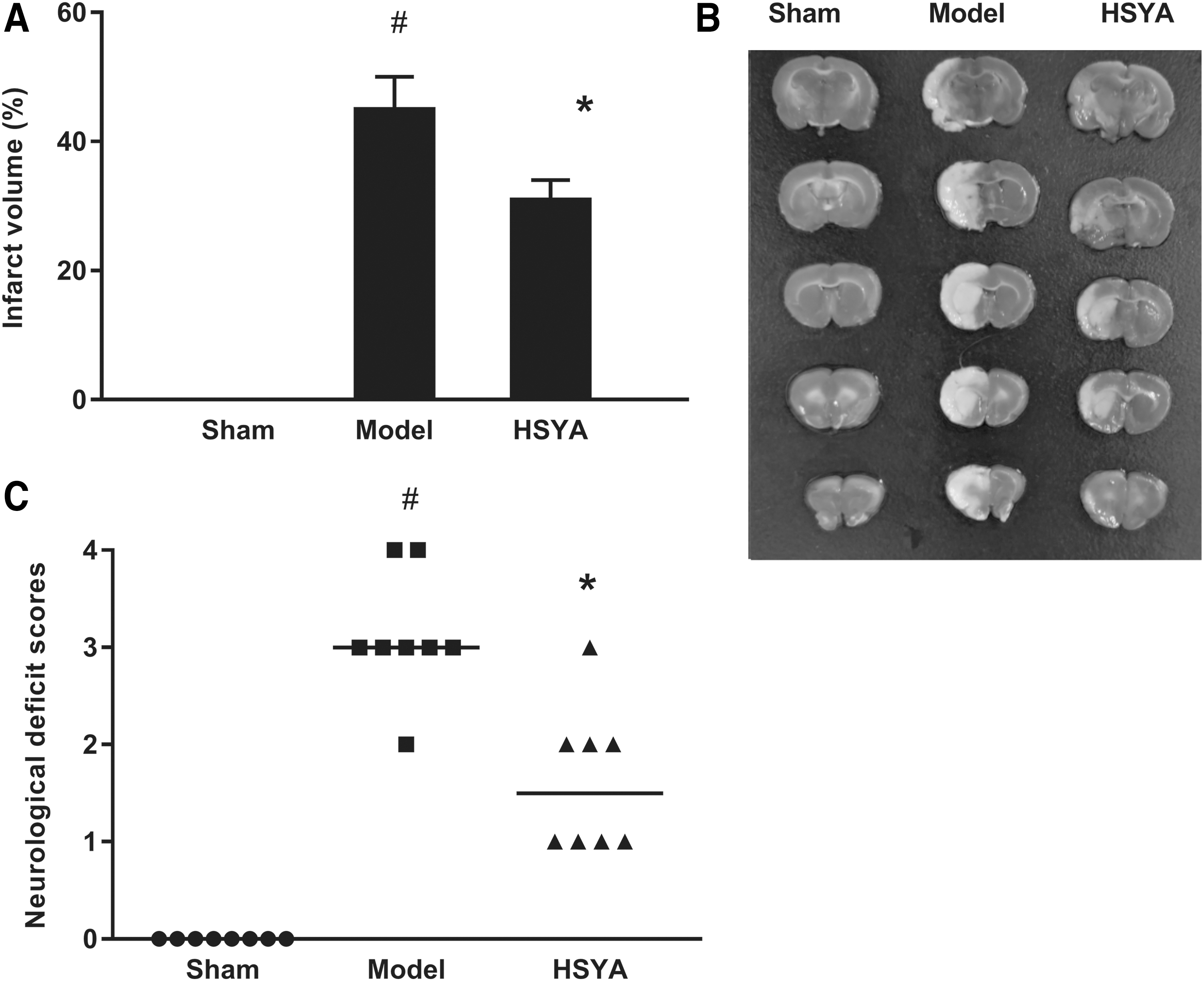
Effect of HSYA on brain injury induced by MCAO/R.
HSYA reduced oxidative stress and inflammatory reaction
The effects of HSYA on oxidative stress and inflammatory reaction were obvious in our experiment. As shown in Figure 2, the release of proinflammatory cytokines in ischemic brain, including TNF-α, IL-1β, and IL-6, was increased markedly in Model group compared with Sham group, which was decreased by HSYA treatment. In addition, the levels of SOD and GSH-Px were significantly decreased, while MDA was increased in Model group, but these changes were inversed by HSYA treatment. These findings demonstrated that HSYA could exert notable effects on CIRI.

Effect of HSYA on proinflammatory cytokines
Identification of DEPs related to HSYA treatment
The label-free analysis results showed that 1982 DEPs were screened from three groups, including 828 proteins in Sham group, 357 proteins in Model group, and 797 proteins in HSYA group. Thirteen of them were overlapped proteins, including DBI, AcsI1, Ppp2r5e, Pabpc4, HK1, Eno2, Gabbr1, Prkcd, Atp1a2, Camk4, mTOR, Rab11b|Rab11a, and Gfap, which were identified based on their t-test values and density difference >1.5-fold (Table 1). Compared with Sham group, five DEPs belonged to the Model group, such as two upregulated proteins (HK1 and Eno2) and three downregulated proteins (mTOR, Rab11b|Rab11a, and Gfap). In addition, compared with Model group, 10 DEPs belonged to HSYA group, such as 5 upregulated proteins (DBI, AcsI1, HK1, Eno2, and Camk4) and 5 downregulated proteins (Ppp2r5e, Pabpc4, Gabbr1, Prkcd, and Atp1a2).
The Overlapping Proteins Identified Based on t-Test Values and Density Difference >1.5-Fold in Three Groups
HSYA, hydroxysafflor yellow A.
GO and pathway analyses of the DEPs
GO analysis was conducted to investigate the main functions of DEPs in three major categories: molecular function, cell components and biological process. Figure 3A shows that the significant GO terms between Model and Sham groups included enzyme binding, anchoring junction, establishment of location, and so on. Figure 3B shows that the significant GO terms between HSYA and Model groups were adenosine triphosphate (ATP) binding, synaptic membrane, behavior, and so on.
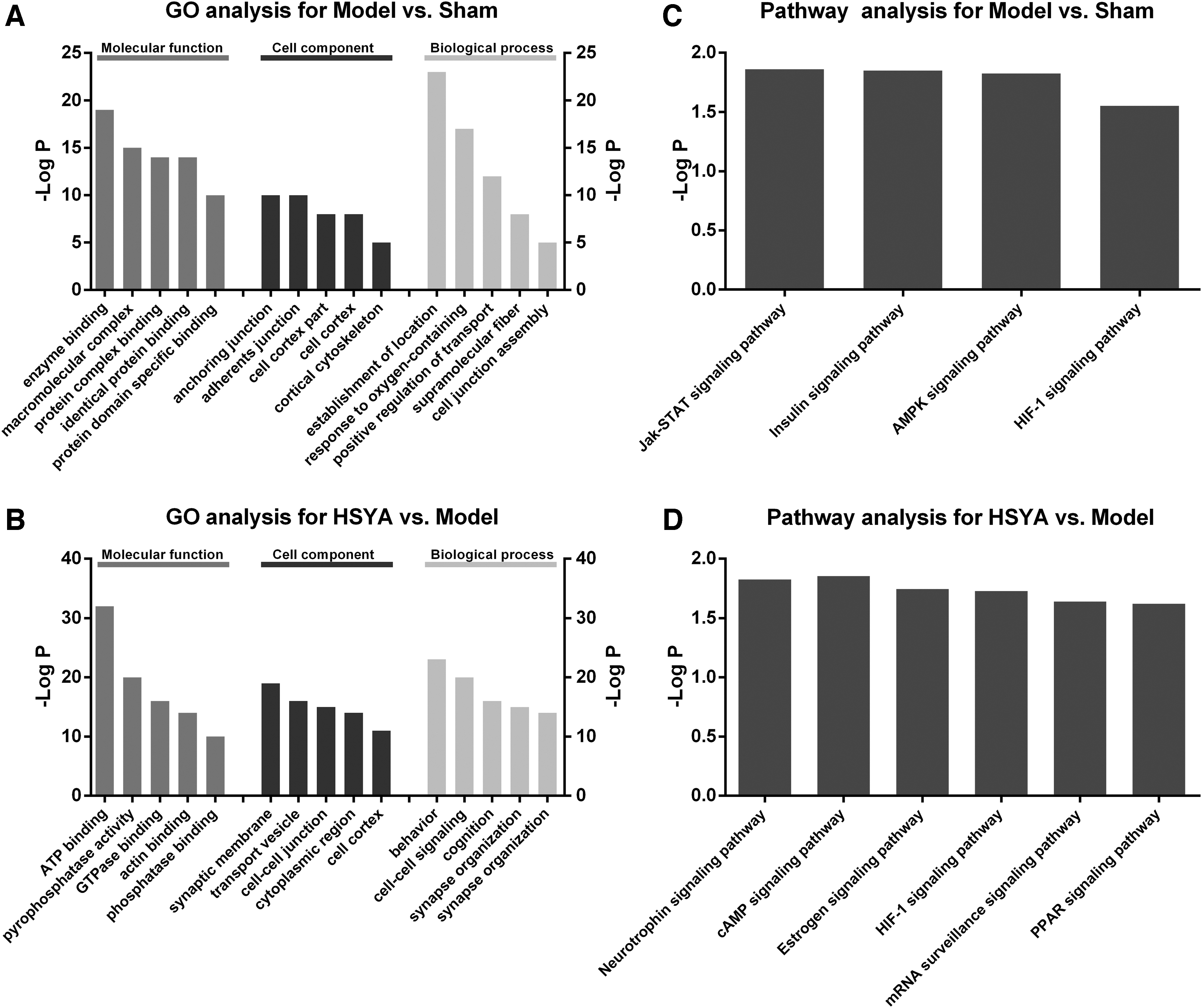
Histogram of GO and pathway enrichment analyses of dysregulated proteins.
The KEGG results indicated that DEPs between Model and Sham groups were significantly related to Jak-STAT, insulin, AMPK, and HIF-1 signaling pathways (Fig. 3C), while DEPs between HSYA and Model groups were closely related to neurotrophin, cAMP, estrogen, HIF-1, mRNA surveillance, and PPAR signaling pathways (Fig. 3D).
Network analysis of the DEPs
The protein-protein interaction (PPI) network was constructed with respect to protein function interactions. To analyze the relationship among those thirteen overlapped proteins, we first constructed a PPI network for these proteins (Supplementary Fig. S1A); the results showed that there were few correlations among these proteins. Therefore, another PPI network was further constructed based on the former network (Supplementary Fig. S1B). Except these overlapped proteins, a few predicted proteins were added in this network. The results suggested that the overlapped proteins mTOR, Ppp2r5e, Eno2, and Rab11b|Rab11a were connected via the predicted protein Akt1, indicating the overlapped proteins as the potential targets of HSYA.
To further seek the hub proteins of the therapeutic mechanism of HSYA, we next focused on the DEPs between different two groups. As shown in Figure 4, 34 nodes representing the DEPs were assigned to the PPI network between Model and Sham groups. Eno2, Gfap, UchI1, Iqgap1, Actn2, Actn4, and Ctnna1 were identified as hub proteins. Besides, the network of HSYA and Model groups consisted of 85 nodes, among which 25 nodes were identified as hub proteins, including Eftud2, Fus, Ddx6, Xpol, Cse1l, Ipo4, Ipo5, Abcel, Farsb, Tars, RGD1563835, RGD1564597, Gart, Rps23, LOC365985, Mpp6, Nedd8, Mut, Mthfd1l, Htra2, Eci2, Ogdhl, Myh14, Aldh7a1, and ENSRNOG00000015290 (Fig. 5).
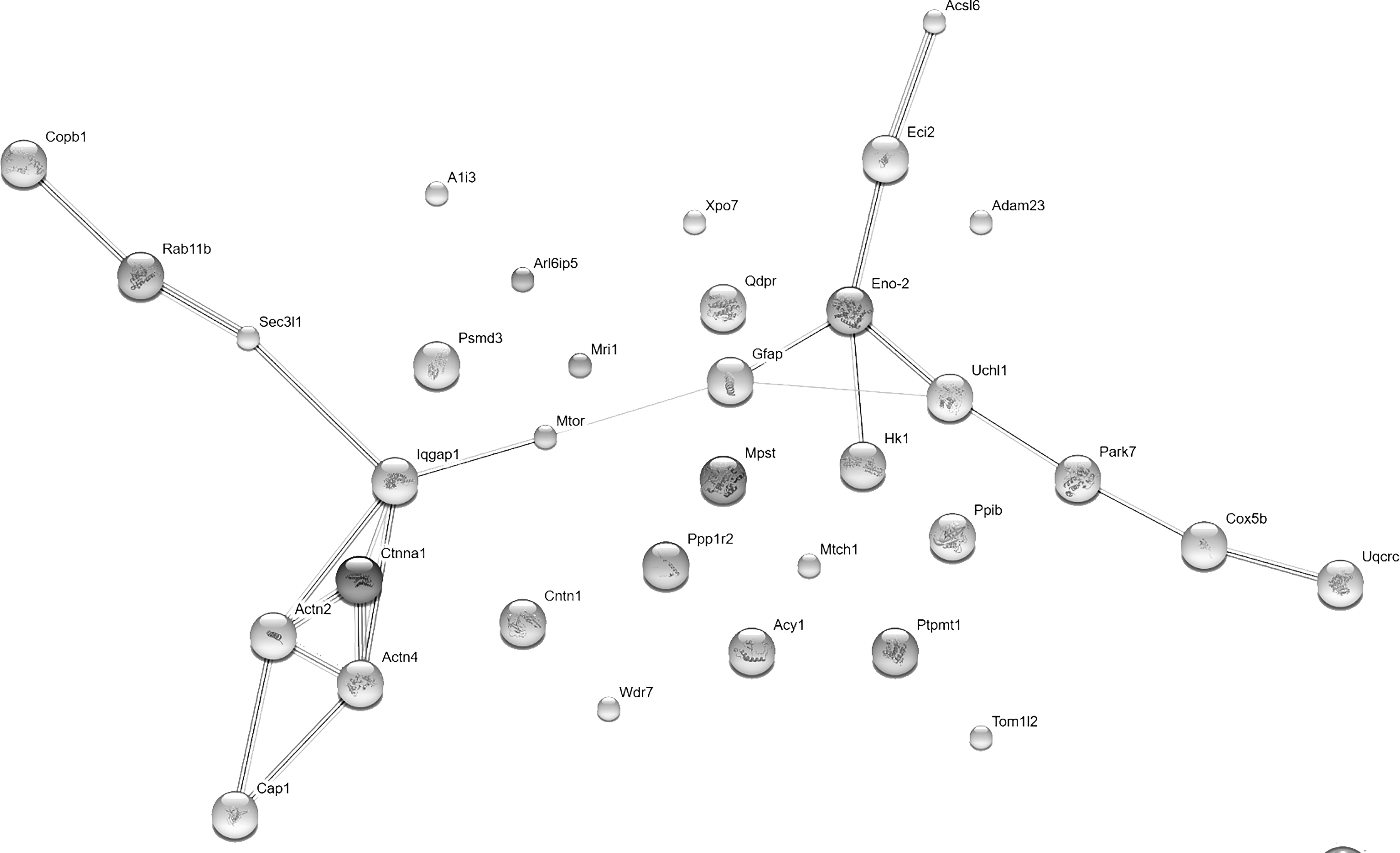
PPI network between Model and Sham groups. Twenty-four proteins were assigned to this network, among which Eno2, Gfap, UchI1, Iqgap1, Actn2, Actn4, and Ctnna1 were identified as hub proteins. PPI, protein-protein interaction.
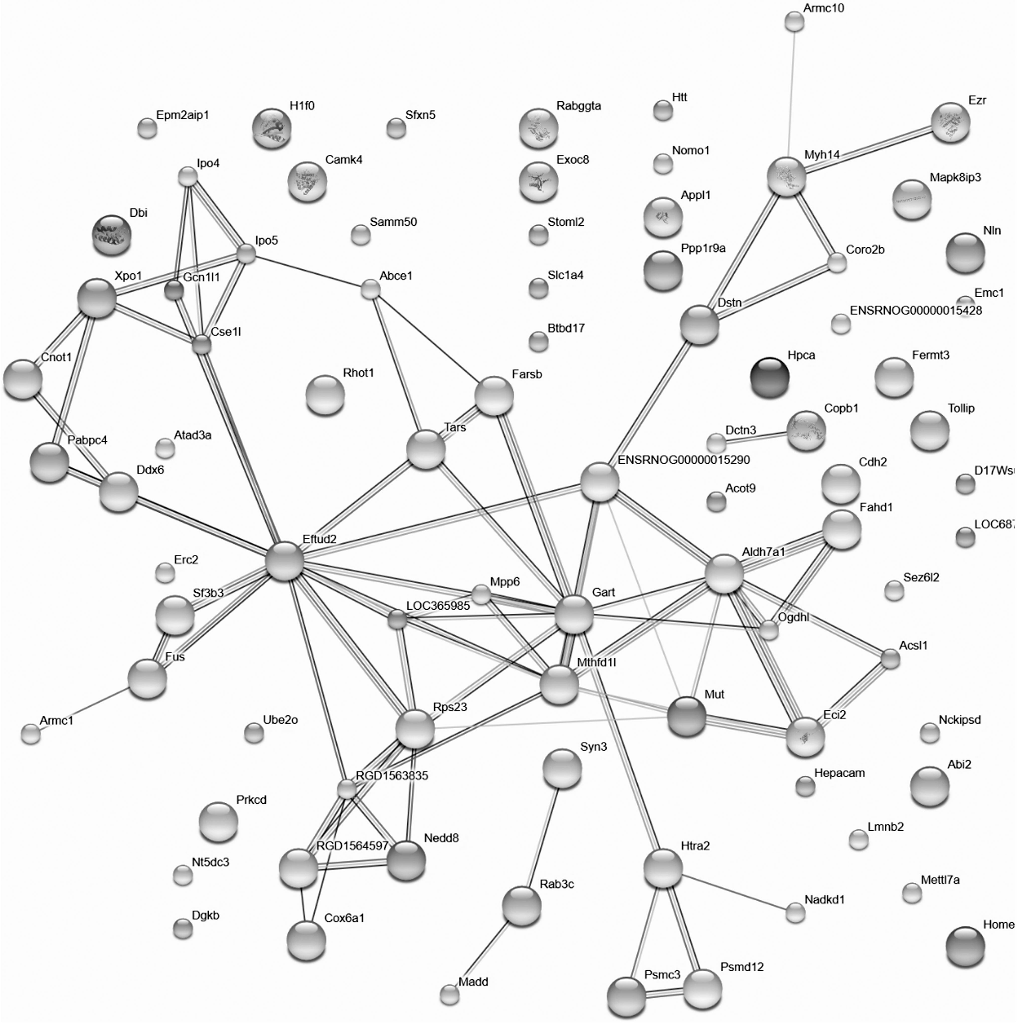
PPI network between HSYA and Model groups. Eighty-five proteins were assigned to this network, among which 25 were identified as hub proteins, including Eftud2, Fus, Ddx6, Xpol, Cse1l, Ipo4, Ipo5, Abcel, Farsb, Tars, RGD1563835, RGD1564597, Gart, Rps23, LOC365985, Mpp6, Nedd8, Mut, Mthfd1l, Htra2, Eci2, Ogdhl, Myh14, Aldh7a1, and ENSRNOG00000015290.
Validation of the identified hub proteins
Western blot analysis demonstrated that the levels of mTOR and Rab11 were decreased, while Ppp2r5e was increased in the Model group compared with Sham group. However, the abnormal expressions of these proteins were significantly reversed in HSYA group (Fig. 6). The differential expressions of mTOR, Rab11, and Ppp2r5e were consistent with the results of label-free analysis.

Validation of the identified hub proteins. The protein expressions of mTOR, Rab11, and Ppp2r5e were investigated by Western blot analysis, which were consistent with the results of label-free analysis.
Discussion
Nowadays, ischemic stroke has become a serious public health threat in developing countries. Some traditional Chinese medicinal herbs have attracted popular attention for their prevention and treatment of CIRI. 24 In addition, the application of proteomic technology in the study of traditional Chinese medicinal herbs in preventing CIRI has made notable progress. 25 Although our recent research demonstrated that HSYA from C. tinctorius L. showed promising neuroprotective effects against CIRI, 16 the systematic mechanism of HSYA against CIRI has not been fully revealed until now.
Since MCAO/R model is an effective stroke model to induce CIRI and study its pathophysiology, 26,27 CIRI has been induced by the MCAO/R model in the present study. Our results revealed that CIRI resulted in severe behavioral and cognitive abnormalities. HSYA could remarkably decrease the infarct volume and improve the neurological outcome compared with Model group. Inflammatory reaction and oxidative stress are important pathogenic factors and damage mechanisms in the pathological process of ischemic brain injury. 28 In this study, the level of both oxidative stress and proinflammatory cytokines was reduced by HSYA treatment, indicating the protective effect of HSYA against CIRI. The significant neuroprotective effects of HSYA on CIRI warranted our further study on the underlying mechanisms.
With the rapid development of proteomics and its wide application in medical sciences in recent years, proteomic analysis was first conducted to evaluate the systematic changes in protein expressions in response to CIRI with HSYA treatment in our study. It is through label-free techniques that 1982 significant proteins were screened, with 13 of them identified as overlapped proteins, including mTOR, Rab11b|Rab11a, and Ppp2r5e. The 5 DEPs between Model and Sham groups revealed the possible pathological features of CIRI, while the 10 DEPs between HSYA and Model groups indicated the potential therapeutic mechanism of HSYA against CIRI. Moreover, GO analysis has been conducted to explore the main functions of the screened 1982 DEPs. The results revealed that the functions of these DEPs were mainly related to enzyme binding, anchoring junction, ATP binding, synaptic membrane, and so on, suggesting that the mechanism of HSYA against CIRI might relate to the dysfunction of these biological functions.
Furthermore, pathway enrichment analysis has been conducted to further investigate the functions of DEPs. We found that hypoxia-inducible factor 1 (HIF-1) signaling was enriched as a significant pathway. HIF-1 is a key transcription factor in response to hypoxia/ischemia and is implicated in cerebral vascular disorders in various pathological conditions, such as ischemic stroke. 29 Increasing evidence has indicated that HIF-1 pathway plays an important role in neuroprotection in ischemic stroke. 30,31 A recent study has proved that the Buyang Huanwu decoction, a classic traditional Chinese medicine with HSYA as a major component, could protect against CIRI in MCAO rats through inhibiting the activation of HIF-1 pathway. 32 Thus, HIF-1 pathway may be considered a target for CIRI treatment.
In addition, PPI network has been constructed to further explore the hub proteins related to the neuroprotective effects of HSYA on CIRI based on the protein function interactions. Our findings have demonstrated that Eftud2 is one of the key proteins considered to be a novel innate immune regulator. It was reported to be related to the production of TNF-α and IL-6, 33 which has been confirmed in our finding that HSYA could inhibit the release of proinflammatory cytokines. Thus, it can be inferred that Eftud2 was involved in the mechanism of CIRI.
Finally, Western blot analysis has been conducted to verify the expression of three main dysregulated proteins, mTOR, Rab11, and Ppp2r5e. The results were consistent with the findings of proteomic analyses, suggesting that the data obtained through label-free techniques are reliable. mTOR, Rab11, and Ppp2r5e have been reported to be implicated in cell growth, proliferation, apoptosis, autophagy, and transcription. 34 –36 Numerous studies have demonstrated that these proteins could play an important role in the repair of ischemic brain tissue. 37,38 A recent research also found that HSYA protects brain microvascular endothelial cells against hypoxic injury through mTOR pathway. 39 mTOR belongs to a family of phosphatidylinositol kinase-related kinases, 38 a central regulator of cellular metabolism, growth, and survival. 35 mTOR could significantly attenuate the inflammatory response in cardiomyocytes by suppressing lipopolysaccharide-induced secretion of IL-6. 40 A new study suggested that mTOR regulated intestinal inflammation and oxidative stress injury via nuclear factor-kappa B pathway. 41 Consistently, our findings showed that HSYA could significantly inhibit inflammatory reaction and oxidative stress. Therefore, our study has proved that mTOR, Rab11, and Ppp2r5e may be associated with the mechanism of HSYA against CIRI.
It should be noted that the overlapped proteins and hub proteins from PPI network were not absolutely consistent in this study, and this may be due to the different research perspectives and algorithms. The 13 overlapped proteins were obtained from an intersection of the 1982 DEPs among three groups, while the PPI networks were constructed based on the intersection between Model and Sham groups or between HSYA and Model groups. In the ordinary way, the number of proteins obtained from the former is much less than the latter, and there is no absolute correlation between these proteins. In addition, the proteins displayed in the PPI network were distributed based on protein/protein interrelationships, and the hub proteins were identified also based on the complex interrelationships; so, the hub proteins were obtained from a different algorithm with the overlapped proteins.
Undeniably, there are some limitations in the current study. These potential hub proteins were identified through proteomic and bioinformatic analysis, but they have not been validated through larger scale samples of clinical patients. Besides, our research is mainly focused on the acute stage of ischemia, but the long-term therapeutic effects of HSYA on CIRI remain to be studied in the future.
In the present study, proteomic analysis has been applied to explore the potential targets and highlight the key roles of the signaling pathways in HSYA against CIRI. Eftud2, mTOR, Rab11, Ppp2r5e, and HIF-1 signaling pathways have been detected as the key proteins and pathways. Our research has provided convincing explanations for the mechanism of HSYA against CIRI and the identified key proteins and pathways might provide novel therapeutics for CIRI.
Footnotes
Acknowledgments
This work was funded by the National Natural Science Foundation of China (No. 81603385, 81573549, 81503285, 81703737, 81600738) and the China Postdoctoral Science Foundation (2018M643843).
Author Disclosure Statement
No competing financial interests exist.
Supplementary Material
Supplementary Figure S1
References
Supplementary Material
Please find the following supplemental material available below.
For Open Access articles published under a Creative Commons License, all supplemental material carries the same license as the article it is associated with.
For non-Open Access articles published, all supplemental material carries a non-exclusive license, and permission requests for re-use of supplemental material or any part of supplemental material shall be sent directly to the copyright owner as specified in the copyright notice associated with the article.