
Abstract
Ischemic stroke is a typical cerebrovascular illness with high morbidity and mortality worldwide. Nevertheless, strategies for the prevention and treatment of cerebral ischemia/reperfusion injury (CIRI) are limited. Gallic acid (GA) is a plant polyphenol that has been used against CIRI. However, the pharmacokinetic (PK) properties of GA, such as its low absorption, poor bioavailability, and quick elimination, have negative effects on its application. To strengthen its effectiveness, a delivery system of GA-loaded o-carboxymethyl chitosan nanoparticles (GA-NPs) was synthesized in our study. In PKs study, GA-NPs apparently increases the area under the curve of plasma concentration–time and prolonged half-life of GA. Then, we measured the in vitro and in vivo effects of the GA-NPs in the oxygen glucose deprivation model and the middle cerebral artery occlusion model. The results from our pharmacodynamic studies, including assessment of neurological deficit, cerebral infarction, levels of inflammation, and oxidative stress, showed that GA-NPs possess better neuroprotection compared with GA. In conclusion, GA-NPs may be used as an efficacious delivery vehicle for GA in the treatment of CIRI.
Introduction
Ischemic stroke (IS) is a common fatal cerebrovascular disease with substantial morbidity and mortality worldwide. 1 It has been estimated that ∼23 million people will have suffered their first stroke, resulting in 7.8 million deaths and costing roughly US$ 240.67 billion, by 2030. Cerebral ischemia/reperfusion injury (CIRI) is the tissue damage caused when the blood supply returns to tissue after a period of ischemia. CIRI creates a condition in which the restoration of circulation results in inflammation and oxidative damage through the induction of oxidative stress causing significant neuronal death and neurological dysfunction. 2 Therefore, anti-inflammatory agents can diminish the damage of ischemic brain tissue by removing inflammatory factors and repairing the damaged tissue. 3 In addition, the antioxidative enzyme system, which is mainly modulated by nuclear factor erythroid 2-related factor 2 (Nrf2), can evoke a neuronal cell defense against toxic reactive oxygen species (ROS) insult. 4,5
Great efforts have been made to explore for potential effective compounds against CIRI in recent years. A large number of studies have found that many plant nutrients or polyphenols have a good effect in preventing IS, such as resveratrol, corilagin, and gallic acid (GA), due to their anti-inflammatory and antioxidant effects. 6 –8 GA is widely distributed in natural nutrients, including in fruits and many common herbs, so it has bright prospects for development and application in the prevention and treatment of IS. GA is an active molecule with the simplest chemical structure of natural polyphenol compounds. 8 Previous studies have demonstrated that GA is capable of increasing the brain's anti-inflammatory and antioxidant capacity. 9 –11 Although GA has been regarded as a promising therapeutic polyphenol for cerebral and cardiovascular diseases, its pharmacokinetic (PK) properties, such as low absorption, poor bioavailability, and quick elimination, are the main bottlenecks in clinical practice. 12 To overcome these problems, the encapsulation technique has emerged as a successful delivery system for a number of drugs. Moreover, chitosan-based nanoparticles have been exploited as delivery systems for brain targeting. Previous experiments have demonstrated the efficacy of boswellic acid-loaded O-carboxymethyl chitosan (O-CMC), a water soluble chitosan derivative, in a brain injury model. 13 Therefore, we hypothesized that the preparation of GA-loaded o-carboxymethyl chitosan nanoparticles (GA-NPs) could enhance the efficacy of GA against CIRI.
In this study, GA-NPs were successfully prepared by a very simple method. They were nanosized and monodispersed with good encapsulation efficiency and sustained release patterns. The PK studies illustrated that GA-NPs could substantially raise area under the plasma concentration–time curve (AUC), leading to enhanced protection against cerebral ischemic insult. Effects of GA-NPs in an oxygen glucose deprivation (OGD) model and a middle cerebral artery occlusion (MCAO) model were determined, showing improved antioxidant defense (superoxide dismutase, SOD and glutathione peroxidase, GSH-Px) and decreasing inflammation (TNF-α and IL-1β) compared with GA. It can be concluded that O-CMC nanoparticles represent an interesting delivery system for GA and, hence, offer an effective and promising candidate for the treatment of stroke.
Methods
Materials
All chemicals and reagents in our experiments were of analytical grade. Adult male Sprague–Dawley (SD) rats (230 ± 20 g) were provided by the Experimental Animal Center of the Fourth Military Medical University. For all experiments, 80 rats were used in total. The experimental protocols were in strict compliance with the National Institutes of Health guidelines. All experimental procedures were performed under the guidelines approved by the Ethics Committee for Animal Experimentation of Zhengzhou University. Food and tap water were available to the rats, which were housed on a 12-hour dark–12-hour light cycle in air-conditioned animal quarters at 22.0°C ± 1.0°C and 40.0%–50.0% humidity before the experiments.
Synthesis of GA-NPs
GA-loaded O-CMC nanoparticles (GA-NPs) were synthesized on the basis of previously reported methods with slight modifications to improve the redispersibility of the NPs. 14 O-CMC (12 kDa, deacetylation 61.8%, substitution 0.54) was supplied by Haidebei Marine Bioengineering Co., Ltd. Methanolic GA solution was incubated with the O-CMC solution in water overnight, followed by TPP crosslinking (1%) at a volume ratio of 25:1 (O-CMC/TPP). Then, the above formed emulsion was incubated with 0.1 mL of 1% BSA solution for 30 minutes. The drug-loaded GA-NPs were isolated, and the pellets were collected and used for further research.
Particle size analysis and zeta potential
The GA-NPs were dispersed in distilled water at the appropriate concentrations. The mean particle size, size distribution, and zeta potential were measured using a DLS Delsa Nano particle analyzer (Beckman Coulter, Inc., CA). Deionized water was used to dilute all samples before measurements at 25°C ± 0.5°C (in triplicate)
Morphology of GA-NPs
The shape and surface morphology of the GA-NPs were observed by a transmission electron microscopy (TEM) (JEOL JEM Dos electron microscope, JEOL, Tokyo, Japan). Magnifications of up to 1,000,000 × were used to view samples.
Drug-loading capacity and entrapment efficiency
The entrapment efficiency (EE) and loading efficiency (LE) of the GA-NP sample were quantified using the High Performance Liquid Chromatography (HPLC) method previously described. 15 EE was calculated as the percent of GA recovered from the GA-NPs in comparison with the initial drug amount. LE was defined as the amount of entrapped GA compared with the total GA-NPs.
The release of GA from the GA-NPs in vitro was performed by dialysis sacs. The GA-NPs were placed in pretreated dialysis sacs, which were immersed in 100 mL of phosphate buffer solution (pH 7.4) under mechanical agitation at 37°C. The medium was withdrawn and replaced by the same medium at regular intervals. All measurements were taken three times in the experiments.
Pharmacokinetics
Rats were divided into a GA group and a GA-NP-treated group (n = 8 each group) for the PK experiment. The native GA (50 mg/kg) solved in CMC-Na solution (0.5%, w/v) and GA-NPs (containing GA 50 mg/kg) dispersed in saline solution were administered orally. This dosage was chosen based on previous studies on GA. 16,17 Blood samples were collected through the caudal vein at predetermined time points. Serum was obtained by centrifugation and stored at −80°C for use. Then, HPLC/MS-MS analysis was performed as previously described. 15 GA content was determined with an Agilent 1100 liquid chromatographic system equipped with a reversed-phase column (Dikma, Beijing, China). PK parameters were calculated with the noncompartmental model using WinNonlin 6.2 (Pharsight Corporation, Mountain view, CA). The AUC was calculated with the trapezoidal rule extrapolated to infinity. The terminal elimination half-life (t1/2) and the systemic clearance (Cl) were obtained.
Oxygen-glucose deprivation and reoxygenation
The OGD model was employed to study the in vitro effects of the GA-NPs on CIRI. Cortical neurons were prepared from the brains of one-day-old rats as described previously. 18 Neurons were cultured at 37°C in a humidified 5% CO2 atmosphere. The primary cultured cortical neurons were subjected to transient OGD for 60 minutes to model ischemia-like conditions in vitro and then incubated in a 95% and 5% CO2 air box for 24 hours. Five groups were designed to simulate the blood deficiency in vitro: control group (no OGD), OGD group, OGD+void NPs group, OGD+GA group (50 μM dissolved in DMSO with dose decision on the basis of previous studies), 19 and OGD+GA-NPs group. The DMSO concentration in the cultured medium remained <0.1% w/v, with the medium-treated cells used as controls.
Then, the cell viability in each group was observed by MTT analysis as previously described. 20 The release of lactate dehydrogenase (LDH) was detected using a commercially available kit (Jiancheng Biological Engineering Institute, Nanjing, China). Data were expressed as the percent of control levels.
Induction of cerebral ischemia and treatment
Rats were randomly assigned to five groups: sham group, vehicle group, NPs group, GA group, and GA-NPs group (n = 8 each group). Native GA was dissolved in CMC-Na solution (0.5% w/v) and GA-NPs were dispersed in saline solution for administration. Rats in both the sham and vehicle groups received only distilled water p.o. for 7 days. The NP group was treated with nanoparticles alone in saline solution. Rats in the GA group were treated with GA solution (50 mg/kg) per day p.o. for 7 days. Rats in the GA-NP group were treated with GA-NPs containing 50 mg/kg GA dispersed in saline per day p.o. for 7 days.
Focal cerebral ischemia was performed using the method of MCAO with an intraluminal filament. 21 On the eighth day, all rats were anesthetized with 2.0%–3.0% isoflurane and kept with 1.0%–1.5% isoflurane at 70% N2O and 30% CO2. Then, MCAO-induced CIRI was sutured with a poly L-lysine-coated nylon line. Restoration of the MCA blood flow was achieved by withdrawing the line 90 minutes after ischemia. Animals that did not show a CBF reduction of at least 70% and animals that died after ischemia induction were excluded from the groups. All of the above described methods were used in the sham group except for MCAO. The rats were allowed to survive for 72 hours. Mean arterial blood pressure, pH, arterial blood gases, and blood glucose levels were evaluated at preischemia and 90 minutes after reperfusion.
Neurological deficit scores and infarct size
The neurological deficits of the rats were evaluated blindly according to the Zea Longa standard 72 hours after reperfusion. 22 The 5-point scale system is as follows: 0 indicates no symptoms of neurological dysfunction; 1 indicates failure to stretch the contralateral torso and forelimbs completely; 2 indicates movement to the ipsilateral side when caught by the tail; 3 indicates falling to the affected side; and 4 indicates no spontaneous walking.
To measure the infarct size, rats in each group were cut into coronal sections (2 mm) 72 hours after MCAO, and the sections were incubated in 2% TTC solution for 30 minutes. Then, the images of the TTC-stained slices were observed. Infarct size was expressed as a percent according to the formula: ([total contralateral hemispheric volume]-[total ipsilateral hemispheric stained volume])/(total contralateral hemispheric volume) × 100%.
TUNEL staining
After tissue sections with 5-μm-thick paraffin were prepared, apoptotic cells were detected using a TUNEL Assay Kit (Roche, Germany) according to the manufacturer's instructions to evaluate DNA damage. 5 Green staining showed the nucleus of apoptotic cells in positive cells. TUNEL-positive neurons in the ipsilateral hemisphere were counted in four different fields of each section by a fluorescence microscope (Olympus, Tokyo, Japan).
Antioxidant and anti-inflammatory indicators
Rats in each group were sacrificed 72 hours after MCAO, and the tissues of the right cerebral cortex were homogenized in 2 mL of phosphate buffer (10 mM). The levels of tumor necrosis factor (TNF)-α and IL-β, as important anti-inflammatory indicators in the tissues of the right cerebral cortex, were detected with ELISA Kits (Jiancheng Biological Engineering Institute, Nanjing, China). 23 In addition, since the activities of SOD and GSH-Px were regarded as important indicators of the antioxidant effects, quantitative analysis was conducted using an ELISA Kit according to the manufacturer's instructions (Jiancheng Biological Engineering Institute, Nanjing, China). The protein concentration was assessed by an ELISA reader (Model ELX800, BioTek).
Statistical analyses
The statistical analyses were conducted with SPSS 19.0 software (IBM SPSS, Chicago, IL). Data are expressed as the mean ± standard deviation (SD), except for the neurological deficit scores. Neurological deficit scores were expressed as the median (range) and then were analyzed using the Kruskal–Wallis test followed by the Mann–Whitney test and Bonferroni post hoc correction. A p-value of <0.05 was considered statistically significant.
Results
Characterization of GA-NPs
To confirm the solubility of GA-NPs, we found that nanoparticle GA dissolved in aqueous solution gave a clear and dispersed formulation (left one), while native GA is poorly soluble in aqueous media (right one) (Fig. 1A). The TEM images showed that the prepared NPs had a smooth spherical surface (Fig. 1). The size of the GA-NP particles was 173 ± 18 nm (Fig. 1C), the polydispersity index was 0.106 ± 0.087, and the value of zeta potential was positive (21.3 ± 2.2) mV. For GA-NPs, the drug interception efficiency was 83.5% ± 6.0%, and the drug loading was 30.0% ± 6.3% (Fig. 1D).
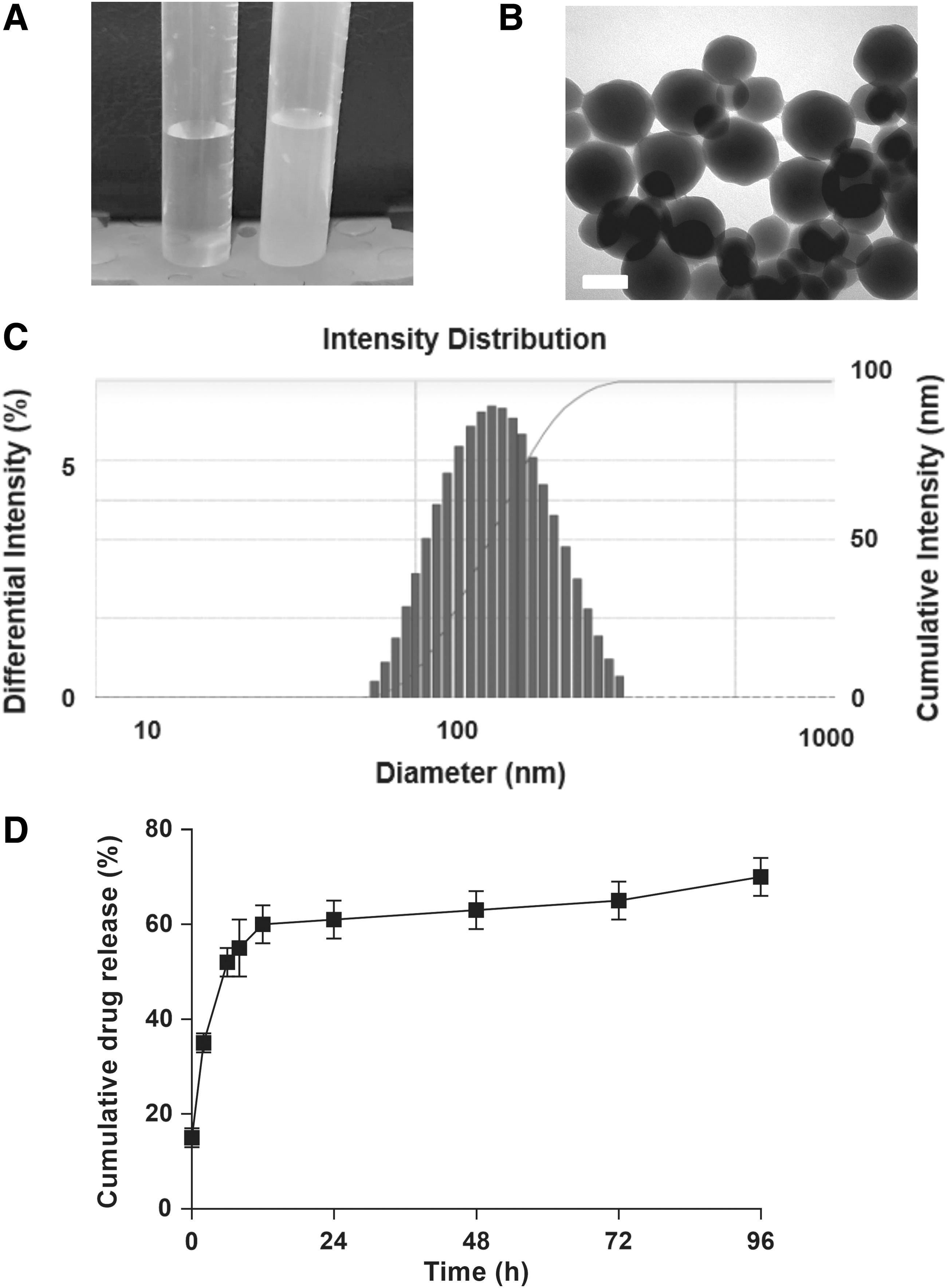
The in vitro release curve of the GA-NPs is shown in Figure 2D. GA-NPs demonstrated 50% drug release in the original 6 hours solution with a slow, gradual release rate. The amount of cumulative drug release after 4 days was more than 70%. The observed initial release amount might be caused by the dissociation of the surface absorption of the GA-NPs in the polymer matrix.
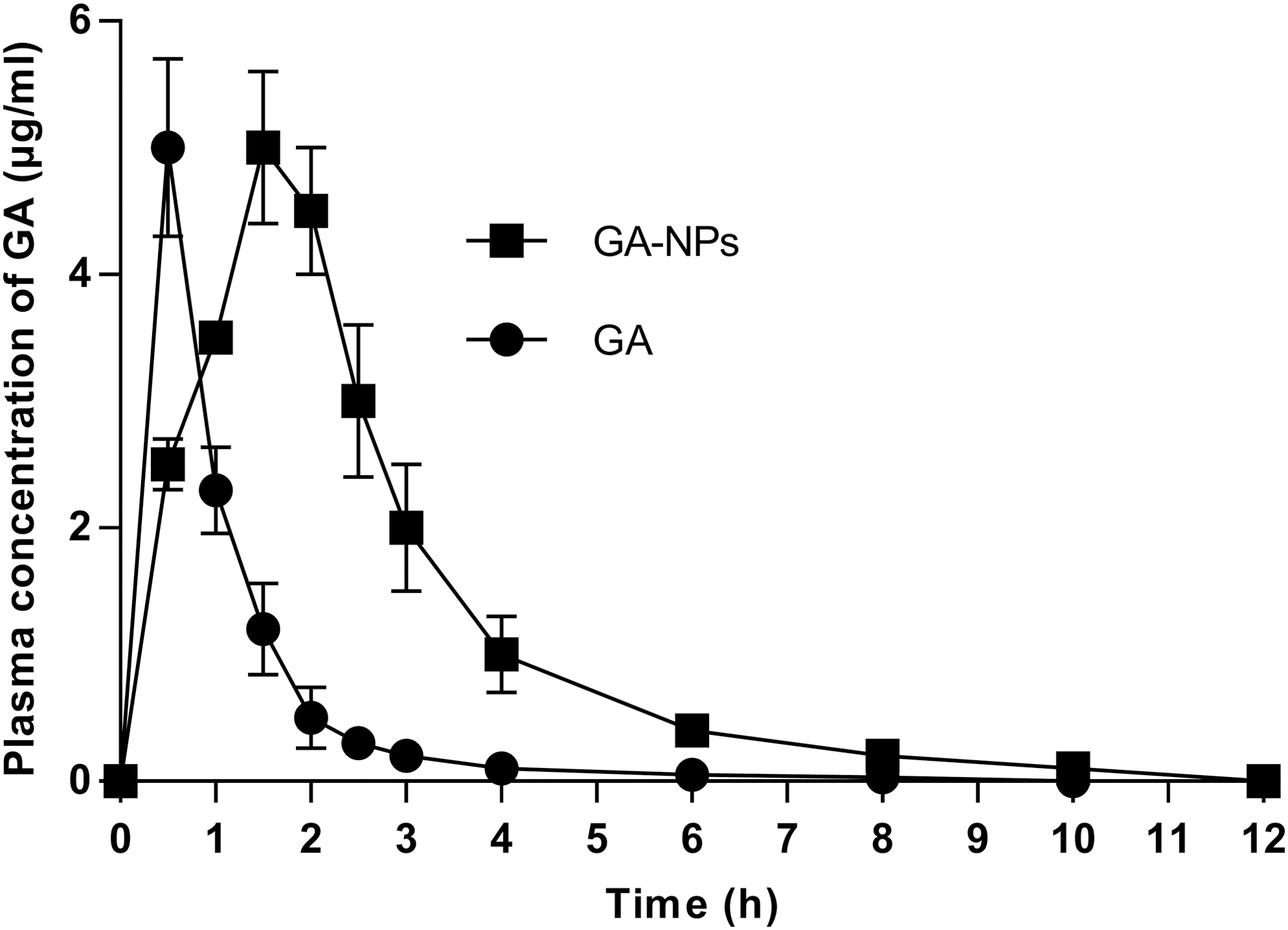
The plasma concentration–time curves after administration of GA and GA-NPs (n = 8).
PKs of GA-NPs
Figure 2 shows the plasma concentration–timetime curves for the GA-NPs and GA after oral administration. The PK parameters of the GA-NPs are shown in Table 1. The plasma AUC for the GA-NPs (13.8 mg min/mL) was enhanced compared with that of GA (5.2 mg min/mL) (p < 0.05). The slow release of GA in the nanoparticle system increased the AUC of GA and the half-life of circulation (t1/2) (p < 0.05).
Pharmacokinetic Parameters of GA and GA-NPs After Administration to Rats
n = 8
AUC, area under the plasma concentration–time curve; GA, gallic acid; GA-NP, GA-loaded O-carboxymethyl chitosan nanoparticle.
GA-NPs protected neuronal cells against OGD-induced injury
Figure 3 shows the cell viability of five groups. The proportion of cell apoptosis in the OGD treatment group was ∼75%. There was no significant difference in the cell survival rate between the NP group and the OGD group (p > 0.05). The cell viability of the GA-NP group (60% ± 5%) was higher than that of the OGD group (45% ± 3%) (Fig. 3) (p < 0.05). LDH testing was used to confirm the protective effects of GA-NPs and demonstrated that GA-NP treatment reduced OGD-induced LDH release by ∼47% (p < 0.05) (Fig. 3). To assess cytotoxicity, cell viability was assessed using MTT assay after exposure to the GA nanoparticles for 48 hours (1–100 μM), and no significance was found between treated groups and the control (Supplementary Fig. S1).

Effects of GA-NPs on cell viability and LDH release in a primary culture of rat cortical neurons exposed to OGD.
GA-NPs improved the neurological deficit and cerebral infarction
No significant differences were found in physiological variables between treated groups (Supplementary Table S1). Figure 4A shows the neurological scores of five groups 72 hours after reperfusion. The score in the GA-NP group was significantly higher than that of the vehicle group (p < 0.05). However, TTC staining showed that the infarct size in all treatment groups was significantly reduced compared with the vehicle group, with the best protective effects were observed in the GA-NP group (p < 0.05). The results of both the neurological dysfunction and TTC staining of infarct size indicated the effects of GA-NPs against ischemic injury.
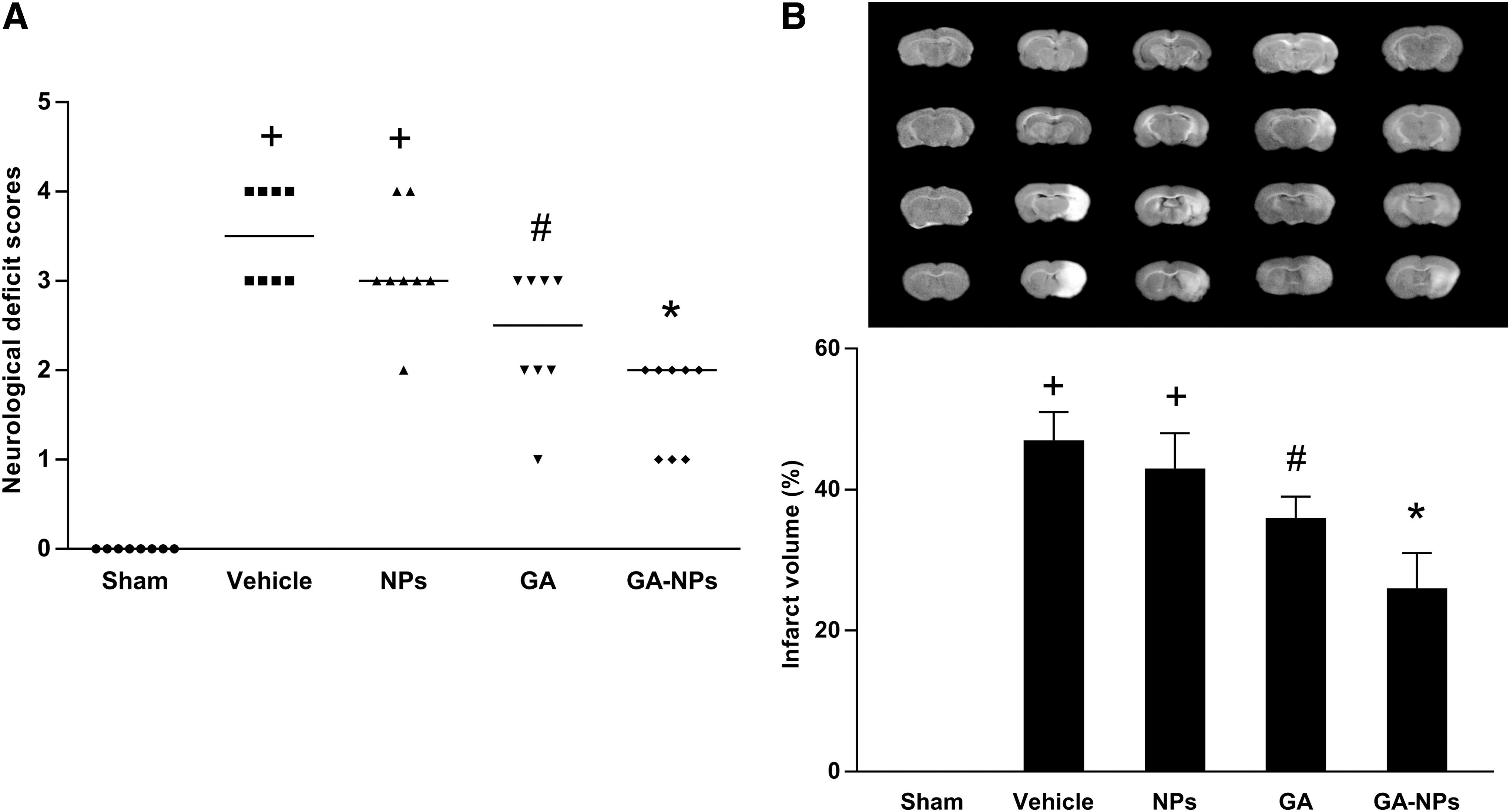
Effects of GA-NPs on brain infarct volume and neurological deficits 72 hours after cerebral I/R in rats.
Apoptotic cells were detected by terminal deoxynucleotidyl transferase-mediated dUTP-biotin nick-end labeling (TUNEL) (Fig. 5). The number of TUNEL-positive cells significantly increased in the cortex of the vehicle group compared with the sham group (p < 0.05). In addition, the number of TUNEL-positive cells were significantly reduced in the treatment groups compared with the vehicle group (p < 0.05). There were fewer TUNEL-positive cells in the GA-NP group than in the other groups (p < 0.05).
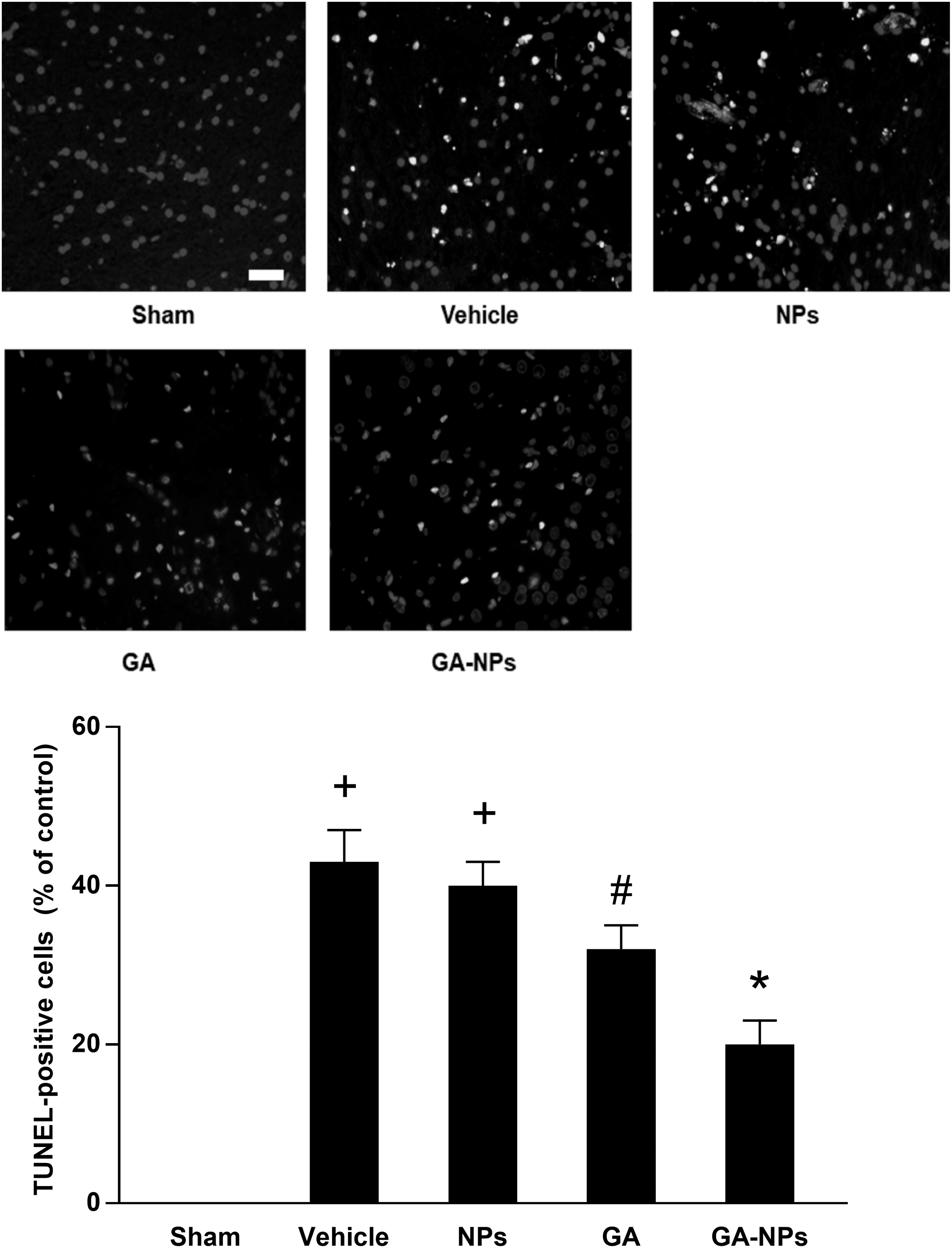
Effects of GA-NPs on neuronal apoptosis of brain tissues from stroke rats. Representative fluorescence micrographs of TUNEL staining (400 × magnification). Scale bars = 20 μm. Representative photomicrographs of the sham group, vehicle group, void NPs group, GA group, and GA-NPs group. Statistical analysis of the number of TUNEL-positive neurons in each group. (n = 8). +p < 0.05 versus sham group; #p < 0.05 versus vehicle group; *p < 0.05 versus GA group.
GA-NPs attenuated inflammation and oxidative stress
The levels of SOD, GPx, TNF-α, and IL-β in the cortex of each group are depicted in Table 2. The levels of TNF-α and interleukin 1 beta (IL-1β) significantly increased in the vehicle group compared with the sham group (p < 0.05). The significant reduction in the TNF-α and IL-1β levels indicated better anti-inflammatory effects of the GA-NPs compared with GA (p < 0.05). In addition, the activities of SOD and GSH-Px were significantly decreased in the vehicle group compared with the sham group (p < 0.05). GA-NPs showed a significant enhancement in the SOD and CAT levels when compared with the pure GA group (p < 0.05).
Levels of SOD, GPx, TNF-α, and IL-β in the Cortex at 72 Hours After MCAO in Each Group
n = 8.
+p < 0.05 versus sham group.
#p < 0.05 versus vehicle group.
p < 0.05 versus GA group.
Discussion
In light of previous reports, GA has demonstrated obvious pharmacological effects on cardiovascular diseases and nervous system diseases. 8,24,25 It is quite evident that the rapid metabolism of GA is a factor for its low bioavailability and fast elimination. 26,27 Therefore, an improvement in the bioavailability and a reduction in the elimination of GA is required for desirable therapeutic activity. Some experiments have been performed on nanoformulations, such as chitosan, which can be used as a carrier for brain protective drugs. 28 Our study confirmed the neuroprotective effects of GA-NPs in the MCAO model. These effects may be associated with the anti-inflammatory and antioxidant functions of GA-NPs. The results of TTC staining, neurological deficit scores, and TUNEL staining demonstrated that GA-NPs could decrease the damage of CIRI compared with the vehicle group.
The TEM images indicated that the GA-NPs were uniform, segregated, spherical, and subspherical in shape. The desired diameter of intravascular long-circulating NPs has been suggested to be within a relatively narrow range (70–200 nm). 29 The mean value of zeta potential for GA-NPs was positive 21.3 mV, which is in agreement with the adsorptive endocytosis of the cationic nanoparticles and permits absorptive-mediated transcytosis across the blood–brain barrier. Additionally, after the initial burst drug release over 6 hours (50%), the controlled release pattern of the GA-NPs was characterized in our in vitro release experiments and was suitable for stroke treatment.
Previous findings have demonstrated that chitosan nanoparticles can significantly prolong the t1/2 and AUC of phenolic compounds, such as chlorogenic acid, resulting in retained antioxidant activity and enhanced bioavailability. 30 Consistently, in this study GA-NPs showed better therapeutic effects due to the improvement in the PKs. Our results of the pharmacodynamics study, such as assessment of neurological deficit, cerebral infarction, levels of inflammation, and oxidative stress, were consistent with the PK experiment, demonstrating that the loading of GA in the NPs was relatively higher in the protective effects against CIRI injury.
In the MCAO model of rats, GA-NPs played an anti-inflammatory and antioxidant role in improving the neurological deficit, alleviating neural dysfunction, reducing cerebellar infarction size, and decreasing apoptosis (p < 0.05). Inflammation is an important pathogenic factor of CIRI. 31 In the process of disease, CIRI induces the production and secretion of apoptosis and inflammatory-related cytokines, such as TNF-α and IL-β, 32 which are used as markers of inflammation of CIRI. It is worth noting that the GA-NPs effectively alleviated the increase in proinflammatory cytokine content, including TNF-α and IL-β, in this study. In particular, the GA-NP formulation reduced the TNF-α and IL-β levels significantly more compared with GA.
Increasing evidence has shown that oxidative stress is another important pathological factor of CIRI. In the process of CIRI, ROS are released, which can cause oxidative stress damage to body cells. SOD and GSH-Px are the main enzymes that remove free radicals in organisms, and they reflect the ability of organisms to remove oxygen-free radicals. 33 Therefore, the elimination of free radicals by SOD and GSH-Px can combat oxidative stress damage caused by IS. 34 Our results showed that CIRI increased lipid peroxidation in the brain and decreased the activity of SOD and GSH-Px. It is through the treatment of GA-NPs that this situation significantly improved. There were also notable increases in the activities of both SOD and GSH-Px in the GA-NP-treated group compared with the GA group.
There are some limitations in our studies. First, we focused on the protective effects of GA against ischemic injury, and some experiment still not conducted in this study according to the ARRIVE guidelines, 35 such as toxicity study in animals. Second, the PK data from rats administered with GA and GA-NPs illustrated that GA-NPs could substantially raise the AUC, improve the bioavailability, and reduce the elimination of GA. But it is still unclear for the uptake of GA by the brain through this formulation, and the brain levels of NPs should be determined in future.
Conclusion
In conclusion, the GA-NPs prepared in our study are nanosized and monodispersed with a high efficiency of encapsulation and a sustained release mode. GA-NPs have significant therapeutic effects on IS by reducing inflammation and enhancing the antioxidant defense. These findings suggest that GA with an effective delivery system is a promising candidate for the treatment of CIRI.
Footnotes
Acknowledgments
This work was supported by the Medical Science and Technology project of Henan province (201303026) and the Foundation of Beijing Medical and Health (YWJKJJHKYJJ-B16239).
Author Disclosure Statement
The authors declare no conflicts of interest.
Funding Information
No funding was received for this article.
Supplementary Material
Supplementary Figure S1
Supplementary Table S1
References
Supplementary Material
Please find the following supplemental material available below.
For Open Access articles published under a Creative Commons License, all supplemental material carries the same license as the article it is associated with.
For non-Open Access articles published, all supplemental material carries a non-exclusive license, and permission requests for re-use of supplemental material or any part of supplemental material shall be sent directly to the copyright owner as specified in the copyright notice associated with the article.