
Abstract
Stem cells hold great promise in tissue engineering for repairing tissues damaged by disease or injury. Mesenchymal stem cells (MSCs) are multipotent cells able to proliferate and differentiate into multiple mesodermal tissues such as bone, cartilage, muscle, tendon, and fat. We have previously reported that the low-affinity nerve growth factor receptor (L-NGFR or CD271) defines a subset of cells with high proliferative, clonogenic, and multipotential differentiation ability in adult bone marrow (BM). It has been recently shown that adipose tissue is an alternative source of adult multipotent stem cells and human adipose-derived stem cells, selected by plastic adherence (PA hASCs), have been extensively characterized for their functional potentials in vitro. In this study, immunoselected L-NGFR+ and CD34+ subpopulations have been analyzed and compared with the PA hASCs. Phenotypic profile of freshly purified subpopulations showed an enrichment in the expression of some stem cell markers; indeed, a great percentage of L-NGFR+ cells co-expressed CD34 and CD117 antigens, whereas the endothelial-committed progenitor markers KDR and P1H12 were mainly expressed on CD34+ cells. Differently from PA hASCs, the immunoseparated fractions showed high increments in cell proliferation, and the fibroblast colony-forming activity (CFU-F) was maintained throughout the time of culture. Furthermore, the immunoselected populations showed a greater differentiative potential toward adipocytes, osteoblasts, and chondrocyte-like cells, compared to PA hASCs. Our data suggest that both CD34+ and L-NGFR+ hASCs can be considered alternative candidates for tissue engineering and regenerative medicine applications.
Introduction
I
Adult mesenchymal stem cells (MSCs) are a population of multipotent cells able to proliferate and differentiate into multiple mesodermal tissues. MSCs have been initially identified in bone marrow (BM) [1], but have been subsequently isolated from many other tissues [2 –10]. Among them, human MSCs derived from adipose tissue (hASCs) show stem cell key features such as the ability to form fibroblast-like colonies (CFU-F), the expression of several common cell surface antigens, the capacity of extensive proliferation, and the potential to differentiate in vitro and in vivo into multiple lineages of mesodermal origin, and also to transdifferentiate into neurogenic and hepatic lineages [4,11 –21]. Similarly to BM-derived MSCs (BM MSCs), hASCs are able to suppress immunoreactivity, suggesting a possible overcoming of histocompatibility limitations in allogeneic transplantation [22,23]; furthermore, they are easily accessible in large amounts by liposuction [4,24,25]. Several studies have demonstrated the efficacy of these cells in clinical applications; in particular, the cell-assisted lipotransfer technique has been successfully used for the treatment of facial lipoatrophy and breast augmentation [26,27] and a clinical trial is actually in progress to evaluate the efficacy of the reimplantation of autologous hASCs purified through the Celution® system (Cytori Inc., San Diego, CA) for breast reconstruction. Moreover, 2 clinical trials (Phase I and Phase II, respectively) were recently performed using in vitro-expanded hASCs for the treatment of perianal fistula [28,29], with satisfactory results.
These cells have been extensively characterized for their functional potential; however, the heterogeneity of the starting cell population and the variability in stem cell recovery among different donors and subcutaneous adipose tissue depots [4,30 –33] cause some difficulties to reproduce and compare the results, underlining the need of specific markers able to identify primitive and multipotent adipose-derived stem cells.
We have previously shown that monoclonal antibodies (MoAbs) to the low-affinity nerve growth factor receptor (L-NGFR or CD271) stain primitive MSCs with high specificity and purity in adult BM, defining a subset of cells with higher clonogenic efficiency, proliferative and differentiative potential in comparison to the whole MSCs population [34]. Furthermore, some authors have highlighted the presence of CD34+ cells in the freshly isolated stromal vascular fraction (SVF) from human adipose tissue [18,19,35,36]. Isolation of CD34+/CD31− cells has revealed that this population differentiated in vitro into endothelial cells and promoted in vivo angiogenesis, participating in the revascularization of ischemic hind limbs of nude mice [18].
In this study, we compare the whole hASCs population, purified by plastic adherence (PA hASCs), with 2 immunoselected subpopulations (L-NGFR+ and CD34+ hASCs), evaluating their phenotypic stem cell profile, their proliferative and clonogenic potential, and their ability to differentiate toward adipocytes, osteoblasts, and chondrocyte-like cells.
Our results show that L-NGFR+ and CD34+ hASCs are 2 distinct purified populations, with a higher clonogenic and differentiative ability compared to the PA unselected population.
Materials and Methods
hASCs isolation
Subcutaneous adipose tissue was collected from 12 female patients (mean age: 40.4 ± 6.6 years, BMI < 30, without any pathological obesity) who underwent aesthetic liposuction of the abdomen (3–4 mm diameter hollow blunt cannula connected to the syringe) at the Plastic and Reconstructive Surgery Unit of the Galeazzi Orthopaedic Institute. All the procedures were approved by the internal IRB and were performed by a senior plastic surgeon of the institute; all patients gave their consent for the research use of the waste tissue deriving from surgical interventions.
Adipose stem cells were isolated as previously described [11]. In brief, the raw lipoaspirates (50–100 mL) were washed at least 3 times with phosphate-buffered saline (PBS) and the matrix was digested with 0.075% type I collagenase (Worthington, Lakewood, NJ) at 37°C with continuous agitation for 30 min. The SVF was centrifuged (1,200g, 10 min) and filtered through a sterile medication lint; the collected cells were centrifuged, resuspended in control medium consisting of DMEM (Sigma-Aldrich, Milan, Italy) supplemented with 10% FBS (fetal bovine serum; Lonza, Milan, Italy), 50 U/mL penicillin (Sigma-Aldrich), 50 μg/mL streptomycin (Sigma-Aldrich), and 2 mM
Positive selection of CD34 + and L-NGFR + hASCs
Positive selection of CD34+ and L-NGFR+ cells was performed using the Direct CD34 Progenitor Cell Isolation Kit and the CD271 Microbead Kit, respectively (Miltenyi Biotech, Calderara di Reno, Italy), according to the manufacturers' instructions. Cells were then counted and assessed for viability; their purity was determined by flow cytometry.
Flow cytometry analysis
To analyze the expression of specific surface markers, 2 × 10⁵ hASCs were incubated with the following anti-human primary MoAbs: CD34-FITC (8G12), CD117-APC (YB5B8), CD45-PerCP (2D1) (Becton Dickinson, Milan, Italy), AC133/2-PE and AC133-APC (293C3) (CD133) (Miltenyi Biotech), CD146-FITC and -PE (P1H12; Chemicon International Inc., Temecula, CA), CD105-FITC (SN6; Serotec Inc., Raleigh, NC), NGFR-PE (C40-1457; BD PharMingen, Milan, Italy), KDR-APC (89106; R&D Systems, Minneapolis, MN) and glycophorin A-FITC (Immunotech SAS, Marseille, France). Samples were then evaluated by FACSCanto II Instrument and Diva software (Becton Dickinson).
hASCs cultures
After the isolation, PA, CD34+, and L-NGFR+ hASCs were cultured in control medium at 37°C in a humidified atmosphere (5% CO2), with weekly refeeds. Cells were grown until 90% confluency, detached by 0.25% trypsin/1 mM EDTA (Sigma-Aldrich) for 5 min at 37°C, counted to assess their proliferation, and tested for CFU-F efficiency and differentiation potential; 10% of the collected cells were re-expanded.
MSCs progenitor assays
Colony-forming units-fibroblast (CFU-F) assay
The clonogenic ability of PA, CD34+, and L-NGFR+ hASCs fractions was determined by a low-density CFU-F assay performed at different passages in culture [37]. The cells from the 3 populations were resuspended in control medium supplemented with 20% FBS and seeded in 6-well plates at different cellular density: 1-3-6-12-24-48 cells/cm2. After 5 days of culture, half of the medium was removed and replaced by fresh medium; after 9 days, the wells were rinsed with PBS, fixed with methanol, and stained with Crystal Violet (Sigma-Aldrich). The colonies consisting of ≥50 cells were counted and the frequency of colonies was expressed on 1 × 10³ plated cells.
Hematopoietic progenitor assays
The colony-forming unit-granulocyte/macrophage (CFU-GM) and burst-forming unit-erythroblast (BFU-E) assays were carried out by plating 1 × 10⁵ CD34+ cells in a methylcellulose culture medium (MethoCult GF H4434; StemCell Technologies Inc., Vancouver, Canada). Triplicate dishes were incubated at 37°C and 5% CO2 in a fully humidified atmosphere. After 14 days of culture, aggregates formed by ≥50 cells were scored as colonies and counted.
Functional assays
Adipogenic differentiation
In order to induce adipogenic differentiation, 5 × 10⁵ hASCs from the different fractions were cultured in hMSC Mesenchymal Stem Cell Adipogenic Differentiation Medium (Lonza) in T-25 flasks, with weekly refeeds. After 21 days of culture, the cells were fixed in 10% formalin for 10 min and stained with fresh Oil Red-O solution (Sigma-Aldrich). Oil Red-O was extracted from the fixed samples incubating for 10 min with 100% isopropanol. Absorbance was read at 490 nm with Wallac Victor II plate reader.
Osteogenic differentiation
In order to differentiate cells toward the osteogenic lineage, 2.5 × 10⁵ hASCs from the 3 populations were plated in T-25 flasks and cultured in hMSC Mesenchymal Stem Cell Osteogenic Differentiation Medium (Lonza), with weekly refeeds [38]. After 21 days, cells were washed with PBS, fixed with ice-cold ethanol 70% for 1 h, and stained with Alizarin Red S (40 mM, pH 4.1, Sigma-Aldrich) for 10 min in order to detect calcium deposition [39]. Alizarin Red S was extracted incubating samples for 30 min with 10% w/v cetylpyridinium chloride in 0.1 M phosphate buffer (pH 7.0) [40]. Absorbance was read at 550 nm with Wallac Victor II plate reader.
Chondrogenic differentiation
Chondrogenic differentiation was induced in pellet culture conditions: 5 × 10⁵ hASCs from the different fractions were centrifuged (500g, 5 min) in a 15-mL centrifuge tube, the pellets were resuspended either in control or in chondrogenic medium consisting of DMEM supplemented with 1% FBS (Lonza), 50 U/mL penicillin, 50 μg/mL streptomycin, and 2 mM
GAGs content of each sample was normalized with respect to DNA measured by Hoechst 33258 fluorescence assay (355 nm excitation–460 nm emission, Wallac Victor II plate reader) using salmon sperm DNA as standard.
Statistical analysis
The Student's t-test for paired data was used to test the probability of significant differences between paired samples for variables with normal distribution.
Differentiation increments were evaluated by one-sample Student's t-test.
Differences were considered statistically significant at P < 0.05.
Results
Enrichment of pluripotent stem cells in hASCs
We determined the mean percentage of CD34+ and L-NGFR+ cells in the SVF derived from 8 donors by cytofluorimetric analysis. The mean percentage of CD34+ cells was 13.7% ± 19.5% and the purity after immunoseparation was 82.6% ± 12.9% (Fig. 1A). The percentage of L-NGFR+ cells was 4.4% ± 6.3% and the purity of the immunoselected population was 88.5% ± 10.6% (Fig. 1A).

(
Phenotype analysis
The freshly purified L-NGFR+ and CD34+ fractions consisted of small round cells rapidly adhering to the plastic and expressing surface markers associated with a primitive phenotype. Despite the high variability among donors, immunomagnetic separation allowed us to identify 2 distinct subpopulations: a high percentage of L-NGFR+ cells co-expressed the stem markers CD34 (78% ± 10.6%), while CD117 and CD105 were variably expressed (45.9% ± 36.5% and 24.8% ± 32.3%, respectively). The endothelial-committed progenitor markers KDR and P1H12 were mainly expressed on CD34+ cells (12.2% ± 21.9% and 36% ± 23%, respectively) (Fig. 1A). Interestingly, CD34 was always highly expressed on L-NGFR+ cells, whereas a variable but smaller percentage of CD34+ cells expressed the L-NGFR antigen (28% ± 34.7%).
As shown in Figure 1B, both CD34 and L-NGFR expressions were progressively down-modulated during culture and definitively lost within 5–8 weeks.
The L-NGFR− and CD34− populations were also analyzed. Both fractions showed a strong decrease in stem cell markers expression compared to the positive populations: on CD34− cells (CD34 expression: 0.4% ± 0.5%) L-NGFR, KDR, and P1H12 positivity diminished to 0.97% ± 0.76%, 0.64% ± 0.79% and 1.68% ± 1.87%, respectively; on the L-NGFR− cells (L-NGFR expression: 0.73% ± 1.1%) CD34, CD117, and CD105 decreased to 6.75% ± 1%, 7.2% ± 4.5%, and 4.1% ± 3.5%, respectively (Fig. 2A). Flow cytometric analysis of a representative sample is shown in Figure 2B and 2C.

(
Clonogenic potential
The clonogenic potential throughout the time of culture was determined in all the 3 populations (Fig. 3). A great variability was found among different donors. At early passages (<10 weeks), we did not observe significant differences among the 3 populations: for 103 cells, the CFU-F median number was 254 (range 146–362) in PA cells, 230 (range 132–328) in CD34+ cells, and 225 (range 166–285) in L-NGFR+ cells (Fig. 3A).
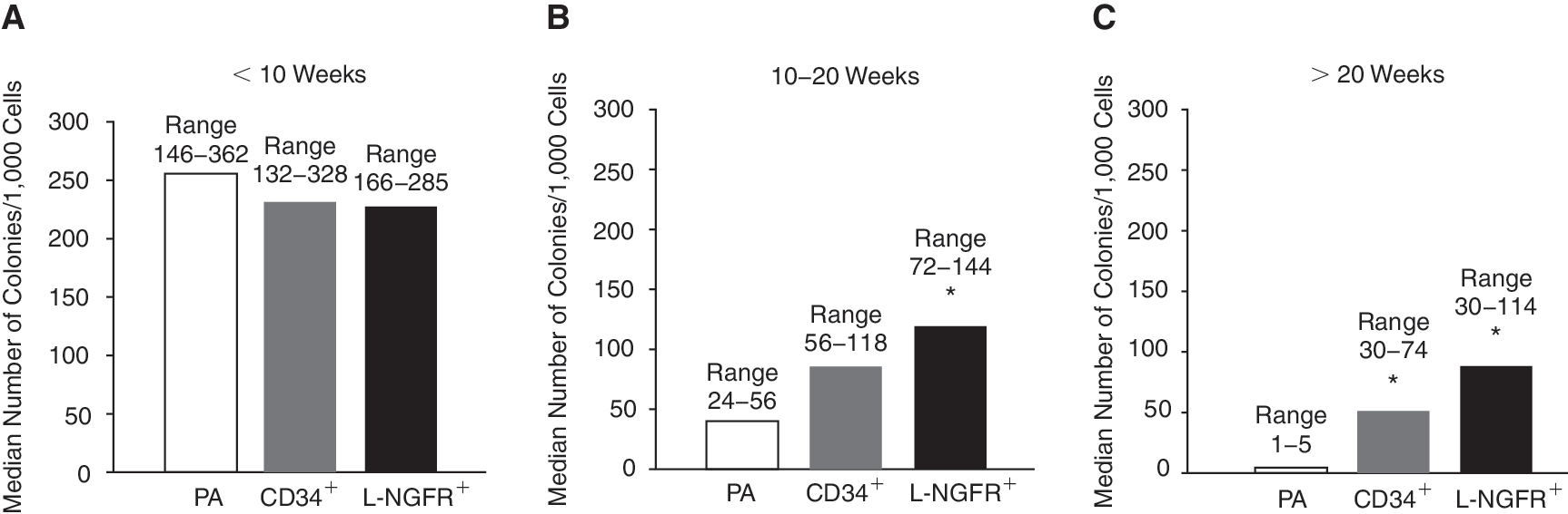
CFU-F activity of the 3 hASC fractions. Median number of colonies for 1 × 10³ cells evaluated at various passages: no significant differences among the 3 populations were observed within 10 weeks of culture (
At late passages, we observed a decrease in the colony-forming ability in all the 3 populations, particularly in PA hASCs. Indeed, between 10 and 20 weeks, the median number of PA colonies strongly decreased to 37 (range 24–56) and was significantly lower in comparison to L-NGFR+ cells (CFU-F median number: 118, range 72–144; P = 0.04), while CD34+ cells median number was 87 (range 56–118) (Fig. 3B). After 20 weeks of culture, PA ability to form colonies nearly disappeared (CFU-F median number: 3, range 1–5) and was significantly lower in comparison to both CD34+ (52, range 30–74; P = 0.04) and L-NGFR+ cells (88, range 30–114; P = 0.03) (Fig. 3C).
Proliferation ability
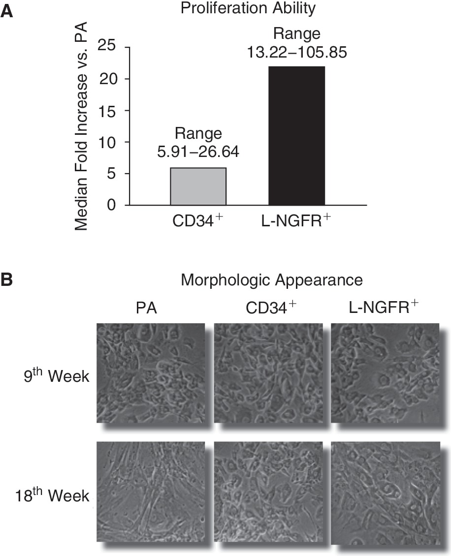
To characterize and compare the expansion ability of the different fractions, the cells were maintained in culture until senescence. The expansion rates varied widely among samples from different donors and exhausted within 20–25 weeks of culture. Differences in proliferation ability among the 3 fractions were not statistically significant, but in the long period PA cells showed a marked reduction in their dividing potential in comparison to the immunoseparated cells. In fact, both CD34+ and L-NGFR+ fractions showed high increments in cell proliferation when compared to PA: PA median fold increase (F.I.) was 39.13 (range 27–70), while the median F.I. versus PA (arbitrarily fixed as 1) was 6.04 (range 5.91–26.64) and 21.71 (range 13.22–105.85) for CD34+ and L-NGFR+ cells, respectively (n = 6) (Fig. 4A).
(
Morphologic appearance
At early weeks of culture, all the 3 populations showed the typical fibroblast-like aspect. After 15–20 weeks, the morphological appearance of the 3 fractions became quite different: PA hASCs grew with a larger and flatten shape, with cells gathered in clusters and cytoplasm rich in granules, in contrast to the immunoselected cells that still appeared homogeneous, spindle-shaped, and with well-defined shape and nuclei (Fig. 4B).
Hematopoietic progenitors evaluation
In order to investigate the presence of hematopoietic progenitors within CD34+ fractions isolated from adipose tissue, hematopoietic culture assays were performed to verify the ability of immunoselected CD34+ cells to form lineage-committed hematopoietic progenitors, according to their growth in semisolid cultures: CFU-GM and BFU-E.
After 14 days of culture, we never observed hematopoietic progenitors growth in none of the samples analyzed (n = 5): on methylcellulose medium, CD34+ cells always gave rise to a monolayer of cells with fibroblast-like morphology.
Differentiation ability
Adipogenic differentiation
When grown in adipogenic medium, hASCs from the 3 fractions showed clear-cut differences, as revealed by Oil Red-O staining and quantification (Fig. 5A). CD34+ and L-NGFR+ cells showed significant increments in differentiation ability compared to the PA unselected cells (arbitrarily fixed at 100%): the average increments were 55% ± 59% (P = 0.032) and 80% ± 86% (P = 0.031) for CD34+ and L-NGFR+ hASCs, respectively (n = 9). We analyzed the modulation of the adipogenic differentiation ability of the 3 populations at different periods during culture: Figure 5B shows a representative sample of the adipogenic potential of PA, CD34+, and L-NGFR+ hASCs isolated from a single donor. At early passages (<10 weeks of culture), the immunoselected populations possessed a greater ability to produce lipid droplets in comparison to PA cells. At later passages (>10 weeks), the adipogenic ability of the immunoselected cells was reduced, even if they still maintained greater levels of lipid vacuoles production compared to the unselected cells.

Assessment of adipogenic differentiation by lipid vacuoles quantification. (
Osteogenic differentiation
The 3 fractions of hASCs showed even more clear-cut differences in their extracellular calcified matrix deposition, as determined by Alizarin Red S staining and quantification. As shown in Figure 6A, CD34+ and L-NGFR+ hASCs showed a significant increase in their differentiation ability compared to the PA cells (arbitrarily fixed at 100%): the average increment was +386% ± 288% (P = 0.0037) for CD34+ hASCs, and +338% ± 235% (P = 0.0024) for L-NGFR+ cells (n = 9). In addition, this difference between the immunoselected and the PA hASCs populations was maintained in the long term, although the calcified matrix deposition strongly decreased for all of them during culture, as reported in Figure 6B where a representative hASCs sample derived from a single donor is shown. At later passages (>10 weeks), the 2 immunoselected populations behaved quite similarly, maintaining more abundant levels of extracellular calcium depots in comparison to PA cells.
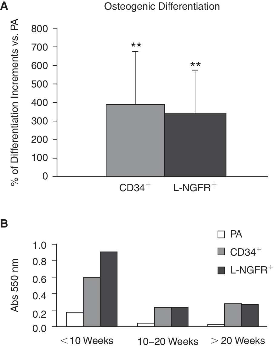
Evaluation of osteogenic differentiation by calcified extracellular matrix quantification. (
Chondrogenic differentiation assessment
The chondrogenic potential of the 3 hASCs populations was evaluated determining GAGs production in cells differentiated in a pellet culture condition. The immunopurified populations showed a similar differentiation potential when compared to PA hASCs, arbitrarily fixed at 100% (CD34+: +2% ± 35%; L-NGFR+: +22% ± 50%) (Fig. 7A) (n = 6). In Figure 7B, we analyzed GAGs production in the 3 hASCs populations isolated from a single donor and differentiated at different times during culture: at early passages (<10 weeks) L-NGFR+ cells produced higher amounts of GAGs in comparison to the other 2 fractions. However at later passages (>10 weeks), we observed a progressive decrease in GAGs production with no relevant differences among the populations.

Assessment of chondrogenic differentiation by glycosaminoglycans (GAGs) quantification. (
Analyzing the DNA content of each pellet to normalize the GAGs levels, we found that DNA in PA and CD34+ chondrogenic micromasses was about 2-fold higher compared to pellets cultured in control medium (Fig. 7C), whereas there was no significant difference between differentiated and undifferentiated L-NGFR+ micromasses (*CD34+ chondro versus CD34+ control: P = 0.027 and *PA chondro versus PA control: P = 0.034) (Fig. 7C).
Interestingly, a significant difference in GAGs production among the 3 populations was observed in micromasses cultured in control medium: indeed, undifferentiated CD34+ and L-NGFR+ cells produced higher levels of GAGs with an increase of +47% ± 32% (P = 0.007) and +81% ± 90% (P = 0.038) (n = 6), respectively, compared to PA undifferentiated micromasses, arbitrarily fixed at 1 (Fig. 7D).
Discussion
The potential utility of human adipose tissue as a source of adult stem cells for regenerative medical therapies needs more stringent isolation procedures and characterization both to improve the homogeneity of the population and to enhance the reproducibility of the results.
Indeed, adipose tissue is a highly heterogeneous tissue: donor-dependent differences have been demonstrated in cell yield, proliferation, and differentiation ability, most likely influenced by age [30,31 –33], sex, hormones, body mass index (BMI) [42,43], inflammatory and pathological conditions like osteoarthritis and diabetes [44,45]. In addition, Jurgens et al. [46] have recently demonstrated that not only differences exist among individuals, but also yield and functional features of hASCs are affected by the adipose tissue-harvesting site.
Despite the extensive characterization, there is still a lack of specific markers and methods to selectively isolate the more primitive cells from adipose tissue.
The results of our study show that the immunomagnetic sorting of hASCs by L-NGFR and CD34 antibodies allows the selection of 2 distinct subpopulations, which are more efficient in clonogenic and differentiative abilities compared to the whole population.
The enrichment of MSCs by a selection with the L-NGFR monoclonal antibody has been previously described in adult BM [34], where it defines a subset of cells with high proliferative, clonogenic, and multipotential differentiation ability. Recently, Yamamoto et al. [47] isolated and analyzed L-NGFR+ cells from mouse subcutaneous adipose tissue, showing that the rate of differentiation into adipocytes, osteoblasts, and neuronal cells was higher than for L-NGFR− ASCs.
In human adipose tissue, L-NGFR and CD34 MoAbs were able to identify 2 fractions expressing surface markers associated with a primitive phenotype. A high percentage of freshly isolated L-NGFR+ hASCs expressed the stem cell marker CD34 on the cellular membrane and variable but consistent expressions of CD117 (c-kit, the stem cell factor receptor) and CD105 (SH2). Fresh CD34+ cells showed a marked positivity for the endothelial-specific markers P1H12 and KDR, and variable co-expression of L-NGFR. Although CD34 is the human surface antigen identifying the population of BM hematopoietic stem cells, commonly used as a marker in clinical settings [48,49], in our experiments the lack of expression of the pan-leukocyte marker CD45 and the incapability of hASCs to give rise to hematopoietic colonies under specific stimuli allows us to exclude the hematopoietic origin of CD34+ adipose stem cells. As already reported, Lee et al. [50] isolated a population of muscle-derived CD34+ stem cells able to improve both muscle regeneration and bone healing, whereas Garcia-Pacheco et al. [51] found that human decidual stromal cells positive for both CD34 and STRO-1 are related to BM stromal precursors. Moreover, expression of CD34 had been already reported in BM MSCs, although it was rapidly lost after in vitro culture [34,52,53]. Accordingly, we observed that L-NGFR and CD34 expressions are progressively down-modulated during culture, in parallel with both the reduction of hASCs clonogenic and multi-differentiative ability and the acquisition of a mature fibroblast phenotype. The developmentally programmed loss of marker expression is reminiscent of what was observed in the case of the CD34 [54], L-NGFR [34], and Stro-1 [55] antigens on BM MSCs, supporting the hypothesis that they are markers of primitive cells.
Both immunoseparated cellular fractions showed higher proliferative ability throughout the time of culture compared to PA hASCs. Due to the wide interdonor variability, differences in proliferation among the fractions are not statistically significant, but reflect what previously observed in BM MSCs [34]. In addition, PA cells prematurely showed morphological signs of suffering and senescence.
At early passages, the clonogenic potential of the 3 populations was similar, but a significant difference was observed at later passages, when the immunoseparated fractions longer maintained the ability to form colonies.
Adipogenic differentiation ability of PA hASCs was pronounced throughout the time of culture, probably due to a precommitment of adipose-derived stem cells. However, during the early weeks of culture, adipogenic differentiation was statistically different between selected and unselected cells.
Differences among the 3 fractions are more evident in the osteogenic differentiation: the immunoselected hASCs showed a clearly higher differentiation ability, particularly during the early passages in culture. Despite a common decrease in calcium deposition during culture, the selected fractions always maintained higher levels than PA hASCs at first passages.
All the populations behaved similarly in term of chondrogenic ability, even if during the first weeks of culture L-NGFR+ cells seemed to possess a greater differentiation potential. Nevertheless, the immunoselected subpopulations better responded both to the physical stimuli and to the ipoxia condition of the 3-dimensional culture, positively affecting the chondrogenic differentiation [56]. Indeed, just the 3D condition induced CD34+ and L-NGFR+ cells to produce similar or even higher levels of GAGs than PA hASCs cultured in chondrogenic medium. No differences in GAGs production were observed between L-NGFR+ micromasses cultured in control and in chondrogenic medium: this could be due to the greater ability of the selected hASCs to respond to the 3D physical stimuli that could partially mask the differentiative effect of chondrogenic medium.
Furthermore, DNA quantification showed a higher proliferative effect of the chondrocyte-inductive TGF-β on PA and CD34+ fractions in comparison to L-NGFR+ cells, maybe depending on the greater heterogeneity of these populations, which contain primitive cells able to differentiate, but also cells only able to respond to the proliferative stimuli provided by the growth factor.
Altogether, our data suggest that the selection with anti-L-NGFR MoAb allows us to obtain a more homogeneous and primitive population: the almost total co-expression of CD34, the expression of the stem markers CD117 and CD105, and the higher proliferative, clonogenic, and differentiative potential, in particular at early passages, seem to support this observation. The role of L-NGFR in MSCs is still unclear, although its expression has been described in BM and in other tissues [34,57] and it has been shown to be involved in several functions including morphogenesis [58 –61], growth factor presentation [62], and apoptosis in response to NGF stimulation [63].
We observed a higher heterogeneity in CD34+ population, with <30% of cells co-expressing L-NGFR and low levels of CD117 and CD105 expression. In the last years, some authors isolated from human adipose tissue CD34+/CD31− cells including adipocytes progenitor cells and CD34+/CD31+ cells defined as capillary endothelial cells [18,64]. They proposed that adipocytes and endothelial cells might share a common precursor, as also suggested by Planat-Benard et al. [19], which could play a determinant role in the excessive development of the adipose tissue by contributing to neo-vascularization and to the apparent adipocyte hyperplasia. Moreover, Traktuev et al. [65] recently described a hASCs subset (CD34+/CD140a+/CD140b+/CD31−/CD45−/CD117−/CD144−) with pericytic properties that participate in vascular stabilization by mutual structural and functional interactions with endothelial cells, maintaining the ability to differentiate into multiple lineages. These evidences suggest that CD34+ hASCs could be preferentially used for bone tissue engineering, due to their ability to promote the neo-vascolarization process, which is known to be a critical point in the healing of bone defects.
These days, in the field of regenerative medicine, physicians prefer one-step surgical procedures in order to reduce patients' discomfort, risks of pathogens transmission, and social costs. In this context, the possibility to rapidly select subpopulations of cells more prone to differentiate into a specific lineage would be very advantageous.
In conclusion, our results suggest that both subpopulations could be useful tools in regenerative medicine applications; nevertheless, best results could be obtained by investigating the interaction between immunoselected hASCs and specific biomaterials, either in a static or in a dynamic culture condition, in order to achieve the most functional cell-scaffold construct.
Footnotes
Acknowledgments
Authors would like to thank Dr. Franz W. Baruffaldi Preis, Dr. Luciano Lanfranchi, and Dr. Matteo Sartori for their contribution to the work.
Author Disclosure Statement
No author has proprietary interest or conflict of interests in connection with the present manuscript.