
Abstract
Therapies based on stem cells have shown very attractive potential in many clinical studies. However, the data about the safety of stem cells application remains insufficient. The present study was designed to evaluate the overall toxicology of human umbilical cord mesenchymal stem cells (hUC-MSCs) in cynomolgus monkeys with repeated administrations. hUC-MSCs were administered by intravenous injection once every 2 weeks for 6 weeks. The dose levels employed in this study were 2×10⁶, 1×10⁷ cells/kg body weight. Toxicity was evaluated by clinical observations (body weight, body temperature, and ophthalmology exams), pathology (blood cell counts, clinical biochemistry, urinalysis, and bone marrow smears), immunologic consequences (lymphoproliferation assay, the secretion of interferon-γ and interleukin-4, the percentage of CD3, CD4, CD8 T cells, and the ratio of CD4 and CD8 T cells) and anatomic pathology. Pharmacodynamics in blood and distribution of hUC-MSCs in the tissues of cynomolgus monkeys were measured by real-time polymerase chain reaction. All animals survived until scheduled euthanasia. No stem cells transplantation-related toxicity was found in this study. hUC-MSCs could be found in the blood of cynomolgus monkeys beyond 8 h. The findings of this 6-week toxicity study showed that the transplantation of hUC-MSCs did not affect the general health of cynomolgus monkeys.
Introduction
M
Preclinical [7 –9] and clinical studies [10 –12] have shown that transplantation of MSCs was an alternative therapy for heart failure. Administration of MSCs has been successful in several clinical applications for bone repairing in fracture and metabolic bone disease [13]. Infusion of MSCs expanded in vitro appeared to be a potentially effective treatment for patients with steroid-resistant, acute graft versus host disease [14]. A study in mice revealed that adipose tissue-derived MSCs facilitated hematopoiesis in vitro and in vivo [15]. MSCs derived from umbilical cord [16 –18] or bone marrow [19,20] were considered as a new therapy for severe and treatment-refractory systemic lupus erythematosus.
Although MSCs derived from different tissues including bone marrow [19,20] and umbilical cord [16 –18] have shown therapeutic potentials in clinical studies, we still required more scientific data to evaluate the risk of human MSCs transplantation. The present study was designed to perform an overall toxicological evaluation of human umbilical cord mesenchymal stem cells (hUC-MSCs) in cynomolgus monkeys with repeated administrations.
Materials and Methods
Preparation of hUC-MSCs
Umbilical cord was obtained following the ethical guidelines after written informed consent from the donor. hUC-MSCs were isolated from the umbilical cord of a healthy donor by the method previously described [3]. The isolated cells were cultured in DMEM/F12 with 10% fetal bovine sera (FBS) at 37°C with 5% CO2 and subcultured when they reached 80% to 90% confluence. hUC-MSCs were harvested from cultures at the second passage (P2) and cryopreserved in 90% FBS plus 10% dimethyl sulfoxide (DMSO) as master cell bank. To produce cells for experiments, cells from master cell bank (P2) were thawed and subsequently cultured through 5 passages (P7, including 2 passages before cryopreservation) and 11 passages (P13, including 2 passages before cryopreservation). At the end of P7 and P13, UC-MSCs were harvested and cryopreserved in a cocktail of 85% Plasma-Lyte A (Baxter), 10% DMSO, and 5% human serum albumin. Immediately before the treatment, hUC-MSCs were thawed out in a water bath at 37°C and suspended in a saline vehicle solution (Plasma-Lyte A containing 2% DMSO, 1% human albumin, and 20.9 IU/mL heparin). Cells were greater than 70% viable by trypan blue exclusion [21].
Animals
We performed the study under the guidelines set by institutional animal care and use committee and also with the approval from the Institutional Animal Care and Use Committee of JOINN Laboratories. Sixteen male and 16 female cynomolgus monkeys were ∼3 years of age and ranged in weight from 2.25 to 3.02 kg. Upon arrival, cynomolgus monkeys were placed in quarantine for 54 days. The animal room was environmentally controlled to maintain a temperature of 18°C–26°C, a humidity of 40%–70%, and a 12-h light cycle [22].
Dosing and experimental design
Thirty-two cynomolgus monkeys were randomly divided into 5 groups: the negative control group, the saline vehicle control group, the UC-MSCs P7 low dose group, the UC-MSCs P7 high dose group, and the UC-MSCs P13 high dose group. Six monkeys were evaluated per group in this study except for the UC-MSCs P7 high dose group. There were 8 monkeys in the UC-MSCs P7 high dose group, of which 2 additional monkeys were evaluated for engraftment 2 weeks after the last administration (Table 1). Cynomolgus monkeys were administrated via i.v. infusion with hUC-MSCs or placebo once every 2 weeks for 4 times at a speed of 6–8×10⁵ cells/min (MSC P7 low dose group) or 3–4×10⁶ cells/min (MSC P7 high dose and MSC P13 high dose group). Animals were euthanatized on the day of the last treatment, 2 weeks after the last treatment (only the UC-MSCs P7 high dose group) and 6 weeks after the last treatment following the schedule.
Negative control group received infusion of 9% sodium chloride.
Clinical observations and pathology
Clinical observations (including recording unscheduled death, behavioral activity, food intake, character of stool, and whether there is erythema, edema, induration, ulcer, purulence on injection sites) were performed every day. Body weights measurements were conducted weekly. Body temperatures and electrocardiograms were monitored prior to the treatment, 2–3 h after the first injection, after the last administration and the end of the recovery period (6 weeks after the last treatment). Ophthalmology exams were performed before treatment and prior to euthanasia [23].
Clinical pathology evaluations concerned blood cell counts (red blood cell, reticulocyte, hemoglobin, hematocrit, mean corpusular hemoglobin, mean corpusular volume, mean corpusular hemoglobin concerntration, platelet, white blood cells, neutrophilicgranulocyte, lymphocyte, monocyte, eosimophil, and basophil), coagulation parameter (activated partial thromboplastin time and prothrombin time), clinical biochemistry (alanine amiotransferase, albumin, albumin/globulin, alkaline phosphatas, aspartate aminotransferase, blood urea nitrogen, calcium, chloride, cholesterol, creatine kinase, creatinine, glucose, kalium, sodium, total bilirubin, total protein, triglyceride, and γ-glutamyl transpeptadase) and urinalysis (acidity, bilirubin, blood, color, glucose, ketone, leucocyte, nitrite, protein, specific gravity, and urobilineogen) [24,25].
Immunologic consequences
Interferon (IFN)-γ and interleukin (IL)-4 in serum were measured by ELISA on the day after the last treatment. The percentage of CD3, CD4, CD8 T cells, and the ratio of CD4/CD8 T cells were analyzed by FACSCalibur at the day after the last treatment. At the time points before the third treatment, the last treatment, and the end of recovery period, blood samples were collected for determining the presence of antibodies to hUC-MSCs by ELISA [26].
Pharmacodynamics in blood and engraftment in tissues
Analysis of human cells in cynomolgus monkeys was based on the detection of DNA sequence specific to homo sapiens by real-time polymerase chain reaction (PCR). Peripheral blood samples (∼1 mL) were collected from cynomolgus monkeys which received hUC-MSCs administrations prior to the first treatment, 5 min, 30 min, 1, 4, 8, 24, 48, 96, 144, 192, and 336 h after the first treatment. Total DNA of blood samples were extracted for real-time PCR. For the UC-MSCs derived from homo sapiens, the samples were analyzed by real-time PCR targeting human specific DNA sequence. If there was no detectable human specific sequence at a certain time point, the samples were not analyzed any more after the time point.
Genomic DNA of brain, heart, lung, liver, kidney, spleen, quadriceps, and iliac marrow were extracted for determining the presence of hUC-MSCs by real-time PCR on the day after the last treatment, 2 weeks after the last treatment (only the MSCs P7 high dose) and the end of the recovery period.
Anatomic pathology
Four cynomolgus monkeys from each group (20 animals in all) were euthanized after the last treatment. Two animals from the UC-MSCs P7 high dose group were euthanized 2 weeks after the last treatment. Six weeks after the last treatment, 2 animals from each group (10 animals in all) were euthanized. Bone marrow was taken from the sternum for smear analysis. The weight of brain, thymus, thyroid gland (included parathyroid gland), liver, kidney, adrenal gland, spleen, lung, heart, testes, epididymis, ovary, womb, and prostate were recorded. Organ coefficients were calculated based on the weights of the organ and the body. Organs or tissues were fixed for histopathological processing.
Statistical analyses
Data are presented as mean and standard deviations. Group variances were compared using Levene's test at the 0.05 significance level. When differences between group variances were not found to be significant, a parametric one-way analysis of variance (ANOVA) was performed. If the variances were unequal, each group was then tested by Games-Howell pairwise comparison test. Changes over time within each group were evaluated by using ANOVA for repeated measurement. A value of P<0.05 was considered statistically significant. When a significant change was detected, each treatment group was then tested for difference from the control group using the Dunnett's test [27].
Results
Clinical observation and general toxicity studies
No unscheduled death was observed during this study. Two animals in the negative control group, 1 of 6 animals in the UC-MSCs P7 low dose group and 2 animals in the UC-MSCs P13 high dose group had short term diarrhea. For the appearance in the negative control group, the diarrhea could not be related to the treatment of hUC-MSCs. Two cynomolgus monkeys in the saline vehicle control group and one cynomolgus monkey in the group of UC-MSCs P7 low dose group vomited after treatment. No other treatment related clinical observation among the animals was found in this study. There was also no significant change in body weight (Supplementary Table S1; Supplementary Data are available online at
Clinical pathology
The results of Dunnett's test showed that the decrease of serum cholesterol in the UC-MSCs P7 low dose group and UC-MSCs P7 high dose group were statistically significant against the negative control group (Fig. 1). There was no any other statistically significant change in blood cell counts (Fig. 2, Supplementary Fig. S1), coagulation (Supplementary Table S7), clinical biochemistry (Supplementary Figs. S2 and S3) or urinalysis.
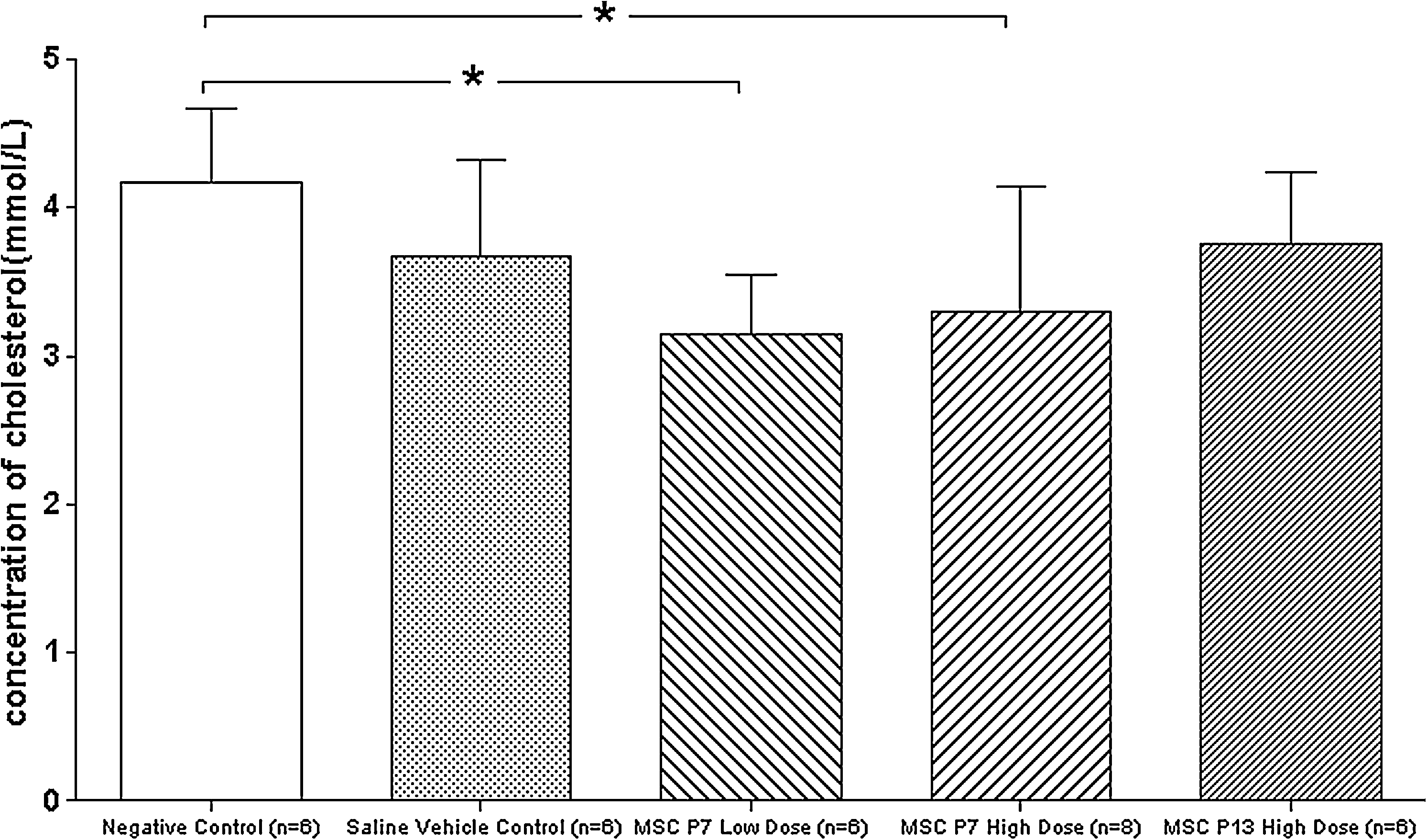
Effect of multiple-administration human mesenchymal stem cells to Cynomolgus monkeys on the concentration of cholesterol in serum. Dunnett t-test treated negative control group as control, and compared all other groups against it. The decrease of serum cholesterol values in the mesenchymal stem cells P7 low (p=0.018) and high dose (p=0.032) group were statistically significant against negative control group. Data were means. Bars were standard deviations. * indicated p<0.05 vs. negative control group.
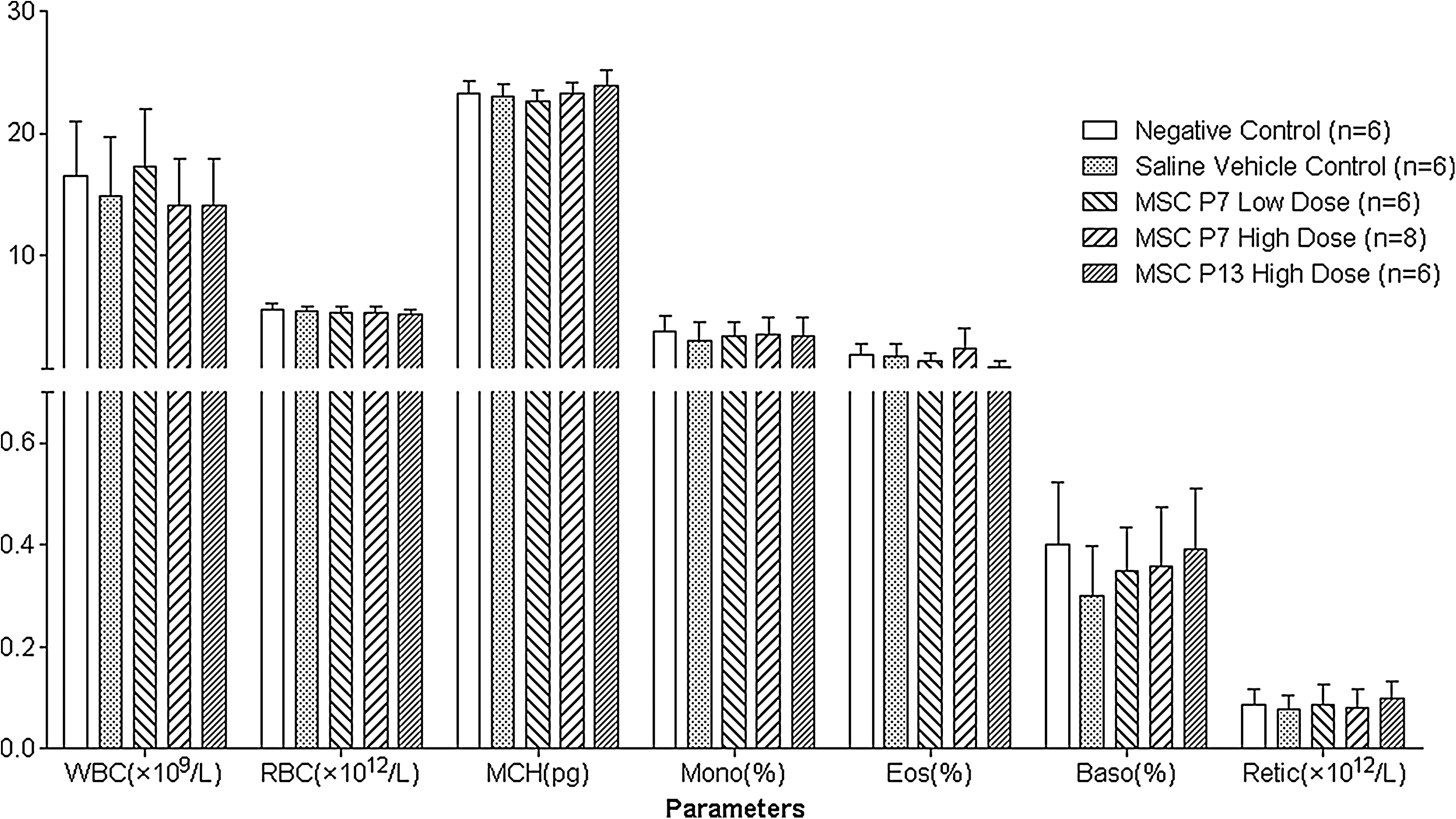
Mean value of blood cell counts from Cynomolgus monkeys following administrations of human mesenchymal stem cells. The mean value of a specific group was calculated from all of the different time points. WBC, white blood cell count; RBC, red blood cell count; MCH, mean corpusular hemoglobin; Mono, monocyte; Eos, eosimophil; Baso, basophil; Retic, reticulocyte. Data were means. Bars were standard deviations.
Immunologic consequences
Sera from all animals were evaluated for the production of antibodies binding to hUC-MSCs. Antibody production to hUC-MSCs is summarized in Table 2. None of the saline vehicle control group was positive for binding to hUC-MSCs, as expected. Less than half of the monkeys which received the administration developed antibody to hUC-MSCs. The incidence of antibody exhibited a dose-dependence tendency.
The incidence of antibody to hUC-MSCs was presented by the no. of monkeys which are positive for hUC-MSCs/the no. of monkeys which are measured in all.
hUC-MSC, human umbilical cord mesenchymal stem cell.
In the saline vehicle control, UC-MSCs P7 low dose and UC-MSCs P13 high dose group, there was one monkey which was positive for IFN-γ in the sera respectively. No monkey developed detectable IFN-γ in the negative control or UC-MSCs P7 high dose group. All of the cynomolgus monkeys in this study were negative for IL-4.
CD3, CD4, and CD8 T cells were analyzed by FASCalibur. There was no significant change in the subpopulation of T-cells on the basis of flow cytometric analysis (Fig. 3).
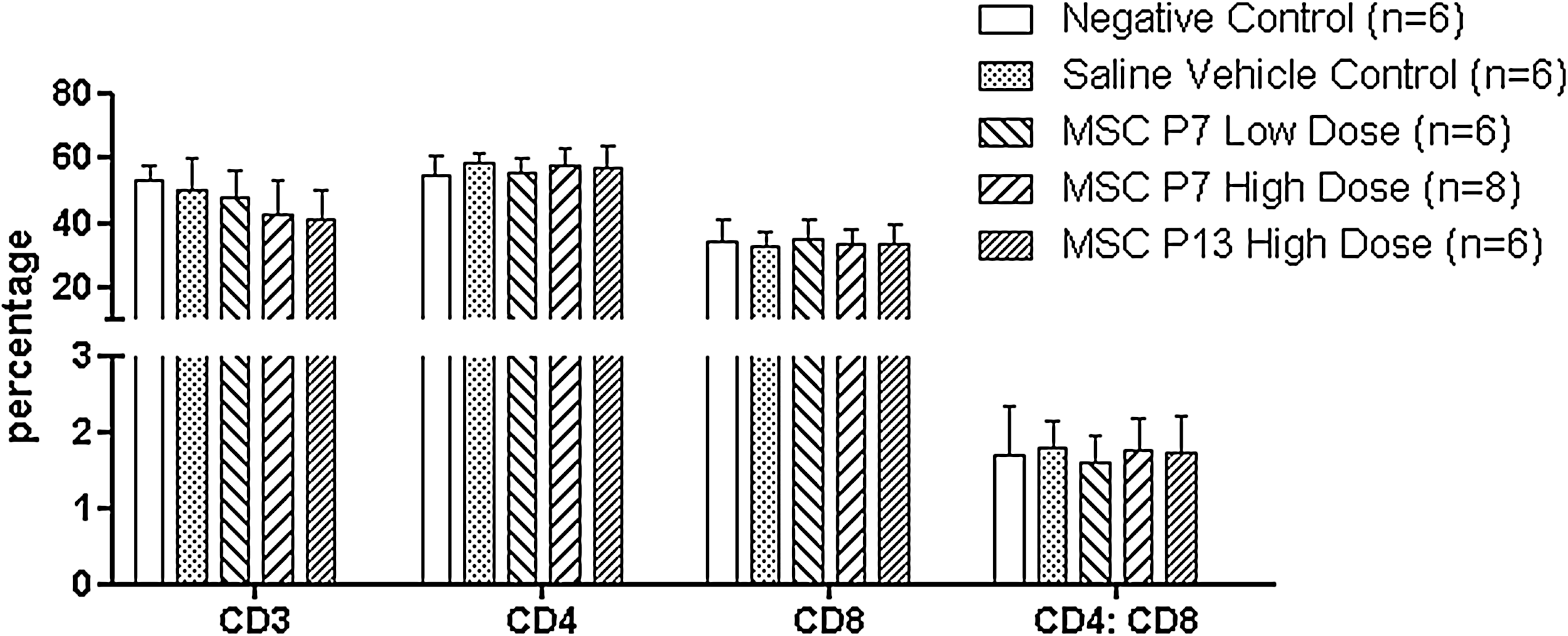
Effect of human mesenchymal stem cells to Cynomolgus monkeys on the subpopulation of T-cells. There was no significant change in the subpopulation of T-cells. Data were means. Bars were standard deviations.
Pharmacodynamics in blood and distribution in tissues
At 5 min after the first treatment, human specific sequence was detected in all of the blood samples from the cynomolgus monkeys which received hUC-MSCs administration. At the time point of 30 min after the injection, the concentration of human cells in the monkey's blood reached the highest point. Pharmacodynamics of hUC-MSCs in the UC-MSCs P7 high dose group is shown in Fig. 4. At the time of 4 h after the first treatment, none of the monkeys in the UC-MSCs of P7 low dose group, one monkey in the UC-MSCs P7 high dose group and 2 monkeys in the UC-MSCs P13 high dose group were positive for human sequence. Eight hours after the first treatment, there was no detectable human specific sequence in the blood of all cynomolgus monkeys (Supplementary Tables S8–S12 and Supplementary Figs. S4 and S5).
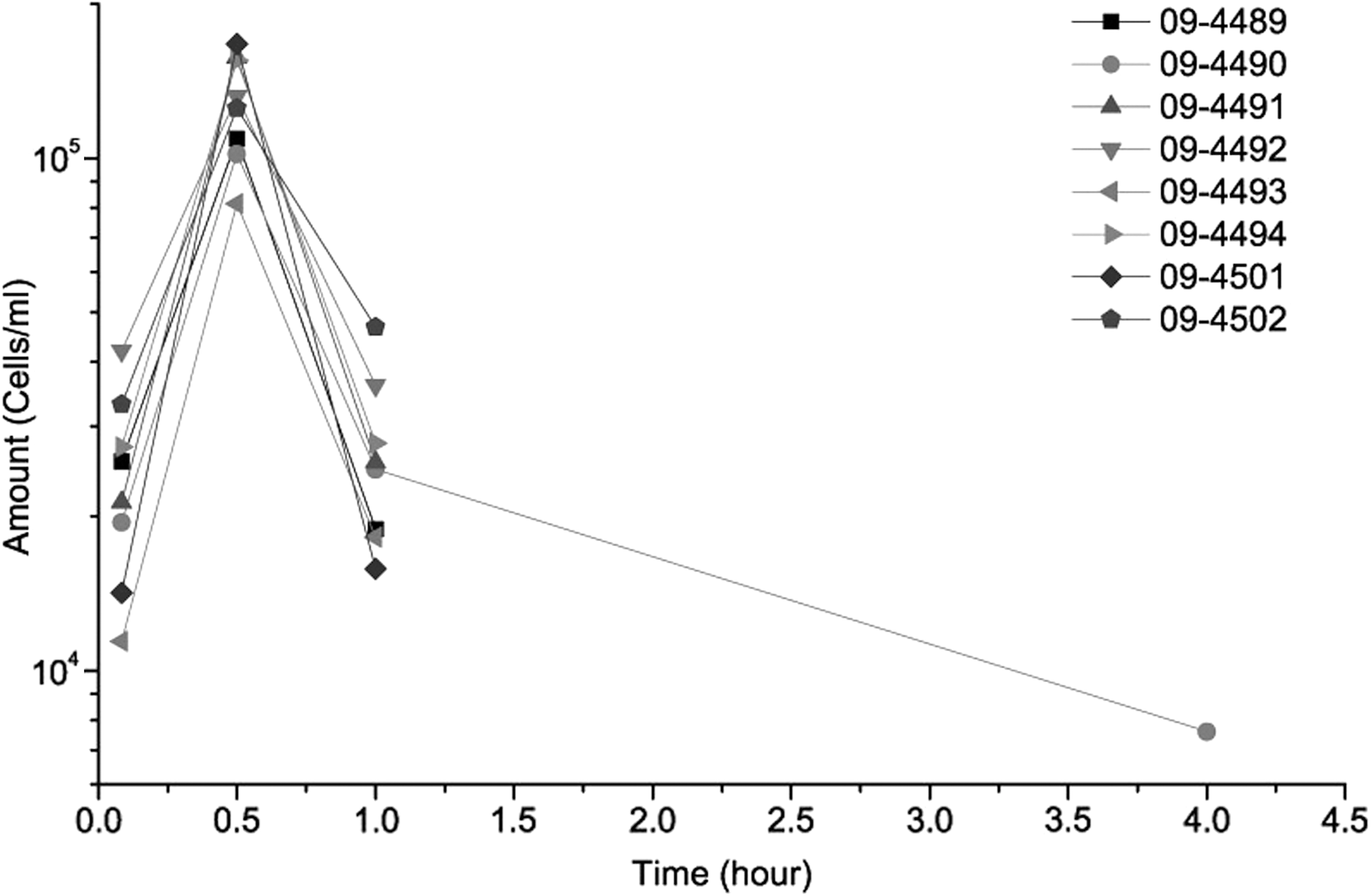
Concentration-time curves of mesenchymal stem cells P7 high dose group human mesenchymal stem cells were eliminated from the blood of Cynomolgus monkeys in 8 hours.
The distribution of the genetic sequence of homo sapiens in the tissue of monkeys which received hUC-MSCs administration was analyzed by real-time PCR. On the day after the last treatment, 4 monkeys were positive for human genetic sequence. Human MSCs were found in heart, brain, liver, lung, quadriceps, and iliac marrow. Two weeks after the last treatment, human genetic sequence was not found in any tissue of the cynomolgus monkeys.
Anatomic pathology
There was no significant change in organ weight or organ coefficient. In the negative control group, a transparent parasitic cyst was found in the lung of one cynomolgus monkey. Another animal in this group got irregular pupil edge. In the saline vehicle control group, a focal brown crater appeared in the right occiput of one monkey. In the UC-MSCs P13 high dose group, a small cyst (0.3×0.2×0.2 cm) was found in the left ovarian of one monkey. Another monkey in the UC-MSCs P13 high dose group got sporadic petechiae in both the lungs. There was not any macroscopic change in the UC-MSCs P7 low dose group and the UC-MSCs P7 high dose group.
A summary of the microscopic finding is given in Supplementary Table S13. Being consistent with the macroscopic findings, lens atrophy was observed in the animal with irregular pupil edge (Fig. 5A). Deposition of brown pigment granules was found in the occipital lobe of the right brain of the monkey with focal occipital depression (Fig. 5B). Spontaneous background pathological findings including mononuclear cell infiltration (into liver, kidney, or heart), cysts (in thymus or ovary) and so on were observed in each group of this study including negative control group [28]. Most of the other cynomolgus monkeys administrated by hUC-MSCs did not show any pathological change (Fig. 5C–M).There was no microscopic finding interpreted to be related to the administration of hUC-MSCs. Changes at the infusion site attributed to the experimental procedures were observed in animals from all groups including the negative and saline vehicle controls.
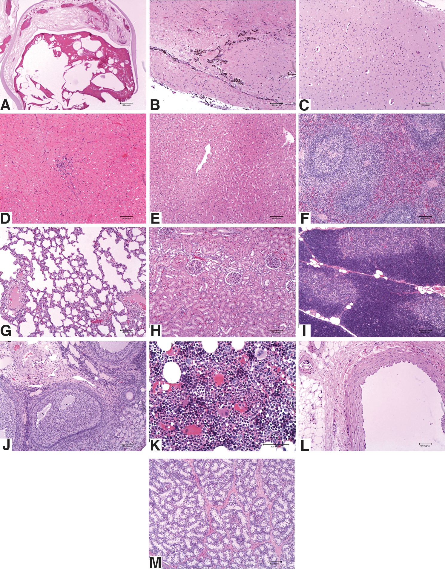
Effect of human mesenchymal stem cells to Cynomolgus monkeys on anatomic pathology. Hematoxylin & Eosin staining revealed that most of Cynomolgus monkeys administrated by human mesenchymal stem cells showed normal histology. The scale bars were 100 micron.
Discussion
In the recent study, we discussed the toxicology of hUC-MSCs transplantation in cynomolgus monkeys to improve the understanding of potential toxicity of human MSCs in primates and enhance our confidence in clinical application of cellular therapies. Cynomolgus monkeys were chosen in the present experiments for their close phylogenic relationship to humans that may potentially more accurately reflect the risk from MSCs transplantation in human than any other animals. The appearance of a homogeneous sample was the main reason why only a single donor was used in this study as a source of UC-MSCs. Based on the results of immunophenotype analysis and differentiation studies, hUC-MSCs used in this study met the basic criteria of MSCs. However, the safety of hUC-MSCs from multiple human donors should be evaluated in further studies. For gaining an overall estimation of the toxicology of human MSCs, we focused on the clinical signs, body weights, body temperature, electrocardiogram, routine hematology, coagulation, clinical biochemistry, urinalysis, ophthalmological examination, percentage of subset of T cells, concentration of IFN-γ and IL-4 in sera, engraftment of hUC-MSCs, and anatomic pathology after euthanasia.
MSCs in this study were isolated from human umbilical cord. After 2 passages, the culture appeared to be homogeneous and a monolayer formed. We cryopreserved these adherent cells as master cell bank. During extensive expansion, UC-MSCs continued proliferating without change in growth and morphology. Jo et al. have reported that UC-MSCs exhibit no evidence of senescence at least for 9 passages [29]. hUC-MSCs evaluated in this study expressed CD73, CD90, and CD105, as measured by flow cytometry. Additionally, these cells lacked expression of CD19, CD34, CD11b, CD45, and HLA-DR (Supplementary Fig. S6). They were able to differentiate into osteoblasts and adipocytes (Supplementary Fig. S7). hUC-MSCs at the seventh passage were chosen in this study since the number of UC-MSCs obtained after 7 passages was sufficient for clinical application and for the whole in vivo study of 32 monkeys. The dosage used in the studies was based upon published reports concerning the transplantation of UC-MSCs in clinical applications [14,18,30 –33].
Transplantation of hUC-MSCs did not affect the body weight, body temperature, and electrocardiogram of cynomolgus monkeys. A few of animals in this study had short term diarrhea and vomiting after treatment. Since the symptoms were also found in negative control and saline vehicle control group, the side effects observed in some monkeys seemed not correlate to UC-MSCs administration. Nausea and vomiting have been previously observed in some clinical transplantation of hematopoietic stem cells [34]. Possible cause for vomiting is the infusion of DMSO [35,36]. However, nausea was also observed in some studies about transplantation of UC-MSCs without the infusion of DMSO [18]. More research is therefore needed to clarify the occurrence of nausea and vomiting following stem cells transplantation.
Previous studies have demonstrated that intracranial injection of UC-MSCs can transiently increase the levels of white blood cells in female infant rhesus macaques. However, the levels of white blood cells diminished back after 30 days posttransplantation, which remained at least 6 months [37]. In this study, we did not find any statistical significant change in blood cell counts. The discrepancy may come from the different statistical methods. We adopt ANOVA for repeated measures which provided ANOVA when the same measurement was made several times on each subject. We found that the decrease of serum cholesterol in the UC-MSCs P7 low dose group and the UC-MSCs P7 high dose group was statistically significant. However, there was no other data related to the decrease of cholesterol in this study. The relationship between decrease of serum cholesterol and administration of UC-MSCs needs to be clarified in further research.
IL-4 is a key regulator in adaptive immunity. The treatment of hUC-MSCs was not sufficient to induce IL-4 synthesis. For the small number of animals with detectable level of IFN-γ and the absence of IFN-γ in the sera of the UC-MSCs P7 high dose group, it is insufficient to make the induction of IFN-γ related to hUC-MSCs administration. Groups treated with hUC-MSCs and control groups shared the same phenotype of the subset of T-cells. The data of immunotoxicity testing indicated that administrations of hUC-MSCs did not affect over all immune system of monkeys, which is consistent with the result of allogeneic transplantation of UC-MSCs in baboons [21]. However, cynomolgus monkeys which received allogenic or xenogenic stem cells transplantation may develop immune-sensitivity toward the transplanted cells. When the same cells are transplanted into the same animals, they are likely to be rejected rapidly. Lymphocytes of cynomolgus monkeys at various time points following hUC-MSCs injection should be taken and mixed culture with human MSCs to see the proliferation responses in further study.
The fate of hUC-MSCs transplanted in cynomolgus monkeys was demonstrated by detecting homo sapiens specific DNA sequence. Eight hours after the treatment, hUC-MSCs were eliminated from the blood of the animals. However, on the day after the last treatment, homo species specific sequences were found in organs or tissues of some monkeys. Two weeks after the last treatment, there was no hUC-MSCs in the tissue of all the treated recipients. Our study demonstrate that hUC-MSCs after intravenous administration were able to migrate into brain, heart, lung, liver, quadriceps, and iliac marrow of cynomolgus monkeys, whereas kidney and spleen from all of animals were negative for human DNA sequence. Being constant with the study of tissue distribution of human umbilical cord matrix stem cells in SCID mice [38], we concluded that hUC-MSCs could cross the blood brain barrier of cynomolgus monkeys. We did not find any pathologic change in the tissue with human DNA sequence. Further studies will be needed to determine whether hUC-MSCs can survive in the local tissue environment and how to improve the migration of hUC-MSCs into the organs of animals with long-term persistence.
Taken together, our study of 6-week toxicity indicates that the transplantation of hUC-MSCs does not affect the general health of cynomolgus monkeys and that transplanted hUC-MSCs could migrate into some organs of recipient monkeys. Our data cannot support that hUC-MSCs survival in the body of cynomous monkeys.
Footnotes
Acknowledgments
This study was supported by 863 projects from the Ministry of Science & Technology of China (2006AA02A110; 2011AA020118) and specialized fund for scientific and technical Innovation in Tianjin (08FDZDSH02900).
Author Disclosure Statement
There is no potential conflict of interest.
References
Supplementary Material
Please find the following supplemental material available below.
For Open Access articles published under a Creative Commons License, all supplemental material carries the same license as the article it is associated with.
For non-Open Access articles published, all supplemental material carries a non-exclusive license, and permission requests for re-use of supplemental material or any part of supplemental material shall be sent directly to the copyright owner as specified in the copyright notice associated with the article.