
Abstract
The therapeutic potential of multipotent stromal cells (MSC) may be enhanced by the identification of markers that allow their discrimination and enumeration both in vivo and in vitro. Here, we investigated the ability of embryonic stem cell-associated glycosphingolipids to isolate human MSC from both whole-bone-marrow (BM) and stromal cell cultures. Only SSEA-4 was consistently expressed on cells within the CD45loCD105hi marrow fraction and could be used to isolate cells with the capacity to give rise to stromal cultures containing MSC. Human stromal cultures, generated in either the presence or absence of serum, contained heterogeneous cell populations discriminated by the quantity of SSEA-4 epitopes detected on their surface. A low level of surface SSEA-4 (SSEA-4lo) correlated with undetectable levels of the α2,3-sialyltransferase-II enzyme required to synthesize SSEA-4; a reduced proliferative potential; and the loss of fat-, bone-, and cartilage-forming cells during long-term culture. In vitro, single cells with the capacity to generate multipotent stromal cultures were detected exclusively in the SSEA-4hi fraction. Our data demonstrate that a high level of surface epitopes for SSEA-4 provides a definitive marker of MSC from human BM.
Introduction
H
Glycolipids are a family of hydrophobic molecules that consist of both lipid and carbohydrate moieties. These membrane-bound molecules participate in the formation of lipid rafts and other cell surface microdomains and are thought to be important for cell adhesion and signal transduction [10]. Changes in surface glycolipid composition have been associated with stage-specific embryonic differentiation, as well as in numerous diseases such as cancers and Guillain-Barré syndrome [11]. More recently, glycolipid epitopes have been associated with pluripotent stem cells and some somatic stem cell types [10,12 –14]. Antibody labeling of the glycolipids SSEA-1, SSEA-3, and SSEA-4, in particular, has been accepted as a method for identifying pluripotency in embryonic and induced pluripotent stem cells. In addition to marking human pluripotent stem cells, epitopes for SSEA-4 have also been associated with neural and hematopoietic stem cells; however, there have been mixed reports of SSEA-4 association with MSC.
We initially hypothesized that glycolipid markers of pluripotent stem cells would also allow the identification and purification of human MSC. To explore this, the surface levels of SSEA-1, SSEA-3, and SSEA-4 were examined on BM cells enriched for MSC. Only SSEA-4 was detectable at high levels (SSEA-4hi) and could be used to FACS-purify in vivo MSC. In vitro, stromal cells with clonal capacity and the ability to maintain multipotent differentiating function in long-term culture were found exclusively in the SSEA-4hi cell fraction. Overall, this study demonstrates that human MSC can be enriched based on the presence of high levels of surface SSEA-4.
Materials and Methods
BM stromal cultures
Human BM was purchased from Lonza Walkersville, an establishment registered under the FDA for the processing of human cells and tissue, as well as cellular- and tissue-based products, in accordance with the U.S. Code of Federal Regulations (21 CFR Par 1271). Human tissues provided by Lonza are obtained from various tissue suppliers and recovery agencies according to the Institutional Review Board-approved protocols and informed consent that allow the use of obtained tissues for general research purposes. Human stromal cell cultures were initiated by plating 1×10⁶ cells per mL either in a low-glucose Dulbecco's modified Eagle's medium (DMEM; Invitrogen/GIBCO BRL) with 15% fetal bovine serum (FBS) qualified for human MSC (HyClone-Thermo-Fisher) [serum-containing (SC) medium] or in a serum- and animal component-free medium provided by Stem Cell Technologies, herein referred to as serum-free (SF) medium.
Flow cytometry and cell sorting
Whole-BM or cultured stromal cells were filtered through a 70-μm cell strainer (BD Bioscience) and resuspended in phosphate-buffered saline (PBS)/2% FBS at 1×10³ cells/μL. Cells were stained with fluorochrome-conjugated monoclonal antibodies (mAb) to human CD105-allophycocyanin (APC) (SN6); CD34-phycoerythrin-Cy7 (PE-Cy7) (4H11); CD45-fluorescein isothiocyanate (FITC) (H130); CD90-PE-Cy5.5 (5E10); SSEA-4-Alexa488 (MC-813-70); SSEA-3-PE (MC-631); SSEA-1-APC (MC-480; eBioscience), and CD73-PE (AD2; BD Bioscience), according to the manufacturer's instructions. Flow cytometric analysis was completed on a minimum of 5×10⁶ (whole BM) or 3×10⁴ (cultured stroma) viable cells using the LSR II instrument (BD Bioscience), and the data were analyzed using FLOWJO™ software (TreeStar, Inc.). Fluorescence-activated cell sorting (FACS) was completed on a MoFlo™ instrument equipped with an Automated Cell Deposition Unit (ACDU; Beckman Coulter). For restaining, purified SSEA-4hi and SSEA-4lo cells were washed with PBS/2% FBS and stained with anti-SSEA-4-Alexa488. Restained cells were analyzed on LSR II.
Multipotent differentiation cultures
The stromal cells were differentiated into adipocytes, osteocytes, and chondrocytes using the Human MSC Functional Identification Kit from R&D Systems. Briefly, to initiate adipocyte and osteocyte formation, stromal cells were cultured in either the SC or SF medium in 24-well plates using 2.1×10⁴ cells/cm2 and 4.2×10³ cells/cm2 cells, respectively. The medium containing supplements to allow the differentiation of adipocytes or osteocytes was added when cells reached 100% or 50% confluence, respectively. The medium was changed every 3–4 days over a 10–28-day period. For chondrogenic differentiation, 1.25×10⁵ of cultured stromal cells were grown in 15-mL polypropylene conical tubes with the DMEM/F12 medium containing chondrogenic supplements. The medium was changed every 3 days for 17–21 days. Adherent cells and pellets were fixed in 4% paraformaldehyde and either stained directly (adipocytes/osteocytes) or cryosectioned before staining (chondrocytes). Sections of 8–10 μm were placed on charged glass slides (VWR). For chemical detection of adipocytes, fixed cells were treated with 60% isopropanol for 10 min and were stained with Oil Red O for 5 min (Sigma-Aldrich). Osteocytes were identified by staining fixed cells with 0.2% Alizarin red stain at pH 6.36–6.4 (Baker) for 60 min. The cryosectioned pellets acquired from chondrocyte differentiation cultures were stained in Alcian blue stain, pH 1.0, for 15 min. Stained cells were washed thoroughly with distilled water and visualized under a light microscope (Zeiss) equipped with an Axiom camera.
Reverse transcriptase–polymerase chain reaction
Total RNA was extracted from a minimum of 40,000 SSEA-4hi and SSEA-4lo stromal cells using the RNeasyPlus Mini Kit (Qiagen) (n=3). The RNA was concentrated using the RNeasyMinElute Cleanup Kit (Qiagen), and equal amounts from SSEA-4hi and SSEA-4lo cells were used to synthesize cDNA with the Superscript III First-Strand Synthesis System for reverse transcriptase–polymerase chain reaction (RT-PCR; Invitrogen). Primers used for PCR were hST3GAL2-F-2 5′-GCACAAGCTCGCCTGACCCAGA-3′ and hST3GAL2-R-2 5′-CTTCACCCGGTGCGTCCCATC-3′. cDNA was amplified over 32 cycles using the AmpliTaq Gold 360 Master Mix (Invitrogen) under the following conditions: 95°C for 5 min followed by 95°C for 30 s; 60°C for 1 min; and 72°C for 1 min, with a final extension 72°C for 7 min. PCR products were run on a 1% agarose gel and visualized with SYBR Safe DNA gel stain (Invitrogen).
Cell expansion and BrdU incorporation
FACS-purified SSEA-4hi and SSEA-4lo cells were seeded at 200,000 cells in T-75-cm2 flasks (n=3). After an 18-h period, cultures were pulsed with 10 μM BrdU once a day for 2 consecutive days. Cells were then harvested, fixed, and stained with a BrdU-specific antibody according to the manufacturer's instructions (BD Biosciences).
Immunohistochemistry
Cells were fixed with 4% paraformaldehyde and treated with a blocking buffer containing PBS/1% BSA containing 10% donkey serum and 0.3% Triton X-100. Fixed cultures and cryosections (chondrocytes) were stained with goat anti-human FABP-4- (adipocytes), osteopontin- (osteocytes), or sheep anti-human collagen II- (chondrocytes) specific polyclonal antibodies as provided in the MSC Functional Identification Kit. Undifferentiated stromal cells were stained with mouse anti-human SSEA-4 (MC-813-70; 1:100) or rat anti-mouse/human SSEA-3 (MC-631; 1:100) for visualization of internal and membrane-bound antigens. Northern Lights™ 557-conjugated secondary antibodies raised in donkey (R&D Systems) were used for fluorescent labeling of primary antibodies. PBS containing 1% BSA, 10% donkey serum, and 10% normal goat serum was used as a negative control for nonspecific secondary binding. To provide a negative control for osteocyte and adipocyte assays, undifferentiated stromal cells, grown in a normal medium, were stained with anti-SSEA-3 or anti-SSEA-4. Samples were mounted with the ProLong Gold antifade reagent containing DAPI (Gibco/BRL) and analyzed using a Zeiss fluorescent microscope equipped with an argon lamp. Photographs of both samples and negative controls were acquired using the same exposure times.
Results
MSC are enriched among CD45loCD105hi BM cells that have high levels of surface epitopes for SSEA-4
To investigate the potential for markers of pluripotency to identify MSC in vivo, the presence of immunoepitopes for SSEA-1, SSEA-3, and SSEA-4 was examined by flow cytometry on the MSC-enriched CD45loCD105hi fraction of BM from 3 separate donors (Fig. 1A). The level of fluorescence detected from antibodies to SSEA-1 and SSEA-3 (blue) was equivalent to that of unstained cells (red) in all samples tested. In contrast, a population of SSEA-4hi cells was consistently detected within the CD45loCD105hi BM fraction in each of the 3 donors. Overall, the mean proportion of SSEA-4hi cells detected within the CD45loCD105hi population was 39%±11% (Fig. 1B). To determine whether in vivo MSC have high or low levels of surface SSEA-4, FACS-purified CD45loCD105hiSSEA-4hi and CD45loCD105hiSSEA-4lo BM cells were cultured in SF culture conditions known to support human stromal cell expansion. Adherent cells with the typical morphology of stroma were detected only in cultures seeded with BM cells derived from the SSEA-4hi subfraction. The same results were obtained when SSEA-4hi and SSEA-4lo cells were purified and cultured from the CD45lo BM fraction alone. These data demonstrate that only SSEA-4hi BM cells are capable of generating stromal cell cultures in the SF medium formulated to maintain MSC.

Multipotent stromal cells (MSC) are enriched in the CD45loCD105hiSSEA-4hi bone marrow (BM) mononuclear cell (MNC) fraction. MNC were isolated from the BM of 4 separate donors and stained with antibodies specific to CD45, CD105, and SSEA-1, SSEA-3, or SSEA-4.
To demonstrate the presence of MSC, single cells were purified from one of the 3 CD45loCD105hiSSEA-4hi-generated cultures and deposited into 96-well culture plates using ACDU. Colonies were identified in a total of 12 wells from 3 single-cell-seeded 96-well plates. Each of these colonies expanded to sufficient numbers for flow cytometric analysis and was found to express the stromal cell markers CD73, CD90, and CD105, but lacks expression of the hematopoietic cell marker CD45 as represented in Fig. 1C.
Of the 12 colonies generated from single-cell cultures, only one was capable of sufficient expansion to allow analysis of multipotent differentiation potential (>1×10⁶ cells). This single-cell-originating culture was found to contain progenitors with the capacity to form mature adipocytes, osteocytes, and chondrocytes under the appropriate conditions (Fig. 1C, side panel). This capacity to detect single multipotent stem cells in stromal cultures derived from CD45loCD105hiSSEA-4hi BM cells demonstrates that MSC can be isolated in vivo based on high levels of surface epitopes for SSEA-4.
Stromal cultures containing MSC are comprised of a heterogeneous population of cells with varied levels of surface SSEA-4
To determine if the quantity of SSEA-4 on the surface of cultured stromal cells was related to MSC function in vitro, BM stromal cultures were derived from 12 separate donors using both SC and SF culture conditions. The presence of MSC was examined in early-passage (P1) cultures using flow cytometric and multipotent differentiation assays. Results from the analysis of cultures derived from 3 of the 12 BM donors are presented in Fig. 2. All cultures consisted of pure stromal cells lacking hematopoietic (CD45-expressing cells) and endothelial [CD34-expressing cells (not shown)] contamination, but expressing CD105, CD73, and CD90 surface markers. Slight variations in the mean fluorescence intensity of these markers were identified between donors and were detectable in both SF and SC cultures. However, in all cultures, >97% of cells were found to express stromal cell markers using gates established from unstained controls (top panel). The presence of MSC was confirmed in all cultures by the detection of adipocyte, osteocyte, and chondrocyte precursors in multipotent differentiation assays. Taken together, this analysis supports previous studies demonstrating that pure stromal cultures containing MSC can be generated from human BM using both SF and SC conditions.
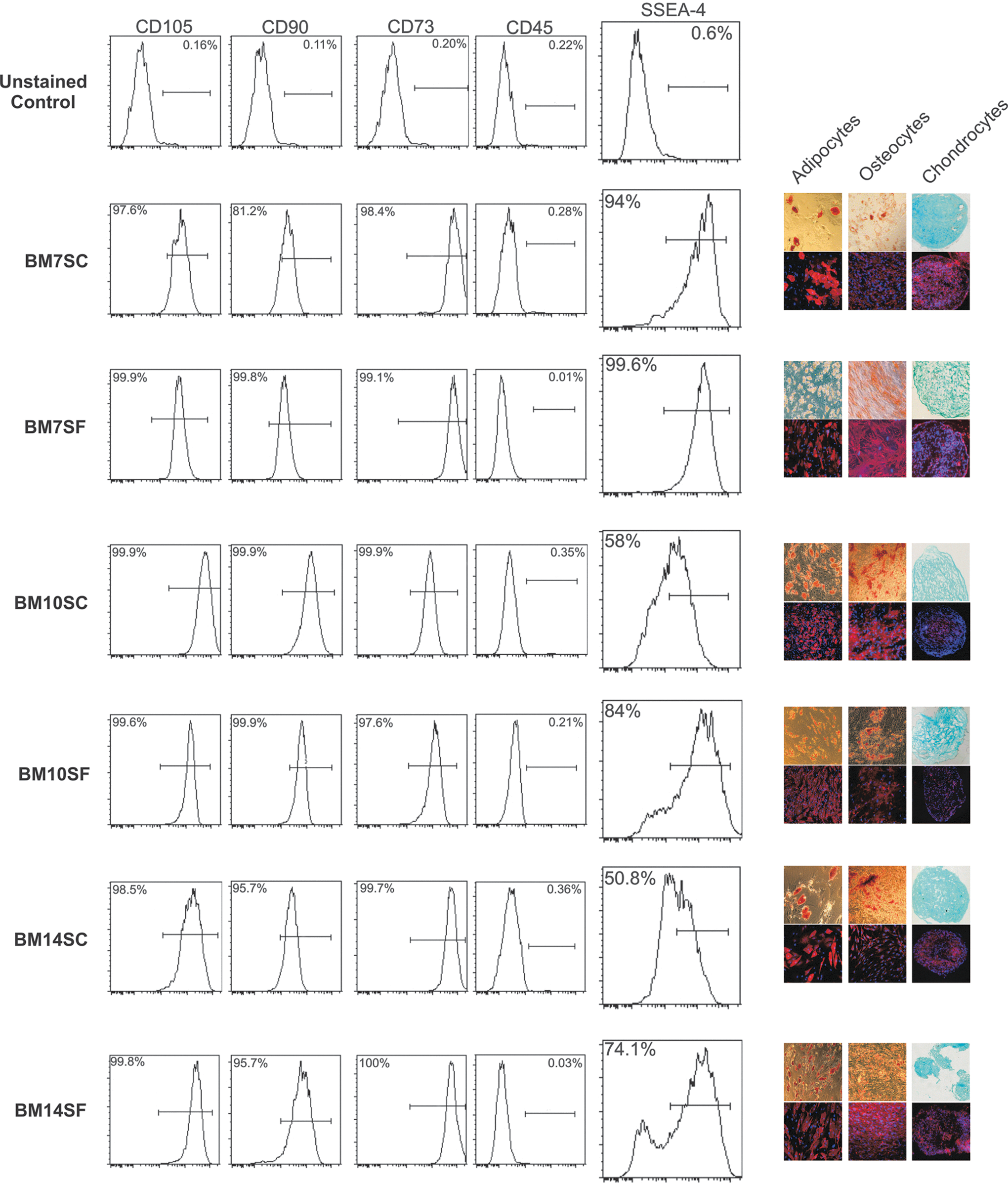
Human BM stromal cultures containing MSC are comprised of both the SSEA-4hi and SSEA-4lo cell populations. BM MNC were extracted from 12 separate donors and cultured in either serum-containing (SC) or SF conditions to support MSC. Early-passage (P1–P2) adherent cells were analyzed by flow cytometry for surface markers of stromal cells and epitopes of SSEA-4 (histograms). Negative controls (top panel) consisting of unstained stromal cells were used to establish positive cell gates. The presence of multipotent progenitors was examined using culture conditions that support the formation of adipocytes, osteocytes, and chondrocytes. Light and fluorescent images (right panel) show the typical level of chemical (Oil red O, Alizarin red, and Alcian blue) and immunohistochemical (anti-FABP, anti-osteopontin, and anti-collagen II) staining detected from each sample. Cell nuclei were visualized with DAPI (blue). Data from 6 representative samples are shown.
To compare the proportion of cells within MSC-containing stromal cultures that carry surface epitopes for SSEA-4, cells from each of the 12 early-passage cultures were stained with anti-SSEA-4 and examined by flow cytometry. All percentages were determined based on gates established from unstained controls (top panel). SSEA-4hi cells were detected in all cultures at proportions ranging greatly depending upon the donor. Percentages ranged from as high as 99.6% (BM7SF) to as low as 11% (BM1SC-data not shown). Cells with a low level of surface epitope were also consistently detected, with some cultures containing a distinct population with an MFI similar to that of unstained controls [BM1SC, BM11SC/SF, BM12 SC/SF (not shown), BM10SF, BM14SF (Fig. 2), and BM 4SF (Fig. 3)]. Interestingly, in SF and SC cultures derived from the same donor, the percentage of SSEA-4hi cells was consistently higher in cultures lacking serum. Overall, flow cytometric analysis determined that SSEA-4hi cells are consistently detected in stromal cultures containing MSC at proportions that vary with culture conditions as well as by donor.

High levels of SSEA-4 epitope identify stromal cells with the capacity to produce SSEA-4. BM-derived stromal cultures from 3 separate donors were harvested and stained with anti-SSEA-4.
High levels of SSEA-4 epitope correlate with the detection of both SSEA-3 and α2,3-sialy-transferase II enzyme necessary for the expression of SSEA-4
SSEA-4 is a glycosphingolipid that is generated from SSEA-3 by the enzymatic addition of N-acetylneuraminic acid by the α2,3-sialy-transferase II enzyme (α-STII) [15]. As such, heterogeneous SSEA-4 epitope levels among stromal cells may be explained by the presence of functionally distinct populations that either lack or maintain the capacity to produce SSEA-4. Alternatively, the presence of surface SSEA-4 epitopes may constantly fluctuate on stromal cells, irrespective of their function. To address these possibilities, we purified the SSEA-4hi and lo stromal populations from 3 SF and 3 SC cultures by FACS and compared both their capacities to express SSEA-4 and differentiate into fat, bone, and cartilage. Cells were consistently isolated at >90% purity, as demonstrated flow cytometric analysis of sorted cells restained with anti-SSEA-4 (Fig. 3A: SSEA-4hi, 97%±3%; SSEA-4lo, 92%±4%). Purity of sorted populations was further examined by immunohistochemistry (IHC). Fluorescence due to antibody staining of the SSEA-4 epitope was detected on purified SSEA-4hi cells (green), whereas neither intracellular nor extracellular epitope was detected in SSEA-4lo-sorted cells.
To examine the potential capacity of SSEA-4hi and SSEA-4lo stromal cells to produce SSEA-4, we compared the presence of SSEA-3 (Fig. 3B) and the expression of α-STII (Fig. 3C) among these purified populations. SSEA-4hi and SSEA-4lo cells were isolated from 3 separate cultures (2 SF and 1 SC) and either deposited directly on 12-well plates containing coverslips or used to isolate total RNA. For immunohistochemical analysis, purified stromal cells were allowed to adhere on coverslips overnight before fixation and staining with anti-SSEA-3. SSEA-4hi cells consistently showed patchy staining for SSEA-3 (red) that was not detected in any SSEA-4lo samples. These data infer that SSEA-4lo cells lack the glycolipid precursor of SSEA-4.
To compare α-STII expression, equal amounts of RNA from the SSEA-4hi- and SSEA-4lo-sorted populations were used for RT-PCR. The transcript encoding the α-STII enzyme was detected only in SSEA-4hi-purified cells as shown in the representative gel shown in Fig. 3C. Together, flow cytometric, IHC, and RT-PCR data suggest that 2 populations of cells exist in BM stromal cultures that can be identified and isolated using SSEA-4 epitopes and differ by the presence or absence of machinery necessary to produce the SSEA-4 glycolipid.
Multipotent progenitor function during long-term culture is maintained by SSEA-4hi stromal cells
To examine potential differences in their multipotent differentiating capacity, stromal cells purified based on surface levels of the SSEA-4 epitope were compared using in vitro differentiation assays. Both the SSEA-4hi and SSEA-4lo cell populations were found to contain adipocyte, osteocyte, and chondrocyte progenitors; however, visual differences in the overall number and size of adipocytes were detectable (Fig. 4A). This difference in adipocyte-differentiating potential was consistent for the SSEA-4hi and SSEA-4lo populations derived from either the SF or SC medium. These data demonstrate that surface SSEA-4 epitope levels do not discriminate adipocyte, osteocyte, or chondrocyte precursors from early-passage BM stromal cultures.

SSEA-4hi and SSEA-4lo stroma represent functionally distinct cell populations in vitro. SSEA-4hi and SSEA-4lo stromal cells were purified from early-passage SC and SF cultures by FACS and either analyzed directly in multipotent differentiation assays
To further compare the functional properties of SSEA-4hi and SSEA-4lo stromal cells, we examined their proliferative potential and capacity to expand and maintain multipotent progenitors in long-term cultures. In our hands, the proliferation rates of early-passage stromal cells differ in the presence and absence of serum (∼24 h in SF; ∼36 h in SC). As such, the proliferation and expansion of SSEA-4hi and SSEA-4lo cells from SF and SC cultures were compared separately.
The proliferative potential was measured in freshly isolated cells by BrdU incorporation over 48 h (Fig. 4B). For this experiment, the SSEA-4hi and SSEA-4lo populations were purified from 3 separate SC cultures, and equal numbers of cells (200,000) were allowed to recover overnight in the culture medium. Adherent cells were pulsed with BrdU immediately after recovery and again 24 h later. Forty-eight hours after the initial pulse, stromal cells were harvested, stained with BrdU antibodies, and analyzed by flow cytometry. BrdU incorporation was detected in 55%±4% of SSEA-4hi cells compared to 25%±5% of SSEA-4lo cells, demonstrating a statistically significant difference in the proliferative potential of these 2 stromal populations (P<0.05). The increased proliferative potential of SSEA-4hi cells correlated with a greater number of cell generations during long-term culture, as represented in Fig. 4B for 2 of the 4 cultures analyzed. Expansion potential was compared between SSEA-4hi and SSEA-4lo cells isolated from early-passage SF and SC cultures by monitoring the number of generations (cell doublings) until the point of senescence. For these experiments, we defined senescence as the point at which <60% viable cells were detected upon harvest. While SSEA-4hi and SSEA-4lo cultures reached senescence at the same time point, SSEA-4hi-seeded cultures consistently underwent a greater number of cell generations over time. At the time of senescence SSEA-4hi cells, isolated from SF and SC cultures, respectively, had undergone 13.7 and 13.5 generations in total (Fig. 4C). By comparison, SSEA-4lo cells underwent 11.0 (SF) and 11.2 (SC) generations. Taken together, these data suggest that increased presence of surface epitopes for SSEA-4 correlates with the proliferative and expansion potential of cultured BM stromal cells.
The ability to maintain a pool of mature progenitor cells with the capacity to differentiate into fat, bone, or cartilage is one purported function of MSC [16]. As such, depletion of MSC function within stromal cultures is likely to result in the loss of multipotent progenitor cells [17]. To examine whether changes in the surface SSEA-4 epitope are associated with enrichment for MSC in vitro, changes in surface SSEA-4 and the presence of adipocyte, osteocyte, and chondrocyte progenitors were compared in long-term cultures derived from unsorted, SSEA-4hi, and SSEA-4lo stromal cells. Figure 4D depicts typical results obtained from the long-term culture of cells in either the SC or SF medium. Flow cytometric analysis of SSEA-4 epitopes determined that purified SSEA-4hi cells had given rise to both SSEA-4lo and SSEA-4hi populations at proportions similar to those seen in unsorted cultures. Purified SSEA-4lo cells were also capable of giving rise to SSEA-4hi cells, but at proportions significantly lower (2–3-fold) than that of SSEA-4hi cells. SSEA-4hi cells became depleted with culture age, regardless of whether cells were derived from unsorted or purified stromal populations. However, in all cases, SSEA-4hi-derived cultures and unsorted stroma showed near-identical SSEA-4 surface epitope profiles until senescence and were divergent from SSEA-4lo-seeded cultures. Specifically, the percentage of SSEA-4hi cells 7 days before senescence (day 28 for SF/day 35 for SC) ranged from 8% to 20% in unsorted and SSEA-4hi-derived cultures and from 2% to 10% in SSEA-4lo cultures. These data demonstrate that the proportion of SSEA-4hi cells within stromal cultures decreases concomitantly with the culture age. In addition, only SSEA-4hi cells are capable of regenerating stromal cultures with a surface epitope profile similar to that of the original culture.
Both the SSEA-4hi and SSEA-4lo populations maintained the capacity to differentiate into adipocytes and osteocytes after 7 days in culture. Chondrocyte differentiation potential was maintained until culture senescence was reached, regardless of the population from which the cells were derived (data not shown). The increased number of adipocytes seen from freshly isolated SSEA-4lo cells compared to SSEA-4hi stroma was also maintained over the first 7 days of culture, but was only slightly increased compared to unsorted stroma (Fig. 4D, panels). However, after 14 days, the overall amount of adipocyte-specific staining (Oil Red O and FABP+) was visibly similar in multipotent differentiation assays derived from SSEA-4hi, SSEA-4lo, and unsorted stromal cultures. Both osteocyte and adipocyte progenitors remained detectable in both unsorted and SSEA-4hi-derived cultures for the entire 28-day culture period. In contrast, adipocytes were only sporadically detected in SSEA-4lo-derived day-28 cultures. In addition, osteocyte differentiation was no longer detectable by either Alizarin red or antiosteopontin staining. Overall, flow cytometric and multipotent differentiation comparison of SSEA-4hi, SSEA-4lo, and unsorted stromal cultures suggests that the cells responsible for maintenance and replenishment of multipotent progenitors in long-term cultures are present in a fraction of cells expressing high levels of the SSEA-4 surface epitope.
In vitro clonal MSC are present in the SSEA-4hi stromal subfraction
As the presence of MSC can only be definitively determined by generating cultures of multipotent progenitors from a single cell, the clonal capacity of SSEA-4hi and SSEA-4lo stromal cells was compared by ACDU purification into 96-well plates. The results of experiments completed on purified populations from 2 SF and SC cultures are presented in Table 1. Overall, an equal number of SSEA-4hi and SSEA-4lo cells was deposited from both culture conditions (SC=1,248; SF=1,536). Cultures were monitored microscopically every 2 days to examine cell growth. Any wells found to contain more than 1 cell at day 0, or that generated more than 1 definitive colony, were not included in further analysis. Single colonies arising in 96-well plates were plucked and expanded sequentially in 24- and 6-well plates. Cultures those reached 70% confluence were further transferred to T-25 flasks, and eventually to T-75 flasks, if at least 200,000 cells were generated. Multipotent differentiation assays were completed on all clonal cultures in which at least 1×10⁶ cells were generated. SSEA-4hi cell-derived colonies were detected and harvested from a total of 30 and 38 wells seeded from SC and SF cultures, respectively. All colonies expanded to >10,000 cells when transferred. One SC-derived and 2 SF-derived colonies were capable of expansion to >1×10⁶ cells. Adipocyte, osteocyte, and chondrocyte progenitors were detected in each of these 3 single-cell-derived cultures. In contrast, SSEA-4lo cells generated only 2 colonies from SC cultures, whereas no colonies were detected from single SF culture-derived cells. Furthermore, the 2 colonies identified from SC SSEA-4lo cells showed only minimal expansion when harvested and transferred, resulting in <10,000 generated cells. The results of clonal culture assays demonstrate that single cells with the capacity to expand and differentiate into fat, bone, and cartilage precursors are present only in the SSEA-4hi fraction. Overall, our work provides strong evidence that, in culture, MSC can be distinguished from other stromal cell types based on the high levels of the surface epitope for SSEA-4.
Less than 10,000 cells generated.
Adipocyte, osteocyte, and chondrocyte progenitors identified by multipotent differentiation assays.
SC, serum containing; SF, serum free.
Discussion
Glycolipids have been detected on the surface of several types of human stem cells, including pluripotent stem cells [18,19], multipotent (somatic) stem cells [20 –23], and cancer stem cells [24 –26]. As such, glycolipids have been proposed as biologically and clinically important markers that will allow the identification and isolation of stem cells [10]. The work presented here demonstrates that antibodies raised to the SSEA-4 glycosphingolipid can be successfully used to purify MSC from both whole-BM and marrow-derived stromal cultures. SSEA-4 has been detected on the surface of several different stem cell types identified in the BM, such as mesodermal progenitor cells [27], marrow-isolated adult multilineage-inducible cells (MIAMI) [28], and multipotent adult progenitor cells [29]. These cell types differ based on the in vitro growth conditions required for their maintenance and expansion as well as in their differentiation potential and growth characteristics. While the relationship between each of the mesenchymal cell types found in BM cultures is currently unclear, it is interesting that SSEA-4 expression is a common characteristic of each. These observations suggest that SSEA-4 is a ubiquitous marker of BM stem cells with a potentially important role in their function.
There have been reports suggesting that antibodies raised to glycolipids are prone to nonspecific binding and recognition of glycoproteins and proteoglycans [10]. In light of this, it is important to note that the anti-SSEA-4 clone utilized for the identification and isolation of MSC in our study (MC-813-70) has been previously shown to specifically recognize SSEA-4 using thin-layer chromatography and mass spectrometry methods for glycolipid detection [12,15,30]. The ability of the MC-813-70 antibody clone to specifically recognize surface SSEA-4 is also supported by our identification of both SSEA-3 and the α-STII enzyme in SSEA-4hi-purified stromal cells. Thus, our use of MC-813-70 epitopes to clonally purify highly proliferative stromal cells with multipotent differentiating function provides strong evidence that SSEA-4 is present on the surface of MSC.
The presence of MSC in the SSEA-4hi fraction of human BM was previously demonstrated by Gang et al. [8]. Our study verifies these findings and further defines the surface phenotype of BM-derived MSC as CD45loCD105hiSSEA-4hi. In addition, the MC-813-70 antibody clone utilized in our study is different than that used in the work presented by Gang et al., thus providing an additional support for the validity of our findings. In the current study, the frequency of BM CD45loCD105hiSSEA-4hi cells was highly variable among the 4 donors characterized. In light of this, it is interesting to hypothesize that SSEA-4 can provide a useful tool for enumerating MSC in vivo. Verification of this will require further studies comparing the CFU frequency with the proportion of SSEA-4hi cells within the CD45loCD105hi fraction from a large number of BM donors. The characterization of SSEA-4 epitopes in conjunction with other known MSC surface markers, such as STRO-1 [7] and CD146 [9], may also provide further insights.
There have been conflicting reports regarding the presence of SSEA-4 on the surface of cultured human stroma ranging from a lack of detectable epitopes [31] to the detection of epitopes on >95% of cultured cells [8]. Our study provides evidence that SSEA-4 epitopes are detectable in human BM stromal cultures, but that the proportion of SSEA-4hi cells varies significantly between donors, culture medium, formulation, and culture age. Thus, the reported discrepancies regarding SSEA-4 on cultured stroma may be due to differences in culture conditions or the analysis of a limited number of cell lines as well as the antibody clones used in each study. Regardless, our work demonstrates that human stromal cultures are comprised of 2 functionally distinct populations that can be distinguished based on the levels of surface SSEA-4. Furthermore, clonal MSC, with the capacity to maintain multipotent progenitors during long-term culture, are found exclusively in the SSEA-4hi cell fraction. It will be interesting to determine whether other functional properties attributed to MSC, such as immune suppression and hematopoietic stem cell maintenance, are also enriched among SSEA-4hi stromal cells. Overall, as a marker of MSC both in vitro and in vivo, SSEA-4 epitopes may provide a useful means of increasing both our biological understanding these stem cells as well as their utility in the clinic.
Footnotes
Acknowledgments
The authors would like to acknowledge Dr. Aaron Farnsworth and Dr. Sean Li for their kind review of the manuscript. As well, we would like to thank Mr. Paul Oleynik for his assistance with cell sorting and ACDU experiments.
Author Disclosure Statement
The authors have no competing financial interests.