
Abstract
Identification and isolation of pluripotent stem cells in adult tissues represent an important advancement in the fields of stem cell biology and regenerative medicine. For several years, research has been performed on the identification of biomarkers that can isolate stem cells residing in neural crest (NC)-derived adult tissues. The NC is considered a good model in stem cell biology as cells from it migrate extensively and contribute to the formation of diverse tissues in the body during organogenesis. Migration of these cells is modulated, in part, by gap junction communication among the cell sheets. Here we present a study in which, selection of connexin 43 (Cx43) expressing cells from human adult periodontal ligament yields a novel pluripotent stem cell population. Cx43+ periodontal ligament stem cells express pluripotency-associated transcription factors OCT4, Nanog, and Sox2, as well as NC-specific markers Sox10, p75, and Nestin. When injected in vivo into an immunodeficient mouse model, these cells were capable of generating teratomas with tissues from the three embryological germ layers: endoderm, mesoderm, and ectoderm. Furthermore, the cells formed mature structures of tissues normally arising from the NC during embryogenesis such as eccrine sweat glands of the human skin, muscle, neuronal tissues, cartilage, and bone. Immunohistochemical analysis confirmed the human origin of the neoplastic cells as well as the ectodermal and endodermal nature of some of the structures found in the tumors. These results suggest that Cx43 may be used as a biomarker to select and isolate the remnant NC pluripotent stem cells from adult human tissues arising from this embryological structure. The isolation of these cells through routine medical procedures such as wisdom teeth extraction further enhances their applicability to the regenerative medicine field.
Introduction
Researchers have worked on the identification and isolation of remnant embryonic-like cells from adult tissues [4 –6]. One of the main focuses has been on tissues arising from the neural crest (NC) [7,8]. The embryonic NC is a transient structure originating at the dorsal region of the neural primordium and serves as a good model in stem cell biology. Cells from the NC migrate extensively, populate various organs and tissues, and give rise to cells from all three embryological germ layers [7 –10]. Modulation of NC cell pluripotency and their highly migratory nature has been an active subject of research for years [9]. NC cells are derived from the dorsal neuroepithelium by an epithelium to mesenchymal cell transformation [9,11] and have been shown to migrate as sheets and streams rather than single or aggregated cell clusters [11 –15]. In prospective migratory NC cells, there is a transition from tight junctions to gap junctions [16]. The migratory conformation of NC cells has been attributed to the ubiquitous expression of gap junctions on the cell membrane and their functional coupling [11,13,14,17]. Xu et al. [11] demonstrated that NC motility is modulated specifically by N-cadherin (N-cad) and Connexin 43 (Cx43) gap junctions by engaging in extensive cross talk with the cells' locomotory apparatus via p120 catenin (p120ctn) signaling [11]. The expression of gap junction protein Cx43α1 in migrating NC cells has been shown at all axial levels [16]. Similarly, using a Cx43 promoter-controlled LacZ transgenic mouse model, Lo et al. [13] showed that Cx43 expression was associated with both cranial and trunk NC cells and showed abundant LacZ expression in the frontal cranium and maxillary/mandibular prominences, where cranial NC contribute ectomesenchyme, which participates in skeletogenesis [13]. Once they reach their destination, NC cells differentiate or aid in the maintenance of the tissues in which they reside. To this effect, Calderone [18] recently identified a Nestin+ stem cell population, which is activated, migrates, and populates the myocardium following an ischemic episode. This cell population was suggested to be the NC remnant that contributes to heart development during organogenesis [18]. Several attempts at isolating stem cells from NC-derived tissues have been conducted over the past decade by means of embryonic and neural marker selection [19 –30]. Special emphasis has been given to the periodontal ligament and other commonly harvested tissue in third molar (aka wisdom teeth) extraction, a routine medical procedure performed on healthy adults [7,31 –34]. These studies have been effective at producing stem cell lines with a high degree of multipotency [23,27,28]. However, heterogeneities found in the marker expression or cellular potency among the different cell lines and clones argue for the lack of an optimal selecting phenotype. Recently, our laboratory reported that a population of cells within the periodontal ligament of adult humans retained some expression of embryonic and pluripotency-associated markers, as well as NC-specific markers [35]. This same observation has since been reported by other researchers in the same periodontal tissues [36]. The idea that a homogeneous population of stem cells expressing pluripotency-associated markers can be isolated from adult tissues is of significant importance to the stem cell and regenerative medicine fields. Researchers have demonstrated the expression of several connexins within periodontal ligament cells [37,38]. Cx43, a major modulator of migratory NC cell function, was among the most predominantly expressed connexin forms [38]. The objective of this study was to investigate whether enrichment of Cx43+ periodontal ligament-derived stem cells (PDLSCs) could enable the isolation of a homogeneous pluripotent NC-derived cell population.
Materials and Methods
Isolation and expansion of PDLSC
PDLSCs were obtained following our IRB approved protocols at the Miami VA Medical Center and the University of Miami. Briefly, impacted third molars were obtained from healthy donors following routine medical procedures requiring their extraction and the middle third of the periodontal ligament was enzymatically digested in a collagenase solution overnight and filtered through a 40-μm cell strainer to obtain single-cell suspensions. Cells were then selected for adherent dependence and cultured in the Dulbecco's modified Eagle's medium (DMEM; Invitrogen) supplemented with 10% fetal bovine serum (FBS; Invitrogen), and 100 U/mL penicillin–streptomycin (Invitrogen) and 0.1% v/v amphotericin B. Passaging of the cells was achieved by enzymatic digestion in Trypsin/EDTA (Invitrogen) and subcultured as mentioned above. For the results presented, three cell lines were investigated [M19: male, 19-years old; F18: female, 18-years old; PDL19.5: male pooled donors, 19- and 20-years old]. All teratoma studies were performed with the M19 parent cell line. Cell lines were passaged at least twice before selection of the Cx43+ fraction was performed.
Isolation of Cx43+ cells by magnetic bead sorting
For Cx43 selection, all cells were used at either passage 3 or 4. Following enzymatic lifting, the cells were maintained at 4°C and incubated in a primary antibody against human Cx43 (abcam #ab11370) diluted to 1:100 in 2% FBS-containing DMEM overnight with constant agitation. Following primary antibody incubation, cells were lightly centrifuged at 1,000 rpm for 10 min, washed with PBS twice, and then marked for magnetic or fluorescence-activated sorting. Following primary antibody incubation and wash, cells were incubated in prewashed Dynabeads® conjugated with the sheep anti-rabbit IgG secondary antibody (Invitrogen, #112-03D) according to the manufacturer's recommended protocol for 4 h at 4°C. The cell–bead suspension was then placed on a magnetic tube rack and allowed to settle for 2 min, after which, the supernate was carefully removed without disturbing the cell–bead conjugates aggregated toward the magnet. The cell–bead suspension was washed twice in PBS following the magnetic aggregation procedure described above, and then resuspended in culture media and seeded onto adherent culture flasks. Cells were then allowed to adhere for a period of 3 days and washed several times with PBS to remove excess magnetic beads still remaining in culture.
Immunohistochemical analysis for pluripotent and NC markers
For immunohistochemical analysis, Cx43+ selected cells were grown on poly-l-lysine-coated glass coverslips at a density of 2,500 cells/cm2 and allowed to reach confluence. Cells were then fixed in 10% neutral-buffered formalin for 10 min at room temperature. Samples were then washed three times in PBS containing 0.05% v/v Tween 20 (Sigma). For nuclear marker staining, further incubation for 10 min in 0.1% Triton X-100 was performed. All samples were then blocked using 5% BSA (containing 0.05% Tween 20) in PBS for 1 h at room temperature. Samples were then incubated in a primary antibody (Rabbit pAb to OCT4, Nanog, or Sox2) diluted in a blocking buffer overnight at 4°C (antibody information available in Supplementary Table S2; Supplementary Data are available online at
In vivo teratoma formation assay
Following isolation of the Cx43+ cell fraction, a teratoma formation service and analysis was performed by Applied StemCell, Inc. Cells provided to Applied StemCell, Inc. were from the parent M19 cell line (Cx43M19) at passage 5. By means of a novel kidney and testis capsule injection technique, 1.5–2 million cells in 30% Matrigel (BD Biosciences) were injected into an air capsule created in the lateral flank of the kidneys and testis (three sites of injection in each) under anesthesia, sutured closed, and allowed to reach homeostasis. Mice used were Fox Chase SCID-beige, male, 6-week-old mice (Charles Rivers), and a total of three mice were used for the procedure. The animals were sacrificed 88 days postinjection of the cells and solid tumors were fixed in 10% formalin overnight, embedded in paraffin, cut into 5-μm serial sections, and processed for hematoxylin and eosin (H&E) staining. Pathological assessments were performed by the contracted company with following assessments of provided tissue sections performed by collaborating pathologists in our institution.
Statistical analysis
All numerical values presented in this study reflect mean±standard deviation. Statistical analyses were performed by means of two-tailed student t-tests and statistical significance was determined by any statistical test returning a P-value less than 0.05 (P<0.05).
Results and Discussion
Whether constrained to the traditional paradigm of stem cells solely as the originators of the various mature tissues in the body, or in combination with the concept of stem cells as regulators of other cells, stem cell potency plays a key role in the regenerative potential and maintenance of the different tissues in the human body. We hypothesized that isolation of Cx43-expressing cells from NC-derived structures could enrich a unique population of pluripotent stem cells in adult tissues. Here we present a unique stem cell population isolated from the adult periodontal ligament that possesses pluripotent capabilities. While the isolated cell population seems to have plasticity greater than that exhibited in normal development by embryonic NC cells, it is impossible to predict how normal embryonic NC cells would behave if introduced into a kidney/testis capsule teratoma assay as the one explored in this study.
Selection, culture, and morphology of Cx43+ cells
Magnetic bead selection (Fig. 1) of Cx43+ PDLSCs yielded a number of cells presenting the correct membrane conformation of the gap junction protein as can be seen by the covering of the cell body with the iron beads immediately after selection (Fig. 1) and remaining after establishment of adherent cultures (Fig. 1). Cultured Cx43+ cells grown in adherent culture appear similar to fibroblastic-like or other mesenchymal cells. Morphologically, however, these cells appear to be thinner, and elongate beyond the traditional spindle-like shape of fibroblastic cultures, with fairly long processes (some >1 mm) that afford them a more neural-like appearance (Fig. 1). Conversely, Cx43-negative fractions have a complete fibroblastic-like appearance and are flatter and much wider than their Cx43+ counterparts (Fig. 1). Furthermore, when grown in nonadherent conditions, the Cx43+ cells readily aggregate into what could be described as neurosphere-like structures, which then create highly branched networks with neural-like morphologies when allowed to adhere (Supplementary Fig. S1).
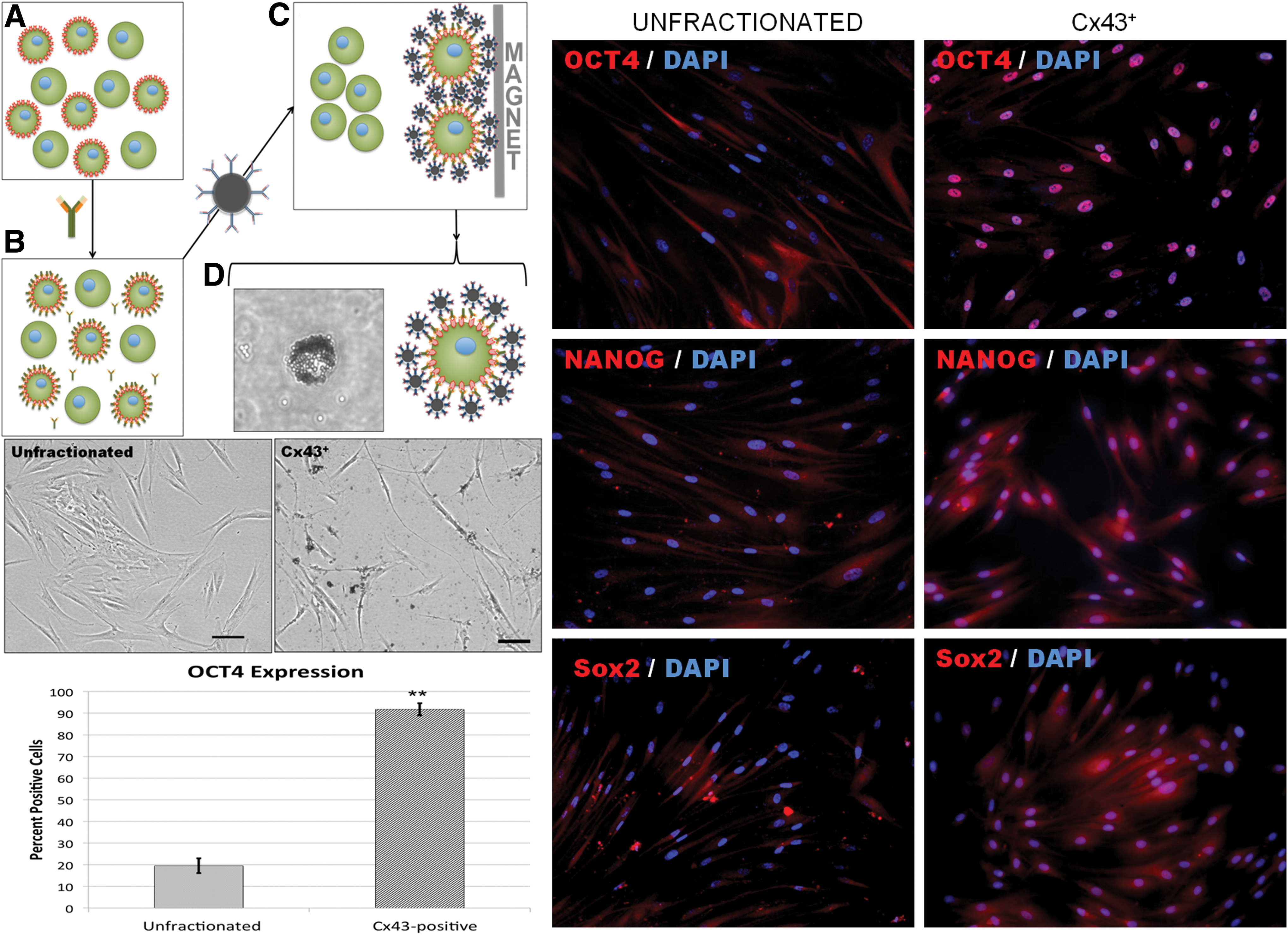
Left, top: Schematic of magentic bead separation technique showing
Cx43+ cells express pluripotency-associated markers
Gene expression analysis for pluripotency markers shows that Cx43+ cells have a statistically significant upregulation of OCT4, Nanog, Sox2, and hTERT expression over commercially obtained bone marrow mesenchymal stem cells (MSC, control) or their unfractionated PDLSC parent line (Supplementary Fig. S2). Immunohistochemical analysis for the expression of pluripotency-associated markers OCT4, Nanog, and Sox2 reveal that Cx43+ selection augments the percentage of cells staining positive for these markers when compared to that seen in heterogeneous cultures of PDLSCs as previously reported by our group [35]. For the expression of transcription factor OCT4, Fig. 1 clearly shows that Cx43+ selection enriches not only the percentage of positively marked cells (19.46%±3.40% for heterogeneous cultures vs. 91.76%±2.81% for Cx43+ cells; **P<0.01), but also the presentation of this marker within these cultures. Heterogeneous cultures of PDLSCs have several positively marked cells for OCT4; however, most of the staining is not located within the nucleus of the cell, but in the perinuclear region—possibly at translational sites for the protein. Conversely, once the cell population has been enriched to a homogeneous Cx43+ phenotype, OCT4 expression is observed mainly in the cell nucleus with positive staining still observed at perinuclear regions of some of the cells. Although not as significant as for OCT4, the selection of Cx43+ cells have a similar augmenting effect on the level of expression and pattern of presentation of Nanog and Sox2 (Fig. 1). Although both Nanog and Sox2 were originally seen in the heterogeneous cultures of PDLSCs, their expression and nuclear translocation is much more pronounced (in intensity and number) when the cells have been enriched to a homogeneous Cx43+ phenotype (Fig. 1).
Enrichment through Cx43+ biomarker expression had a profound effect on the phenotypic expression of pluripotency-associated markers OCT4, Nanog, and Sox2. Cx43+ cells have a significantly greater number of OCT4-positive cells than the unfractionated cell population from the same donors. Moreover, Cx43+ cells presented a functional expression of these markers with nuclear translocation of all three markers seen mainly in Cx43+ cells (Fig. 1). While several connexins had previously been identified in cells arising from the periodontal ligament [37], for the purposes of this project, we decided to focus on Cx43, a known modulator of NC cell migratory activity [11]. This potentially describes a useful biomarker technique for the isolation of NC-derived stem cells in other tissues arising from this embryological structure.
Cx43+ cells express NC-specific markers
Cx43+ cultures are positive for the NC-specific transcriptional factor Sox10 (Fig. 2), a known NC specifier gene [9]. Bronner [9] classify Sox10 as a NC specifier gene in their newly proposed NC gene regulatory network [9]. Similarly, the selected cells are positive for the expression of the p75 neurotrophin receptor, another marker recently proposed to isolate NC-derived stem cells [19]. Analysis of the correlation between Cx43 expression and the neural marker Nestin, as well as the NC modulator N-cad [11], revealed that nearly all cells ubiquitously express both markers as clusters on cell membranes and within cell bodies (Fig. 2). Furthermore, Cx43 immunohistochemical analysis shows that selected cells express this gap junction protein as discrete membrane-bound structures specifically at cell–cell interfaces, indicating the correct functional presentation of the protein within these cells.
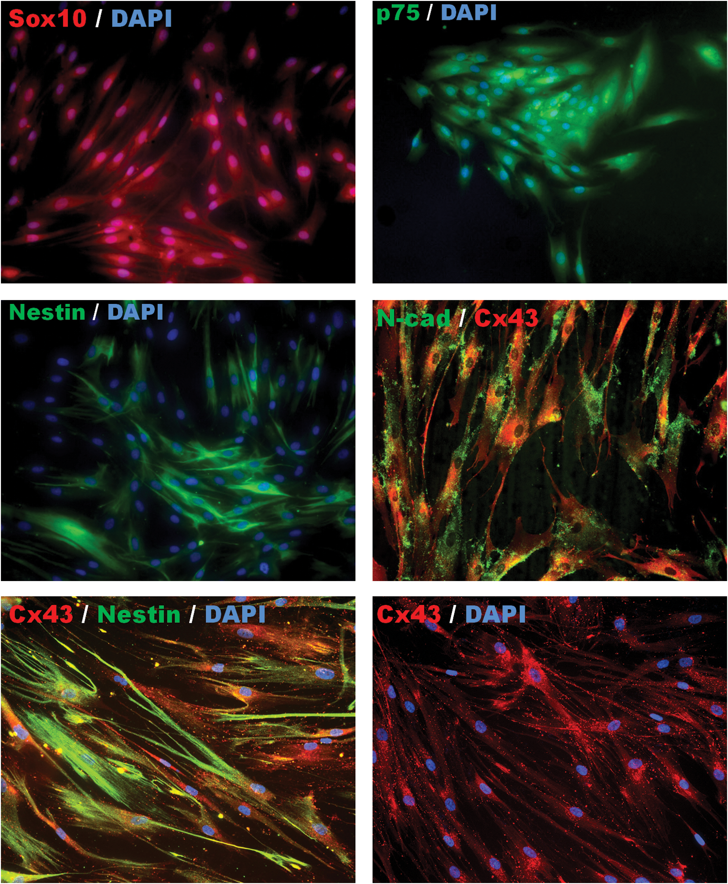
Immunohistochemical images of the expression of neural crest-specific markers Sox10, p75NTR, Nestin, and N-cadherin (N-cad) in Cx43+ cell populations of PDLSCs. Middle right and bottom left images show coexpression and localization of N-cad and Nestin, respectively, with Cx43 in these cells. Images are representative of obtained results for three cell lines established in this study. Color images available online at
Cx43+ cells form teratomas with mature structures in vivo
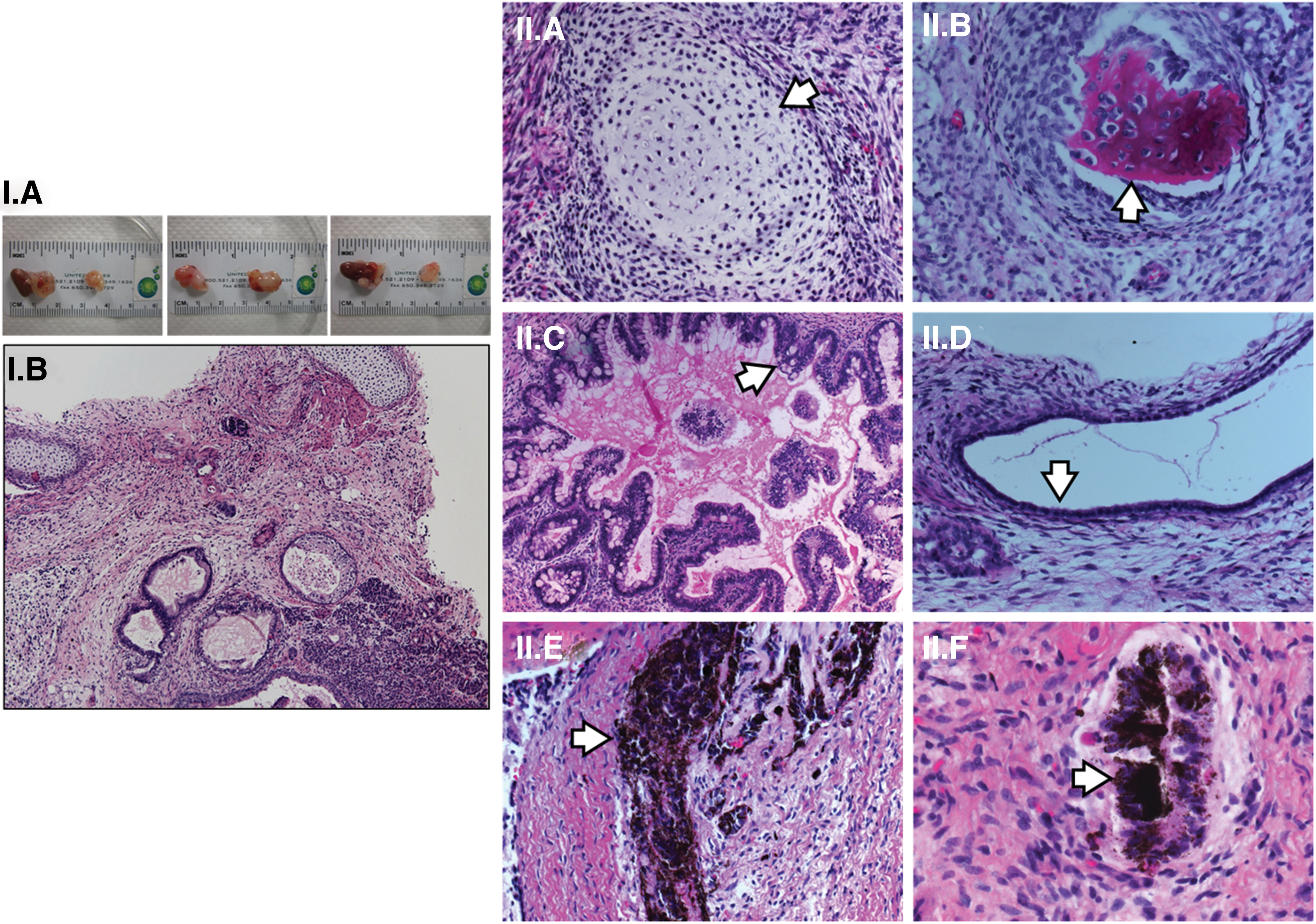
In an in vivo setting, Cx43+ cells were capable of generating teratomas in 100% of the inoculation sites. Immunodeficient mice inoculated with Cx43+ cells in air capsules created in the lateral flank of the kidneys and testis generated neoplastic tumor growth (Fig. 3.I.A). Teratoma tissues consisted of scattered regions of differentiated cells containing tissues from all three embryological germ layers clearly identifiable (supplemental report Applied Stem Cell, Inc). Among the tissues identified are cartilage and ossified cartilage (mesoderm, Fig. 3.II.A, B), glandular and duct structures (endoderm, Fig. 3.II.C, D), and pigmented cells (ectoderm, Fig. 3.II.E, F).
Closer histological analysis of teratoma tissues revealed the presence of various other tissues, including eccrine sweat glands (Fig. 4A), pockets of neural-like tissues (Fig. 4B, C; Supplementary Fig. S3), cells resembling gut epithelial lining containing enterochromaffin cells (Fig. 4D, E), muscle tissue (Fig. 4F, G; Supplementary Fig. S3), cartilaginous tissue (Fig. 4H), and blood vessels (Fig. 4I) in teratomas formed by Cx43+ cells, which further confirms these cells as NC-derived cells. The presence of the double-walled eccrine sweat glands (Fig. 4A) in teratoma tissue confirms the human origin of the tumor cells, since murine skin does not possess endogenous sweat glands and those found in the palmar skin of mice are single-cell walled type glands [39]. Therefore, these observations suggest the isolation of a NC-derived pluripotent stem cell population by means of Cx43 enrichment. The formation of morphologically mature gut epithelial lining deserves a closer examination in this study as this is a tissue normally derived from the endodermal germ layer and the contribution from NC cells, while highly investigated, has been ruled out [40]. NC cells are known to migrate into the gut and form the enteric nervous system. Similarly, NC cells at the vagal and sacral level have been shown to populate the gut mesenchyme surrounding the gut endoderm [40]. Thus, the differentiation of the cells down the enterochromaffin line may be a result of the plasticity of the cells and the microenvironment afforded by the kidney and testis hormonal cues. However, contributions of NC to purely endodermal tissues are still being explored and re-evaluated. Recently, a study presented findings of NC cells contributing to the formation of the epithelial lining of the middle ear (a tissue considered endodermal) [41]. In this study, NC cells were shown to undergo an epithelial-to-mesenchymal transition and form a lining continuous with the endodermally derived auditory tube [41]. Whether the contribution of NC cells to the gut endoderm may follow a similar phenomenon is beyond the scope of this study.
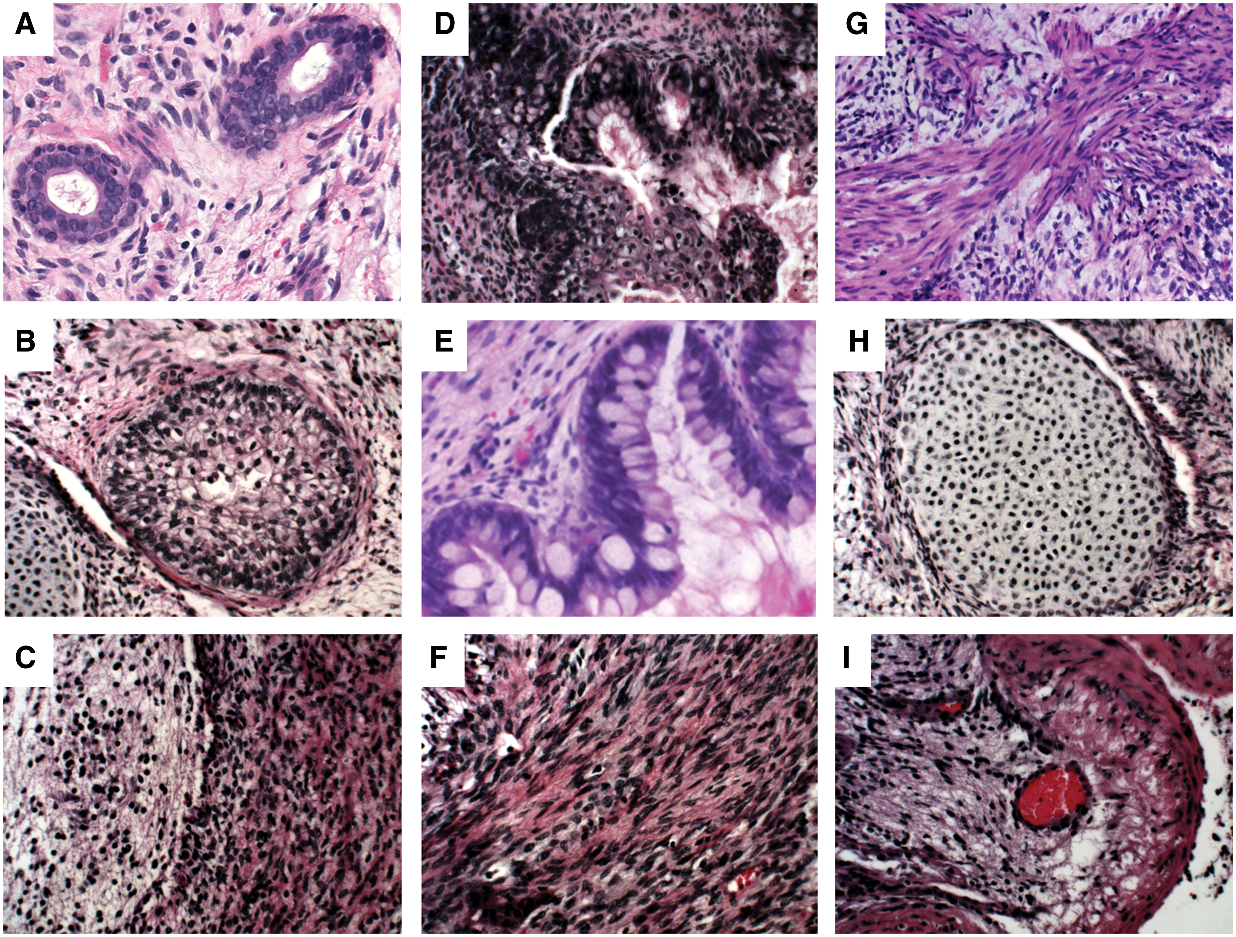
Top: H&E-stained histological section images of diverse tissues/structures within teratomas formed by the Cx43M19p5 cell line in immunodeficient mice. Images show formation of structures of ectodermal
The observations made from the morphological assessment of H&E sections were verified by means of immunohistochemical analysis. Identification of neoplastic cells as being of human origin was confirmed by positive staining with the anti-human nuclear mitotic apparatus protein (NuMA, Fig. 5) in neoplastic cells. Additional immunohistochemical analyses of obtained tissues also showed the existence of neural cells within defined pockets of teratoma tissues (Fig. 5) that stained positive for the glial fibrillary acidic protein. The endodermal nature of what has been identified as epithelial lining cells was confirmed by staining with the N-cad antibody showing specific staining in the structures identified as being of endodermal origin (Fig. 5). Finally, immunohistochemical analysis demonstrated that there are regions of undifferentiated stem cells still expressing the transcriptional factor OCT4 (Fig. 5). The optimal maintenance and differentiation conditions needed to guide these NC stem cells down specific lineages in vitro, however, still need further investigation. All of the cell lines presented in this study were maintained in high serum-containing media under plastic adherent conditions. Whether culture settings can be optimized to maintain the phenotypic characteristics of NC cells over extended passaging of the cells is currently being explored.
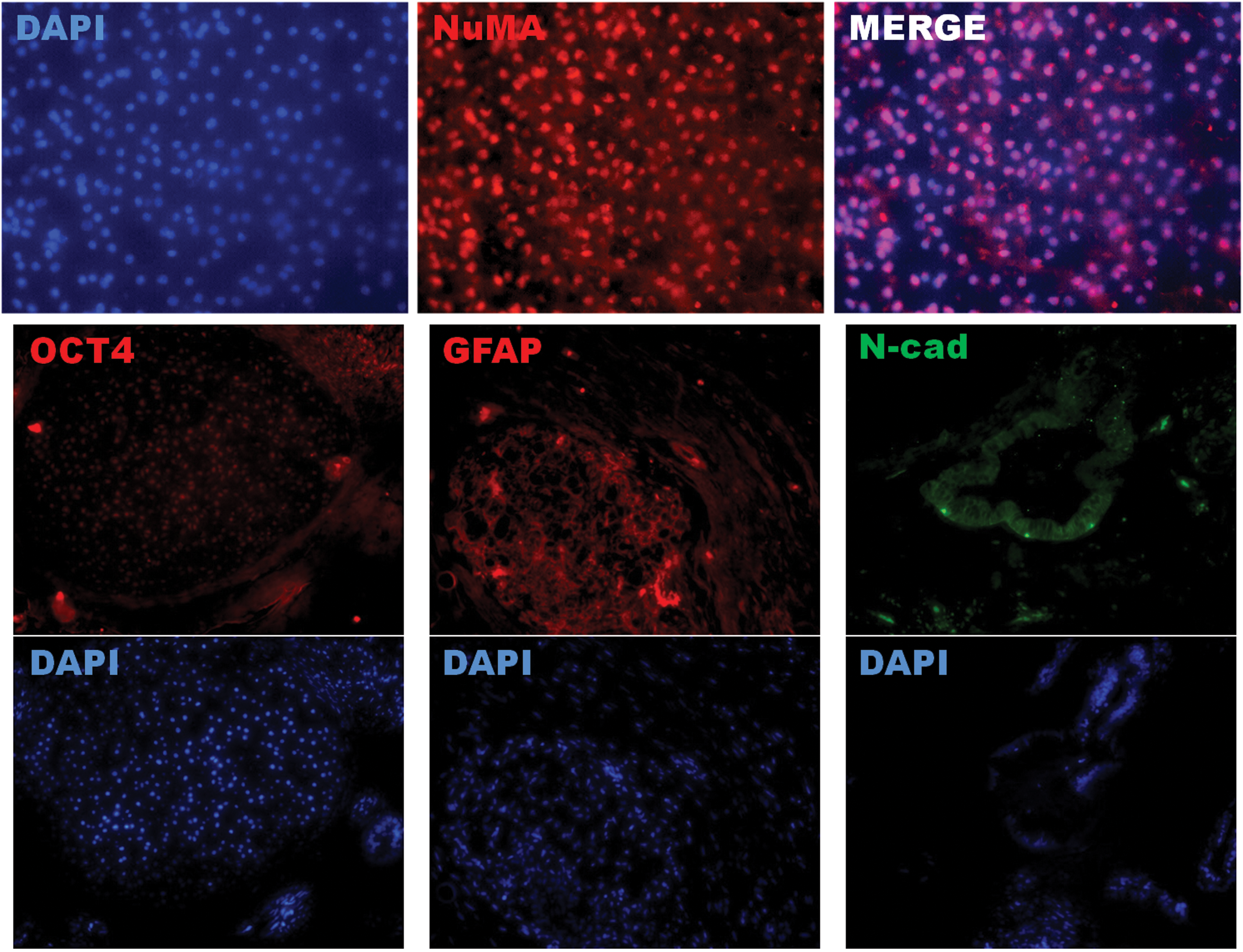
Fluorescent immunohistochemical analysis of neoplastic cells in teratoma assay showing human origin of neoplastic cells (human nuclear mitotic apparatus protein [NuMA], top); pockets of undifferentiated cells still expressing the pluripotency marker OCT4 (bottom, left); neural structures positive for the Glial fibrillary acidic protein (GFAP); and the glandular structure positive for the endodermal marker N-cad. Color images available online at
The identification and isolation of a pluripotent stem cell population within adult tissues is an important advancement for the fields of stem cell biology and regenerative medicine. The significance of this concept is greatly enhanced by the possibility of a patient-specific cell line creation for regenerative medicine. Here we present an isolation technique of a pluripotent stem cell population from tissues obtained during routinely performed medical procedures such as third molar extraction. The isolated cells are related to the migrating cranial NC, which populates the craniofacial mesenchyme and contributes to its development, as evidenced by their expression of NC-specific markers Sox10, p75, and Nestin. These cells present a plasticity that is beyond that of other cell populations isolated from the periodontal ligament or other adult tissues in humans. While several reports have argued for the existence of other pluripotent stem cell reservoirs in the adult body [4 –6], some of these sources are still under investigation [42]. Similarly, several attempts have been made to isolate NC stem cells from other adult tissues arising from this embryological structure [19,26,30]. Whether the presentation of functional gap junctions in Cx43+ cells has a further modulatory effect on the overall biology and epigenetic profile of these cells is work that is currently ongoing. It is possible that this functional interaction further enhances the niche-regulatory nature of the cells to modulate their own undifferentiated state.
Footnotes
Acknowledgments
This study was supported in part by a Veterans Affairs (VA) Merit Review Grant and a VA Senior Research Career Scientist Award. We would like to thank Applied StemCell, Inc. for their services with teratoma assay formation and histological report, as well as Dr. Harold Cuello and Dr. Teresita Reiner for their expertise in histological assessment. We would also like to thank Dr. Yoh Sawatari for his assistance in the procurement of the periodontal tissues.
Author Disclosure Statement
No competing financial interest exists.
References
Supplementary Material
Please find the following supplemental material available below.
For Open Access articles published under a Creative Commons License, all supplemental material carries the same license as the article it is associated with.
For non-Open Access articles published, all supplemental material carries a non-exclusive license, and permission requests for re-use of supplemental material or any part of supplemental material shall be sent directly to the copyright owner as specified in the copyright notice associated with the article.