
Abstract
Mesenchymal stem cell (MSC) transplantation after ischemia/reperfusion (I/R) injury reduces infarct size and improves cardiac function. We used mouse ventricular myocytes (VMs) in an in vitro model of I/R to determine the mechanism by which MSCs prevent reperfusion injury by paracrine signaling. Exposure of mouse VMs to an ischemic challenge depolarized their mitochondrial membrane potential (Ψmito ), increased their diastolic Ca2+, and significantly attenuated cell shortening. Reperfusion of VMs with Ctrl tyrode or MSC-conditioned tyrode (ConT) resulted in a transient increase of the Ca2+ transient amplitudes in all cells. ConT-reperfused cells exhibited a decreased number early after depolarization (EADs) (ConT: 6.3% vs. Ctrl: 28.4%) and prolonged survival (ConT: 58% vs. Ctrl: 33%). Ψmito rapidly recovered in Ctrl as well as ConT-treated VMs on reperfusion; however, in Ctrl solution, an exaggerated hyperpolarization of Ψmito was determined that preceded the collapse of Ψmito . The ability of ConT to attenuate the hyperpolarization of Ψmito was suppressed on inhibition of the PI3K/Akt signaling pathway or IK,ATP. However, protection of Ψmito was best mimicked by the reactive oxygen species (ROS) scavenger mitoTEMPO. Analysis of ConT revealed a significant antioxidant capacity that was linked to the presence of extracellular superoxide dismutase (SOD3) in ConT. In conclusion, MSC ConT protects VMs from simulated I/R injury by its SOD3-mediated antioxidant capacity and by delaying the recovery of Ψmito through Akt-mediated opening of IK,ATP. These changes attenuate reperfusion-induced ROS production and prevent the opening of the permeability transition pore and arrhythmic Ca2+ release.
Introduction
T
Ischemia in the cardiac muscle results in the loss of energy production and changes in the intracellular ion homeostasis [10]. While rapid re-oxygenation is the treatment of choice, reperfusion itself causes further damage to the myocardium. pH recovery through the sodium hydrogen exchanger increases [Na]i and promotes Ca2+ entry through reverse mode sodium calcium exchanger (NCX), which can lead to Ca2+ overload-induced arrhythmia [11]. The mitochondrial membrane potential (Ψmito ) depolarizes during ischemia followed by a loss of ATP production [12]. While mitochondria can repolarize during reperfusion, the reestablished oxygen supply promotes a surge of reactive oxygen species (ROS) production that can lead to oxidation of proteins of the electron transport chain or opening of the permeability transition pore (PTP) and collapse of Ψmito [12 –14]. Prevention of Ca2+ overload as well as delay of Ψmito recovery were shown to be cardioprotective [13,15], and PI3K/Akt signaling has been postulated as a mediator of postconditioning [10,16]. MSC-mediated cardioprotection from ischemia/reperfusion (I/R) injury has been demonstrated through preconditioning of the cardiac tissue [17]. In these cases, postinfarct recovery was enhanced when MSCs expressed increased levels of interleukin-18, and depended on the secretion of the vascular endothelial growth factor [8,18 –20]. However, a postconditioning benefit of MSCs was also demonstrated when cells were injected 2 h after ischemic injury [21].
We have previously demonstrated [22] that conditioned tyrode (ConT) obtained from MSCs enhances cardiac excitation–contraction coupling (ECC) by Akt-mediated activation of endothelial nitric oxide synthase (eNOS) and a subsequent increase in L-type Ca2+ current (ICa,L) and enhances Ca2+ uptake through the sarcoplasmic reticulum Ca2+ ATPase (SERCA). While we could also describe an enhanced survival of isolated mouse ventricular myocytes (VMs) in ConT, the context of this anti-apoptotic mechanism during I/R injury remained to be determined. To test the hypothesis that ConT confers a postconditioning benefit, we used an in vitro model of simulated I/R and determined the physiological properties of isolated VMs by monitoring field stimulation-induced changes in [Ca2+]i and Ψmito .
Methods
Ethical approval
All animal procedures were performed with the approval of IACUC at the University of Illinois at Chicago and in accordance with the National Institute of Health's Guide for the Care and Use of Laboratory Animals.
Cardiomyocyte isolation and MSC-ConT
VMs were isolated from 3–5 month-old C57BL/6 mice by Langendorff perfusion. The mice were anesthetized by inhalation of Isoflurane (3%). Anesthesia was confirmed by lack of response to toe pinch before the aorta was transected and the heart was excised [22,23]. Cells were plated on laminin-coated (Sigma Aldrich; 1 mg/mL) 25 mm coverslips in standard tyrode solution (in mmol/L: NaCl 130, KCl 5.4, CaCl2 1, MgCl2 1.5, NaHCO3 10, Glucose 10, HEPES 25,
In vitro model of simulated I/R
To mimic ischemic conditions in vitro, VMs were superfused with a solution that mimics hypoxia, hyperkalemia, acidosis, and lactate accumulation [25]. The solution contained (mmol/L): NaCl 113, NaHCO3 6, NAH2PO4 0.9, KCl 8, MgCl2 1.5, CaCl2 1, Na/lactate 20, (pH 6.8). The solution was gassed with 95% N2, 5% CO2. Ischemia was maintained for 15 min before reperfusion with either control (Ctrl) or ConT solution. Using a trypan blue dye exclusion assay, the effect of ConT on cell survival was evaluated at the end of reperfusion (15 min). Trypan blue was added for a final concentration of 0.4%, and slides were evaluated for the percentage of surviving cells [26].
Detection of protein carbonylation
VMs were plated on laminin-coated culture dishes at 70% density and exposed to the simulated I/R protocol. During the procedure, the cells were field stimulated (0.5 Hz; multi-channel stimulator; IonOptix, LLC). After 15 min of reperfusion, the myocytes were recovered and processed for sodium dodecyl sulfate–polyacrylamide gel electrophoresis (SDS-PAGE) and immunoblotting as previously described (8). Ninety micrograms of cell lysate was loaded per well. For the detection of ROS-mediated oxidative modification (carbonylation) of proteins, introduced carbonyl groups were derivatized to 2,4-dinitrophenyl (DNP)-hydrazone in a reaction with DNP-hydrazine solution for 5 min by directly treating the blotted nitrocellulose membrane. Western blotting was completed using an anti-DNP moiety antibody (OxyBlot Kit No. S7150; Millipore). To evaluate equivalent sample loading, the blot was stripped and re-probed with primary antibodies against glyceraldehyde 3-phosphate dehydrogenase (GAPDH- No. 5174; Cell Signaling Technology). To determine the induction of apoptosis, cell lysates were probed for cleaved caspase-9 (Asp330; No. 9501; Cell Signaling Technology).
Measurements of [Ca2+]i, mitochondrial membrane potential and ROS production
Measurements of [Ca2+]i were performed as previously described [22]. For recordings of Ψmito, VMs were loaded with tetramethylrhodamine, methyl, ester, and perchlorate (TMRM: 100 nmol/L) for 40 min; after that, 40 nmol/L TMRM was maintained in all extracellular solutions [27]. Dynamic changes in Ψmito were determined by superfusing VMs with (1) oligomycin (OM: 10 μmol/L), a blocker of proton entry via ATP-synthase, which causes a hyperpolarization of Ψmito or (2) fluoromethoxy-phenylhydrazone (FCCP), which induces a collapse of Ψmito [28] (not shown). No significant changes in Ψmito were determined when VMs were superfused with either 15 mmol/L [K]o or pHo 6.7, ruling out changes in TMRM due to experimental conditions alone (not shown). Dynamic changes in mitochondrial ROS production were identified by loading VMs with the mitochondrial dye MitoSox Red (20 min, 0.5 μmol/L). MitoSox fluorescence was measured by taking 2D confocal images (0.5 Hz; excitation: 400 nm; emission: 561 nm). ROS production was quantified as the change in MitoSox fluorescence over time (Δfluorescence a.u./5 min)[29].
Nitro blue tetrazolium in gel assay for superoxide dismutase activity
Superoxide dismutase (SOD; Sigma-S5395) in units of activity and ConT proteins were directly compared using an in-gel assay for enzymatic activity. For this assay, in situ visualization of anti-oxidant/SOD protein activity occurs as an achromatic gel band resulting from the inhibition of nitro blue tetrazolium (NBT) chromogenic reduction within the gel [30,31]. Briefly, protein samples were mixed with 5X-loading buffer (1.5 mol/L Tris, pH 8.3) without 2-mercaptoethanol, SDS, or heating and loaded directly onto precast 4%–20% Novex tris-glycine gels (Invitrogen) for nondenaturing PAGE. After the dye front reached the bottom of the gel, it was transferred to a solution containing 28 μmol/L riboflavin and 28 mmol/L TEMED in potassium phosphate buffer (pH 7.8). A 20 min incubation in the above solution was followed by incubation in 2.45 mmol/L NBT until the visualization of SOD activity was optimal for evaluation and image acquisition.
Chemicals
Fluo-4/AM and TMRM were purchased from Invitrogen, LY294002 from Cayman Chemicals, 5-hydroxydecanoate (5-HD), diazoxide (DZX), oligomycin (OM) and carbonyl cyanide-p-tri-FCCP from Sigma, the Akt inhibitor V (triciribine) from EMD/Calbiochem, and mitoTEMPO from Enzo Lifesciences.
Statistics
Data sets other than for cell survival were statistically evaluated using paired and un-paired t test. Cell survival was shown as a Kaplan–Meier curve, and differences in viability to control were evaluated with a log-rank test [26] (GraphPad Prism software). All averaged data are presented as means±SEM.
Results
ConT enhances cell survival and attenuates arrhythmic activity after simulated I/R
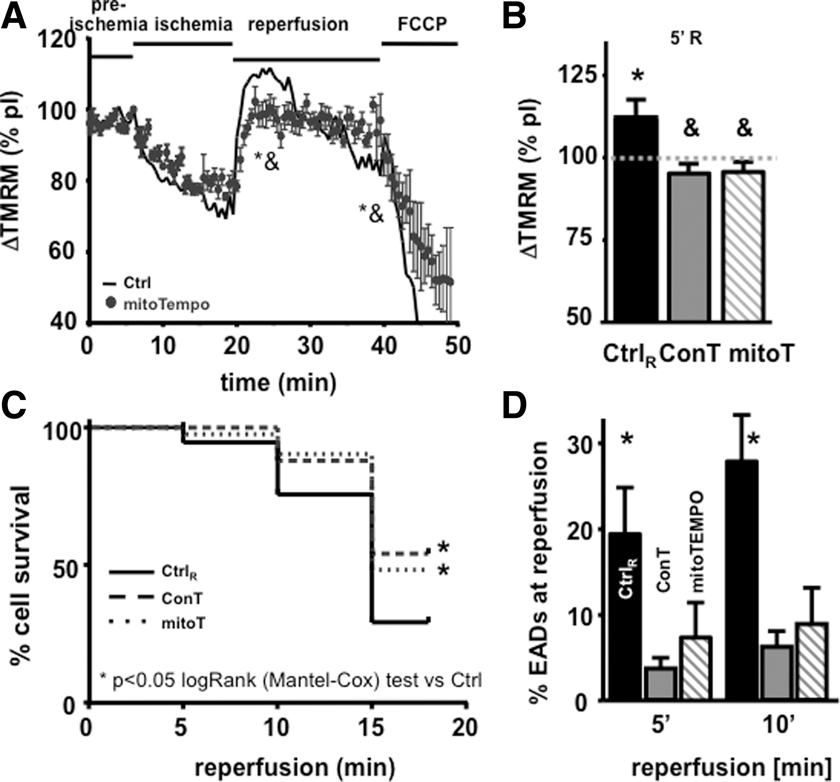
We have previously demonstrated that MSC-derived ConT significantly enhances cell survival of isolated VMs in culture [22]. To determine whether this translates into the protection of VMs during simulated I/R injury, we monitored intracellular Ca2+ transients from isolated, field-stimulated VMs that were loaded with Fluo-4/AM. Figure 1A and B shows representative F/F 0 plots in preischemic (pI) and ischemic conditions (15 min), as well as 1 min, and 5 min after reperfusion. While all VMs were treated identically during pI and ischemic conditions, reperfusion was performed with either Ctrl solution (Fig. 1A) or ConT (Fig. 1B). Cell survival after reperfusion (Fig. 1D) and the occurrence of EAD (Fig. 1C: EADs: No EAD×100/No twitch during 10 s) were quantified [32]. Analysis of VMs 15 min after reperfusion by a trypan blue exclusion assay showed that ConT promoted cell survival after simulated I/R injury (ConT: 56.9%±2.2%, n=9 slides, 2,123 VMs vs. Ctrl: 35.8%±1.3%, n=9 slides, 2,485 cells; P<0.05; Fig. 1D) and suppressed EADs at 5 min and 10 min of reperfusion (Ctrl: 19.8%±5.5% and 28.4%±5%, n=21 vs. ConT: 3.7%±1.4% and 6.3%±2%, n=29). An examination of oxidative protein modifications (carbonylation) in whole cell lysates recovered 15 min after reperfusion showed overall increased levels of detectable carbonylation in different protein bands. Carbonylation levels were comparable to untreated VMs when ConT was used for reperfusion (n=3, 2 shown; Fig. 1E). In addition, ConT reperfusion prevented the increase in cleaved caspase-9 levels following the simulated I/R protocol when Ctrl solution was used for reperfusion (Fig. 1F, n=3). The results indicate that ConT mediates a postconditioning benefit during reperfusion after an ischemic challenge.
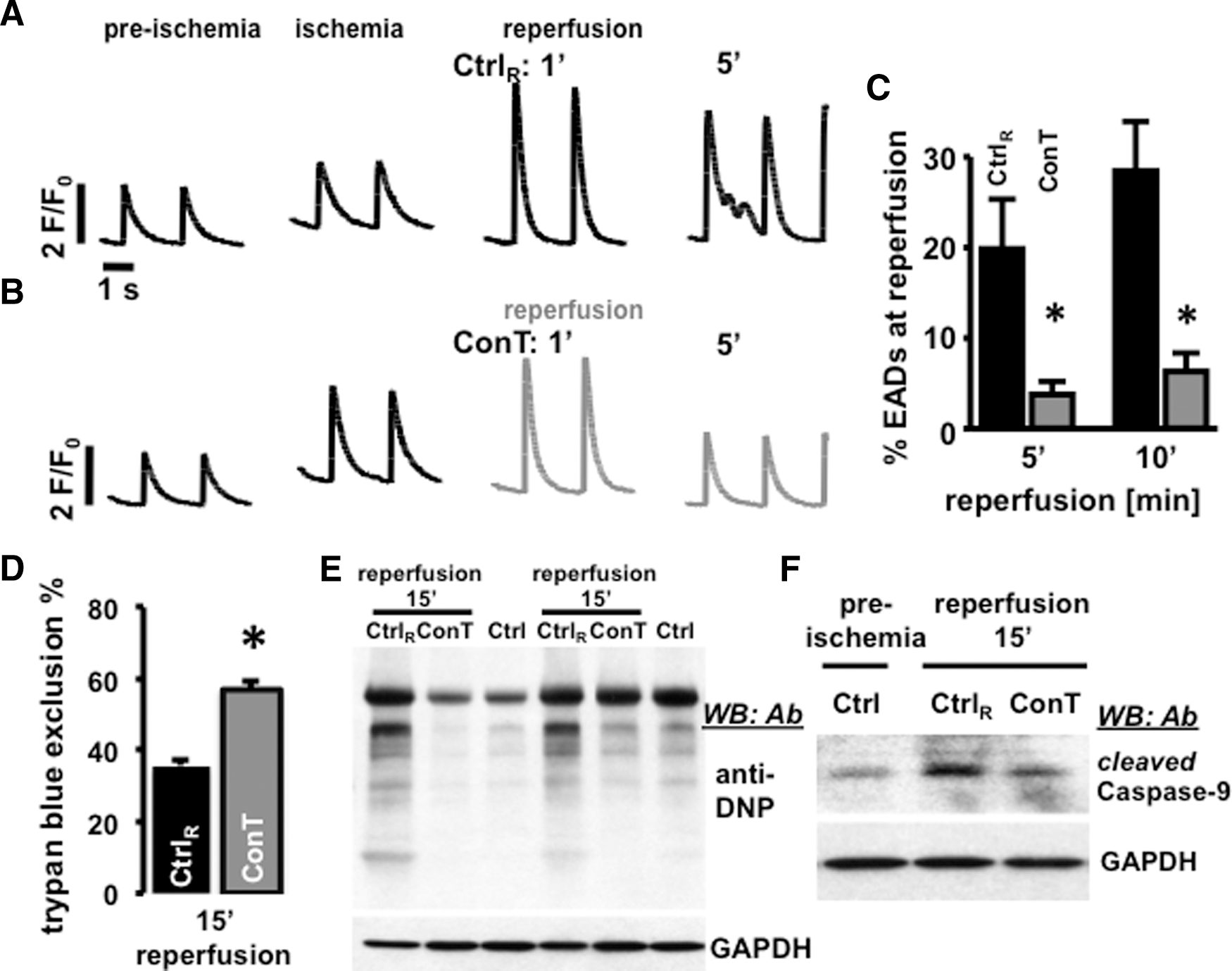
Reperfusion with conditioned tyrode (ConT) protects ventricular myocytes (VMs) early after depolarization (EADs) and cell death. F/F
0 plots show changes in [Ca2+]i in field-stimulated VMs during preischemic (pI) and ischemic conditions and after 1 and 5 min of reperfusion with Ctrl
Changes in stimulation-induced Ca2+ transients during simulated I/R
To determine whether ConT-induced changes in ECC [22] contribute to changes in VM survival [Ca2+]i, cell shortening and SR load were recorded during the simulated I/R protocol. During ischemia, no significant change in Ca2+ transient amplitude (Fig. 2B) or diastolic cell length (Fig. 2D) was determined. However, diastolic Ca2+ (Fig. 2A) and the Ca2+ transient decay constant (τCa, Fig. 2C) increased significantly, and cell shortening almost ceased (Fig. 2E). On reperfusion, the Ca2+ transient amplitudes significantly increased (ΔF/F 0, Ctrl: pI: 2.98±0.2; R1′: 6.26±0.44; n=12, P<0.01) before returning to pI levels. Concomitant with this increase, cell shortening was enhanced [cell length (μm): Ctrl: pI: 1.83±0.27; R1′: 7.2±1.99; n=8, P<0.05), but incomplete relaxation left the cell in a state of contracture (Fig. 2D). ConT reperfusion did not prevent the increase in [Ca2+]i (ΔF/F 0, ConT: 2.05±0.14; R1′: 4.83±0.37; n=12, P<0.01) or decrease in τCa (Fig. 2B, C), but the diastolic cell length remained comparable to pI conditions (Fig. 2D, E) although cell shortening was enhanced during reperfusion [(μm), ConT: preischemia: 1.13±0.29; R1′: 6.12±1.43; n=6, P<0.01; Fig. 2E).
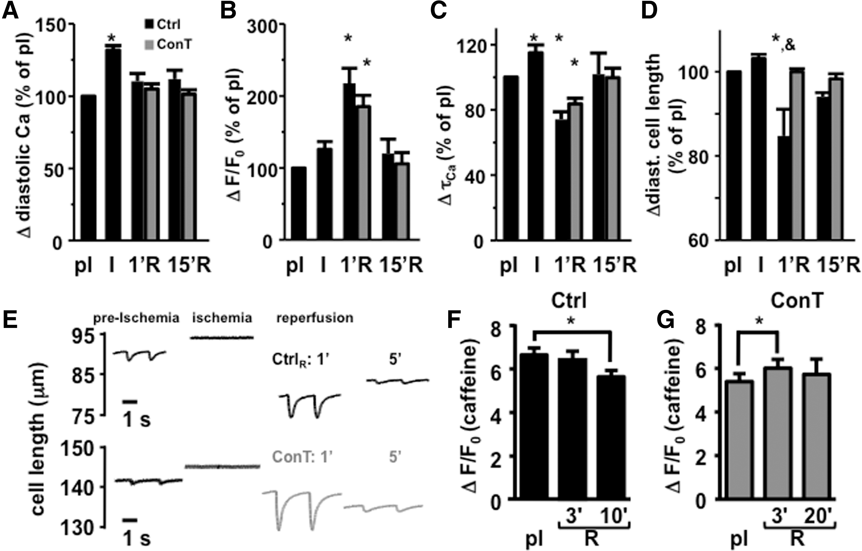
ConT does not change cellular Ca2+ handling during reperfusion. The change in diastolic Ca2+
ConT is known to enhance SERCA-mediated Ca2+ uptake, resulting in an increased SR load [22]. Caffeine (10 mmol/L) was applied to identify differences in SR load between Ctrl and ConT reperfused VMs under pI and ischemic conditions, and during reperfusion. The caffeine transient amplitude was different in ConT and Ctrl-reperfused VMs compared with preischemia. In Ctrl-reperfused cells (Fig. 2F), it decreased over the time of reperfusion (ΔF/F 0: Ctrl pI: 6.62±0.33; R10′: 5.61±0.3; n=4, P<0.05); whereas in ConT-reperfused VMs (Fig. 2G), it was transiently increased at R3′ (ConT: pI: 5.4±0.36; R3′: 6.0±0.4; n=8, P<0.05). Due to increased cell death in Ctrl-reperfused cells, the second caffeine transient had to be induced at 10 min in Ctrl rather than at 20 min for the ConT-reperfused cells. No difference in the caffeine transient amplitude was determined between the two treatment groups. The results indicate that there was no ConT induced change in ECC during reperfusion; however, it preserves SR load likely by the suppression of EADs.
ConT preserves the mitochondrial membrane potential (Ψmito) during simulated I/R
The recovery of VMs from I/R injury depends on the recovery of the cells energy metabolism [14,33]. To determine whether differences in cell survival in ConT and Ctrl-reperfused cells are based on I/R damage of cardiac mitochondria, we measured Ψmito with the voltage-sensitive dye TMRM during the simulated I/R protocol. During ischemia, Ψmito depolarized but rapidly recovered on reperfusion with Ctrl solution or ConT (Fig. 3A). In Ctrl-reperfused VMs, an exaggerated hyperpolarization of Ψmito could be observed that exceeded pI fluorescence by 111.7%±5.9% (Fig. 3D; n=9; at R5′ P<0.05 to pI). During reperfusion with ConT, Ψmito recovered to 95.5%±2.3% (Fig. 3D; n=5; at R5′ P<0.05 to Ctrl) of pI fluorescence. In Ctrl VMs, this transient hyperpolarization was followed by a continuous depolarization of Ψmito, which reached significantly lower levels compared with ConT-treated cells at 15 min of reperfusion (Ctrl: 81.3%±3.1%; n=9; ConT: 101.1%±2.6%; n=5; at R15′ P<0.05). The difference was independent of the cells' contractile properties (not shown). The results indicate that ConT treatment on reperfusion can better preserve Ψmito.
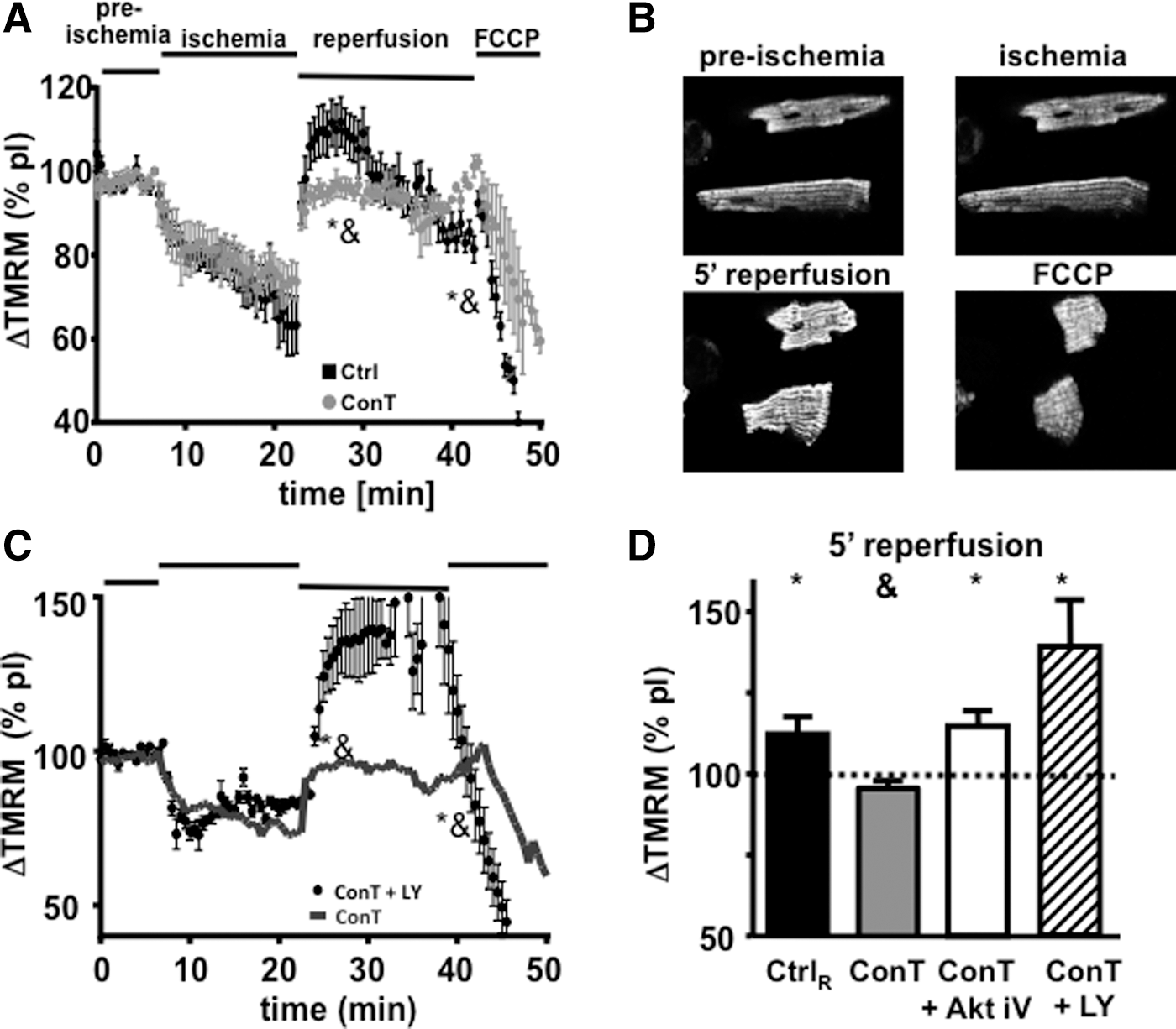
Changes in Ψmito
of VMs during simulated I/R injury.
Mechanism of ConT-mediated protection of Ψmito
ConT was previously determined to be an activator of the PI3K/Akt signal transduction pathway [22]. To determine the role of PI3K/Akt in the protection of Ψmito , we supplemented ConT during reperfusion with inhibitors of PI3K (LY294002, LY: 10 μmol/L) or Akt [Akt inhibitor V (Akt iV): 20 μmol/L] [22]. In the presence of LY or Akt iV, ConT no longer prevented the exaggerated hyperpolarization of Ψmito on reperfusion (Fig. 3C, D: LY: 139.1%±14.9%; n=3; Akt iV: 114.7%±4.7%; n=4, at 5 min of reperfusion: P<0.05). The results indicate that ConT attenuates the hyperpolarization of Ψmito through PI3K/Akt-dependent signaling. In the following, we used the degree of hyperpolarization on reperfusion as a measure of ConT action.
The recovery of Ψmito depends on the functional recovery of proton pumps, which allow for the simultaneous e− flow across the electron transport chain [13]. Potassium influx through the mitochondrial ATP-sensitive potassium channel (IK,ATP) can counteract the recovery of Ψmito . Since IK,ATP is proposed to be activated downstream of the PI3K/Akt pathway [34], we hypothesized that ConT activates IK,ATP. To test this hypothesis, ConT was supplemented with the IK,ATP inhibitor 5-HD (500 μmol/L) [35]. During ConT5-HD reperfusion, the action of ConT was attenuated and Ψmito now exhibited an increased hyperpolarization (Fig. 4A, B; ConT5-HD: 124.4±3; n=6 at 5 min of reperfusion). In addition, five out of seven cells died within the first 3 min of reperfusion. Next, we aimed at determining whether the opening of IK,ATP can mimic the attenuated recovery of Ψmito during ConT treatment. For this purpose, Ctrl solution was supplemented with the IK,ATP opener DZX (200 μmol/L; CtrlDZX) [36]. On reperfusion with CtrlDZX, no increase of Ψmito above pI levels was determined (CtrlDZX: 99.3±4.8; n=7 at 5 min of reperfusion) and now, seven out of seven VMs survived for approximately 20 min of reperfusion. No significant difference of Ψmito between ConT5-HD and CtrlDZX could be determined at 15 min of reperfusion. The experiments support the hypothesis that ConT promotes cell survival by Akt-dependent activation of the uncoupling protein IK,ATP attenuating the rapid recovery of Ψmito .
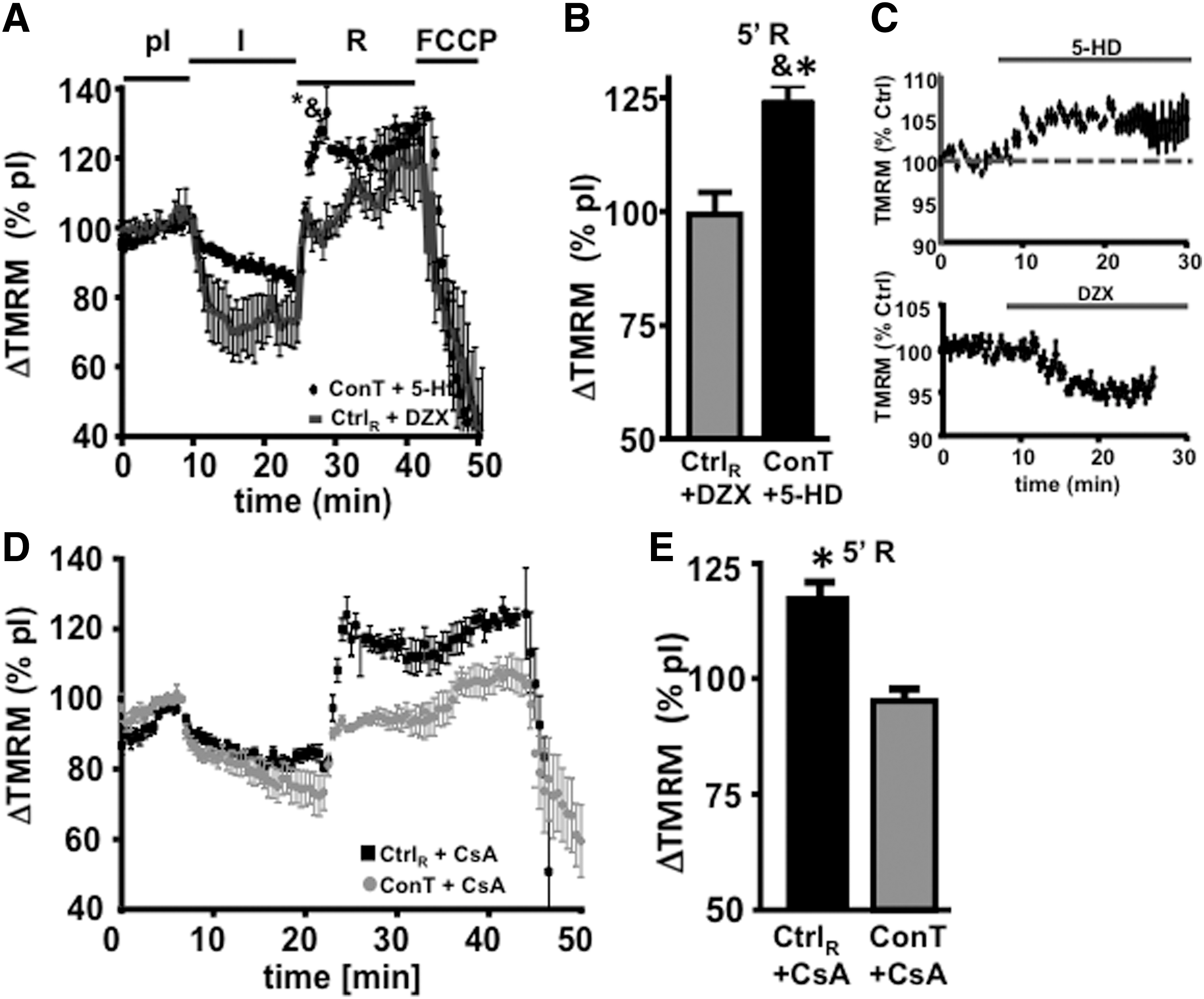
Reperfusion-induced Ψmito
hyperpolarization depends on IK,ATP open probability.
Inhibition of the mitochondrial PTP has been described to protect VMs from I/R injury; however, early and transient opening of the channel could also attenuate Ψmito hyperpolarization [37]. To determine the role of PTP in Ψmito recovery after ischemia, we supplemented Ctrl as well as ConT with the PTP inhibitor cyclosporin A (CsA: 1 μmol/L). CtrlCsA reperfusion could not prevent the exaggerated hyperpolarization at 5 min of reperfusion; however, in contrast to Ctrl-reperfused cells, Ψmito remained constant for the duration of reperfusion (Fig. 4D, E), indicating that the PTP opening might be responsible for the decay of Ψmito after reperfusion. In ConTCsA-reperfused VMs, Ψmito recovery was not significantly different from ConT-reperfused cells. The data support the hypothesis that the PTP opening does not play a role in the delayed hyperpolarization of Ψmito , but the opening contributes to the collapse of Ψmito in the later phase of reperfusion.
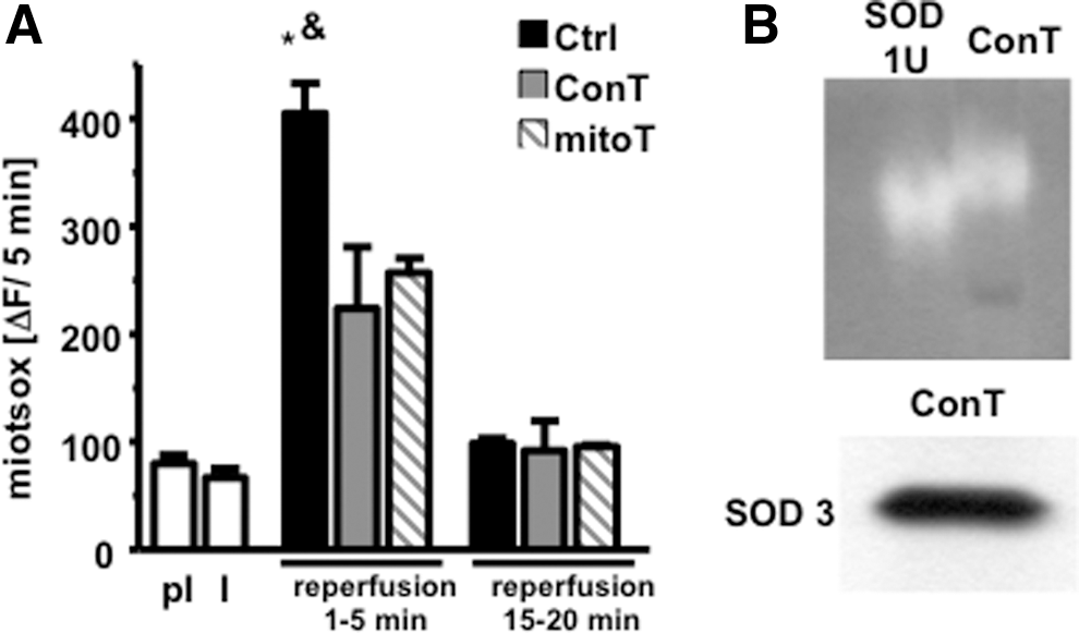
ConT prevents ROS production and concomitant arrhythmic Ca2+ release
ROS production is significantly enhanced during reperfusion when oxygen supply is re-established and Ψmito as well as the intracellular pH return to physiological levels [12,14]. To determine whether ROS production influences the recovery of Ψmito , in subsequent EADs and cell survival, we supplemented Ctrl reperfusion solution with the mitochondrial ROS scavenger mitoTEMPO (5 μmol/L) [12]. As shown in Fig. 5C and D, mitoTEMPO significantly enhanced cell survival and attenuated the occurrence of EADs, thereby reproducing the benefit of ConT. In addition, mitoTEMPO even reduced the exaggerated hyperpolarization of Ψmito (Fig. 5A, B) (significantly different at 15 min compared with Ctrl), suggesting that reperfusion-induced mitochondrial ROS production further enhances Ψmito hyperpolarization. To identify whether ConT changes ROS production during the reperfusion period, we exposed mitoSOX-loaded VMs to the I/R protocol. Constant stimulation led to a steady increase of fluorescence with an attenuated slope during ischemic conditions. Reperfusion in Ctrl, ConT, and Ctrl+mitoTEMPO induced a rapid increase in ROS production; however, the total amount of fluorescent change was significantly reduced in ConT and Ctrl+mitoTEMPO-treated VMs (Fig. 6A), indicating that ROS production and Ψmito hyperpolarization are closely linked events and have an adverse effect on cell survival.
Scavenging of intra-mitochondrial ROS enhances cell survival on I/R.
ConT and mitoTEMPO attenuate reperfusion-induced ROS production.
To evaluate the possibility that ConT can have a direct function as an antioxidant, we separated ConT proteins with PAGE and soaked the gel in NBT (2.5 mmol/L) and riboflavin (28 mmol/L). NBT serves as a substrate for the superoxide-inducing riboflavin so that proteins with antioxidant properties in the gel can be identified as areas in which NBT oxidation was suppressed. Since the presence of SOD in MSC conditioned media had been suggested [38], we ran the antioxidant SOD (1 U) as a control in the lane adjacent to ConT. The gel shown in Fig. 6B displayed areas of oxidized NBT in both lanes at a comparable molecular weight. In addition, western blotting of ConT proteins identified the extracellular copper/zinc-SOD (SOD3) at a molecular weight of 30 kDa, supporting the capacity of ConT as an antioxidant.
Discussion
We demonstrate that ConT from mouse bone marrow-derived MSCs exerts a postconditioning benefit on VMs when applied during reperfusion. ConT prevents an exaggerated reperfusion-induced hyperpolarization of Ψmito , subsequent PTP opening, and arrhythmic Ca2+ release (EADs). Mechanisms of cardioprotection are (1) the SOD3-mediated antioxidant capacity of ConT that reduces ROS and (2) the Akt/PI3K-mediated opening of IK,ATP. Both mechanisms prevent the exaggerated hyperpolarization of Ψmito that further enhances reperfusion-induced ROS and, ultimately, cell death (Fig. 7).

Schematic summary of the experimental results illustrates the proposed signal transduction pathway.
In vitro model of simulated I/R
We used an in vitro model of simulated I/R that was previously used to analyze ischemia-induced changes in protein expression as well as cardiac stunning [25]. Ischemia induced an increase in basal [Ca2+]i, cessation of contractile activity and depolarization of Ψmito . While we observed a 30% decrease in TMRM fluorescence during 15 min of ischemia, more extensive depolarizations of Ψmito are reported in other models [39]; in these cases, a recovery of Ψmito was rarely determined on reperfusion. In our experiments, the rapid recovery of Ψmito on reperfusion indicates that no permanent damage to the mitochondria had been induced. While one could argue that the time for ischemia compared with effective in vivo models is relatively short, one has to remember that ischemic conditions do not develop gradually in the model used. Changes in oxygen, pH, and ionic balance are immediate, and the increased cell death observed indicates that the duration of ischemia applied in this in vitro model is effective. Overall, the described changes in [Ca2+]i [25], contractility, and Ψmito [40] are in good agreement with other models of I/R. The amount of [Ca2+]i increase or Ψmito depolarization during ischemia was previously described to correlate with the cells' propensity for death on reperfusion [39]. In our model, all VMs were treated identically until the onset of reperfusion; ischemia-induced changes in Ca2+ and Ψmito , therefore, should not contribute to the differences in survival, EADs, and Ψmito observed between ConT and Ctrl-reperfused cells.
Reperfusion-induced changes in cardiac ECC
Our experiments demonstrate that the addition of ConT on reperfusion maintains contractility, suppresses EADs, and prevents an exaggerated hyperpolarization of Ψmito . While changes in Ψmito and [Ca2+]i signaling can enhance cell death individually, both parameters are highly interdependent [33,40]. The ischemia-induced change in the ionic balance, particularly the increase in [Na]i, results in enhanced Ca2+ entry on reperfusion. In our experiments, this rise in [Ca2+]i is reflected in increased Ca2+ transient amplitudes (Figs. 1 and 2) [11, 25]. Limiting Ca2+ entry and enhancing Ca2+ extrusion during this period through the block of the Na/H exchanger [13,15], inhibition of reverse mode NCX [11] or enhanced SERCA activity [41], can decrease infarct size and limit the number and duration of arrhythmic events. There is experimental evidence that reperfusion-induced spontaneous Ca2+ oscillations are the initial trigger for the increase in mitochondrial Ca2+ and, consequently, PTP opening [42]; however, other experimental approaches showed that reperfusion-induced ROS production remained elevated even when spontaneous Ca2+ release events were suppressed [43]. We have previously reported that ConT changes ECC in VMs by increasing ICa,L and enhancing SERCA activity [22]; however, on reperfusion, no significant differences in Ca2+ transient amplitude or τCa were determined between Ctrl and ConT-reperfused cells (Fig. 2). These observations could be due to the fact that (1) ConT-mediated changes in ICa,L and SERCA are delayed; (2) the ConT-induced increase is masked by the reperfusion-induced Ca2+ entry through reverse mode NCX [11]; and (3) the ConT-induced nitrosylation of SERCA is masked by Ca2+, CaMKII-induced phospholamban phosphorylation [44]. The lack of difference in [Ca2+]i between Ctrl and ConT-treated groups implies that the ConT-mediated survival benefit does not occur through a change of the Ca2+ handling properties on reperfusion. The difference in contractility was observed at 1 min of reperfusion between Ctrl and ConT-treated cells. Since no change in [Ca2+]i occurs, it is likely based on an increased myofilament Ca2+ sensitivity in ConT-treated cells. At this point, it is difficult to evaluate the mechanism, as significance between the two treatment groups is only transient.
Reperfusion-induced ROS production and its consequences
Reperfusion restores the availability of nutrients as well as the supply of oxygen to the myocytes. The concomitant reactivation of the electron transport chain promotes the recovery of Ψmito (Fig. 3A), which can lead to a hyperpolarization that exceeds pI values in VMs [45]. This enhanced hyperpolarization of Ψmito can be induced experimentally by H2O2 [46], high glucose, and increased cytosolic Ca2+ [47] and is followed by mitochondrial depolarization and cell death [33]. This is in agreement with our results, where the prevention of Ψmito hyperpolarization enhances cell survival during reperfusion. The role of Ψmito hyperpolarization in reperfusion-induced cell damage is further supported by the fact that a delay in Ψmito recovery through reduction in the extracellular pH, inhibition of Na/H-exchanger [13], or opening of mitochondrial ion channels such as IK,ATP (Fig. 4A, B) [36,48] or Cx43 [49] reduces ROS production and mitochondrial Ca2+ loading.
The mechanism by which this exaggerated hyperpolarization occurs is not entirely understood. The delayed recovery of Ψmito in the presence of mitoTEMPO indicates a major role for ROS in this process (Fig. 5A). In brain tissue, ROS was shown to enhance cytochrome C oxidase (COX) activity directly [46,50] or decrease its phosphorylation [51]. Here, we have identified SOD3 in ConT, indicating secretion from MSCs, which is consistent with previous reports by Kemp et al. [38]. The anti-oxidative effect of ConT determined in a SOD activity assay was equivalent to 1U of purified and activity-assayed SOD1 (U/mg protein), which would be compared with approximately 200 pg SOD in ConT that is again comparable to the previously reported release of 0.046 pg/cell [38]. SOD3 is actively secreted by cells through the trans-golgi network and while it only constitutes approximately 1% to 5% of the total cellular SOD, its loss has been shown to result in vascular dysfunction and during trans-aortic constriction, to enhance cellular remodeling and fibrosis [52]. SOD3 deletion was further shown to impair tissue recovery after renal ischemia, which is consistent with the proposed benefit of SOD3 during reperfusion.
Our experimental results further support the fact that ConT induces postconditioning by activation of the PI3K/Akt pathway. Akt-induced postconditioning has been described to lead to the opening of IK,ATP through eNOS/NO-mediated activation of guanylate cyclase and protein kinase G [34,53]. The sensitivity of the ConT benefit to blockers of IK,ATP and the ability of IK,ATP openers to mimic its effect underline the involvement of this mitochondrial ion channel in the regulation of Ψmito on reperfusion. However, in addition, an NO-dependent inhibition of COX [51] could contribute to an attenuated hyperpolarization of Ψmito. There are multiple factors described in ConT that could induce the activation of the PI3K/Akt pathway; however, it is interesting to point out that aSOD3-dependent increase in Akt signaling was described in neuronal cells [38].
Our experimental results show that addition of the membrane-permeable ROS scavenger mitoTEMPO attenuates EADs and cell death in VMs (Fig. 5). mitoTEMPO allows ROS scavenging in the extracellular solution, the cytoplasm, and the mitochondria; while superfusion of an anti-oxidant such as SOD3 would only target extracellular ROS. In this case, the location of ROS scavenging does not indicate the site of ROS production. For example, superoxide has limited membrane permeability, but can be released from mitochondria by PTP opening, or diffuse through the plasma membrane after intracellular SOD-dependent conversion to H2O2. The increase in mitochondrial ROS (Fig. 6A) that we observed on reperfusion, however, indicates mitochondria as one source of ROS in our model. The membrane-bound NADPH oxidase 2 (NOX2) could be another potential source, but experiments on NOX2-deficient mice indicate that lack of NOX2 does not confer a benefit for I/R injury [54]. However, recent data obtained from an in vitro model demonstrate NOX2-dependent ROS production on reperfusion. Nevertheless, our data indicate that independent of the source of ROS, extracellular anti-oxidant activity supplements the overburdened cell mechanisms of ROS removal. Therefore, ConT-dependent extracellular ROS scavenging at the time of reperfusion can attenuate the downward spiral of ROS-induced mitochondrial ROS production, mitochondrial ROS release, and ROS-dependent oxygenation of cardiac proteins, thereby preventing arrhythmic Ca2+ release events such as EADs [55]. This is further supported by the reduced oxidative protein modifications determined in ConT-reperfused myocytes. As indicated by the activation of Akt, ConT contains an array of signaling components. At the current point, we cannot rule out the fact that other intracellular anti-oxidative mechanisms are activated or up-regulated by ConT as previously described for the conditioned media isolated from endothelial progenitor cells [56]. Nevertheless, the immediate effect of ConT on ROS production limits the role of ConT-induced changes in protein expression.
Conclusion
We demonstrate that tyrode conditioned by bone marrow-derived MSCs mediates a postconditioning benefit which protects cardiomyocytes from simulated I/R injury. Primary mechanisms involve the SOD3-dependent extracellular scavenging of ROS, the PI3K/Akt-mediated activation of IK,ATP, and, subsequently, the attenuation of Ψmito hyperpolarization. The changes in Ψmito prevent excessive ROS production and subsequent damage through PTP opening and ROS release. The mechanism of MSC-mediated cardioprotection indicates that the use of MSC-ConT can enhance cell survival in the acute phase of I/R. The experiments presented, therefore, bridge a gap in knowledge between the benefits of MSC transplantation seen in vivo and the understanding of the cellular mechanisms by which these benefits are induced.
Footnotes
Acknowledgments
The work was supported by grants from the National Institutes of Health (HL089617 and HL089617-03S1 to K.B.).
Author Disclosure Statement
No competing financial interests exist