
Abstract
Embryonic stem cells (ESCs) can differentiate into all cell types of the body and, therefore, hold tremendous promise for cell-based regenerative medicine therapies. One significant challenge that should be addressed before using ESCs in the clinic is to improve methods of efficiently and effectively directing the differentiation of this heterogeneous cell population. The work presented here examines the potential of harnessing naturally derived extracellular vesicles to deliver genetic material from mature cells to undifferentiated ESCs for the purpose of manipulating stem cell fate. Vesicles were isolated from preosteoblast cells and were found to be ∼170 nm in diameter and to express the CD40 surface marker. Multiple interactions were visualized between vesicles and ESCs using confocal microscopy, and no significant difference in cell viability was noted. Incubation with vesicles caused significant changes in ESC gene expression, including persistence of pluripotent gene levels as well as increased neurectoderm differentiation. Genetic cargo of the vesicles as well as the cells from which they were derived were examined using a small microRNA (miRNA) gene array. Interestingly, ∼20% of the examined miRNAs were increased more than twofold in the vesicles compared with preosteoblast cells. Together, these results suggest that extracellular vesicles may be utilized as a novel method of directing stem cell differentiation. Future work examining methods for controlled delivery of vesicles may improve the clinical potential of these physiological liposomes for therapeutic applications.
Introduction
E
Another method of directing stem cell differentiation involves manipulation at the genetic level via ectopic expression of genes of interest [7,8] or suppression using RNA interference [9 –11] to influence the direction of cell fate [12,13]. However, such genetic manipulation can be less than ideal for clinical applications, as exposure to these added genes or expression vectors may have long-term negative consequences [13]. In recent years, another method of altering recipient cell behavior at the transcript level has emerged that involves horizontal gene transfer using particles naturally shed from cells. These extracellular vesicles have been implicated in cell-to-cell communication in a variety of mammalian cell systems, including cancer cell lines [14,15], stem cells [16 –18], and primary cells [19 –21]. Subclasses of these vesicles can be categorized based on their biogenesis pathways and can sometimes be identified based on enrichment of specific surface markers as well as particle size. For example, exosomes are formed via the endolysosomal pathway, are typically 40–120 nm, and are enriched in tetraspanins; microvesicles are formed through the outward budding of the cell membrane, are 50–1,000 nm, and can be identified by expression of the CD40 ligand [21]. Both types of vesicles have been shown to contain a variety of genetic information, including RNA, microRNA (miRNA), and other non-coding RNA [22]. A wide array of literature supports the presence of these vesicles in cancer cells [23,24], but there has also been evidence that indicates their presence in a variety of other cell systems as well, including ESCs [17,18,25], mesenchymal stem cells [26 –31], and dendritic cells [32,33]. In several of these cases, research has also shown that cells shedding these vesicles are able to transfer their own genetic material to distant cells and that this genetic material can be incorporated and translated to protein in the recipient cell population [22,34]. Such a transfer of genetic material holds tremendous promise for the devised manipulation of a targeted cell population, and, indeed, is already known to play roles in human physiology, including in stem cell maintenance [17] and tissue repair [21,35].
The aim of the work presented here was to determine whether vesicles derived from a non-embryonic source could be used to manipulate cell fate in a pluripotent stem cell system. It was hypothesized that a preosteoblast cell line would be of interest as a candidate for the vesicle source, as its own potential for differentiation suggests an active signaling base which could result in vesicle-bound paracrine cues, potentially leading to quantifiable changes in the ESCs. In this study, vesicles were derived from a murine preosteoblast cell line and characterized using several techniques, including nanoparticle tracking analysis (NTA), flow-activated cell sorting (FACS), and immunogold transmission electron microscopy (TEM). The ability of these cells to alter the behavior of differentiating ESCs was then examined using confocal microscopy, viability studies, and quantitative polymerase chain reaction (PCR). Finally, the genetic content transferred from the donor cell to its vesicles was examined in depth using a miRNA array. Results from this work suggest that preosteoblasts can reproducibly produce vesicles containing genetic information which can effectively alter ESC fate. This novel means of manipulating stem cell differentiation may signify future possibilities of altering cell behavior, thereby improving the potential of using heterogeneous pluripotent stem cell populations for cell-based clinical therapies.
Materials and Methods
Cell culture
Mouse preosteoblasts derived from bone/calvaria (MC3T3-E1 subclone 4) were purchased from ATCC and cultured using minimum essential medium (MEM) alpha without ascorbic acid (Invitrogen) that was supplemented with 10% (v/v) fetal bovine serum. Cells were trypsinized [0.05% (v/v) Trypsin EDTA] routinely once they reached ∼90% confluence and were fed every 2–3 days. The preosteoblasts were grown to confluence and were switched to serum-free MEM alpha 48 h before particle collection.
Undifferentiated mouse ESCs (D3 line, purchased from ATCC) were cultured feeder free on tissue culture-treated polystyrene plates coated with 0.1% (w/v) gelatin in Dulbecco's modified Eagle's medium (DMEM; Invitrogen) that was supplemented with 10% (v/v) fetal bovine serum, 0.1 mM non-essential amino acid solution, 2 mM
Particle isolation
Conditioned media from preosteoblasts grown in serum-free conditions for 48 h were collected and spun down at 4,000 rpm (3,220 rcf) for 30 min at 4°C. The supernatant was then transferred to 25 mL sterile Konical™ ultracentrifuge tubes (Beckman) and spun at 100,000 g for 2 h at 4°C in a Beckman Coulter Optima L-100XP ultracentrifuge. Particles were concentrated at the bottom of the conical ultracentrifuge tubes and could then be resuspended in phosphate-buffered saline (PBS) or ESC medium depending on the application.
Transmission electron microscopy
Particles were fixed in paraformaldehyde directly after isolation and incubated on a TEM grid (copper grid with 300 mesh, carbon coated; Electron Microscopy Sciences) for 10 min. For samples to be immunostained, excess sample solution was gently wicked off using filter paper, and the grid containing the particles was blocked in a 1% (v/v) bovine serum albumin (BSA)/PBS solution for 10 min. The sample was then incubated with anti-CD40 primary antibody (diluted 1:100 in BSA/PBS; Abcam) for 1 h and washed five times [1 min each, in 1% (v/v) BSA/PBS]. Particles were then incubated with immunogold solution (diluted 1:1 in PBS; 7.5–10 nm particles synthesized according to [36,37]) for 1 h, washed five times (1 min each, in PBS), fixed in 4% (v/v) gluteraldehyde for 5 min, and finally washed again five times (1 min each, in water). Samples were imaged at 80 kV using a JEOL 2010 TEM.
Nanoparticle tracking analysis
Particles were resuspended in 10 mL PBS immediately after isolation. After ensuring even distribution of the particles within PBS through repeated pipetting, 1 mL of the solution was drawn into a syringe and inserted into the NanoSight LM10. Particle movement was tracked for 60 s where particle size distribution was monitored using Nanoparticle Tracking Analysis software. Within one batch of isolated particles, this process was repeated an additional two times, and all readings were averaged to obtain n=1. NTA readings were taken from four separate batches of isolated particles (n=4).
Flow-activated cell sorting
Isolated particles were suspended in 50 μL PBS/0.1% (w/v) BSA. Samples were then labeled with CD40 (5 μL, FITC hamster anti-mouse CD40; BD Pharmingen™) or isotype control (FITC hamster IgM, λ1 isotype control, BD Pharmingen) and incubated for 20 min in the dark. Particle populations were analyzed in a three laser, 11-color Becton Dickinson Fortessa instrument equipped with DiVa software; ten thousand events were collected for each sample.
Confocal microscopy
ESCs were plated at a density of ∼9,000 cells/cm2 onto u-Slide eight-well ibiTreat chamber slides (Ibidi) and were allowed to adhere overnight at 37°C and 5% CO2. Immediately after isolation, particles were suspended in a solution containing CellMask™ Orange (1:1,000 dilution in PBS; Invitrogen) in order to label their membranes. Undifferentiated ESCs that had been allowed to attach overnight were switched to differentiation media (ESC medium without LIF) containing the stained vesicles and were then returned to growth conditions (37°C and 5% CO2). After six and 24 h, wells were rinsed with PBS to remove traces of medium and non-adherent particles, fixed for 30 min at room temperature in 4% (v/v) paraformaldehyde, and blocked for 1h with 4% (w/v) BSA. Wells from each time point were then incubated for 3 min with 300 μL 3 mM DAPI solution (1:1,000 in water), rinsed in PBS, incubated with 300 μL 488 Phalloidin (1:350 in PBS; Invitrogen) for 20 min, and finally rinsed thrice in PBS before coverslipping. The samples were analyzed with a Leica SP5 inverted confocal microscope with 63×(1.4n/a) oil immersion objective under standard imaging conditions.
Viability (alamarBlue® and LIVE/DEAD®)
ESCs were plated onto 24-well plates at a density of ∼5,500 cells/cm2 and allowed to adhere overnight at 37°C and 5% CO2. Immediately after isolation, particles were suspended in ESC medium without LIF and distributed to wells; each experimental group was given vesicles derived from 6 mL of conditioned MC3T3 medium. Cells were then returned to growth conditions (37°C and 5% CO2). At 24 and 48 h, cells were rinsed in PBS and replenished with a solution of ESC medium without LIF containing a fresh batch of isolated vesicles. After 4 and 6 days, wells were rinsed with PBS and incubated in 10% (v/v) alamarBlue (Invitrogen) solution for 2.5 h at 37°C and 5% CO2. Fluorescent readings of the wells containing cells alone and cells incubated with particles were taken in a Spectramax M5 plate reader (Molecular Devices) (excitation 570 nm, emission 585 nm) to determine the relative levels of metabolic activity of the two populations.
In parallel, separate wells were incubated with LIVE/DEAD solution (Invitrogen; 3 μL 1 mg/mL calcein+3 μL ethidium homodimer-1 in 1.5 mL PBS) for 20 min at 37°C and 5% CO2. Cells were then rinsed in PBS and imaged using a 1×51 inverted microscope (Olympus) and DP70-BSW software (Olympus). Cells fluorescing green are considered live, as the calcein (excitation 495 nm, emission 515 nm) is well retained in live cells; those fluorescing red are considered dead because the ethidium homodimer-1 (excitation 528 nm, emission 617 nm) is able to penetrate the membrane.
RNA and DNA extraction and quantification
ESCs were plated onto 24-well plates at a density of ∼5,500 cells/cm2 and allowed to adhere overnight at 37°C and 5% CO2. Immediately after isolation, particles were suspended in ESC medium without LIF and distributed to wells; each experimental group was given vesicles derived from 6 mL of conditioned MC3T3 medium. Cells were then returned to growth conditions (37°C and 5% CO2). At 24 and 48 h, cells were rinsed in PBS and replenished with a solution of ESC medium without LIF containing a fresh batch of isolated vesicles. After 0 (the day of initial particle distribution), 3, and 7 days, wells containing treated and untreated cells were rinsed with PBS, trypsinized [0.05% (v/v) Trypsin EDTA], and spun down (300×g, 5 min). The resulting pellet was rinsed in PBS and resuspended in 350 μL RNeasy lysis buffer (Qiagen). RNA was then extracted according to the manufacturer's protocol. Briefly, the lysed cells were homogenized using a Qiashredder and incubated with an equal volume of 70% ethanol. Samples were then transferred to the RNeasy spin column and rinsed with Buffer RWI and Buffer RPE before final elution in water. RNA was then quantified using a Nanodrop 2000c (Thermo Scientific).
Isolated particles were examined for the presence of DNA and RNA (see earlier protocol). For DNA analysis, particles were dissolved in 6 M guanidine hydrochloride immediately after isolation. DNA was then quantified using the Quant-iT™ PicoGreen® assay (Invitrogen) according to the manufacturer's instructions. Briefly, samples as well as calf thymus DNA standards were incubated with PicoGreen buffer TE and solution containing PicoGreen dsDNA reagent, a fluorescent nucleic acid stain, and read on a Spectramax M5 plate reader (excitation: 485 nm, emission: 528 nm). A linear equation was generated from DNA standards and used to determine DNA concentrations in the samples.
Quantitative real-time PCR
To explore alterations at the gene expression level of ESCs incubated with the particles, cDNA was first synthesized from isolated RNA using the QuantiTect Reverse Transcription kit (Qiagen) according to the manufacturer's protocol. Briefly, RNA diluted to 40 ng/μL using RNase-free water was incubated with 2 μL gDNA Wipeout Buffer for 2 min at 42°C. Samples were then incubated with Reverse Transcription (RT) Master Mix (at a ratio of 1 volume Reverse Transcriptase:4 volumes RT Buffer:1 volume RT Primer Mix) at 42°C for 15 min, followed by 3 min at 95°C. Freshly synthesized cDNA samples were then combined with QuantiTect SYBR Green and PCR primers and tested under the following cycle conditions: hold at 95°C for 15 min, cycle (40 cycles) at 94°C for 15 s, 58°C for 30 s, and 72°C for 30 s. A melt curve was then run between 70°C and 95°C. For annealing temperatures and primer details, see Supplementary Table S1 (Supplementary Data are available online at
miRNA array and analysis
To further explore genetic content contained within the vesicles, a miScript™ miRNA PCR array (Qiagen) was run on both particles and the cells from which they were derived. Samples were isolated through ultracentrifugation (particles) or trypsinization (cells) and analyzed according to the manufacturer's protocols. Briefly, total RNA (including small RNAs) was isolated using the miRNeasy Micro Kit (Qiagen) in which samples were lysed using QIAzol Lysis Reagent and homogenized via vortexing. Samples were then incubated at room temperature for 5 min, and chloroform was added to induce phase separation. Ethanol was then added to the aqueous phase, and total RNA, including small RNAs, were bound, washed, and eluted in 14 μL RNase-free water. To synthesize cDNA, 250 ng RNA from each sample was incubated with miScript™ 5× HiSpec buffer, 10× Nucleics Mix, and Reverse Transcriptase Mix for 60 min at 37°C, then 5 min at 95°C. To run the array, cDNA samples were combined with 2× QuantiTect SYBR Green PCR Master Mix, 10× miScript™ Universal Primer, and RNase-free water and dispensed equally (10 μL per well) into Rotor-Disc® 100 Mouse Cell Differentiation and Development arrays. Arrays were then sealed using a Rotor-disc heat sealer (Qiagen), placed into the RotorgeneQ PCR machine (Qiagen), and run using the following settings: initial activation step of 95°C for 15 min, three-step cycling (40 cycles) of 94°C for 15 s, 55°C for 30 s, and 70°C for 30 s (fluorescence data collection was performed at this step).
Using the miScript miRNA PCR Array Data Analysis Web Portal provided by SABiosciences, a normalization gene was selected among the examined miRNAs, as the housekeeping genes typically used for quantitation were dissimilar between the cells and vesicles. Software analysis assessed the most tightly regulated gene among those examined and selected miR-1a-3p as the normalization gene. Fold changes in gene expression were subsequently analyzed using the ΔΔCt method of quantitation, by which vesicles were compared relative to the cells from which they were shed after individual samples were normalized to internal miR-1a-3p levels. The Data Analysis Web Portal was also utilized to generate a two-dimensional hierarchical clustering of the data using average linkage clustering. Clustering results were represented visually by a heat map, with green indicating decreased expression and red indicating increased expression. The degree of color intensity corresponds to the magnitude of fold change (either an increase or a decrease). A scatter plot examining log10(2^Delta Ct) of the vesicles v. cells was also generated to highlight fold regulation, with the red dots and text indicating an up-regulation in gene expression in vesicles compared with cells and green indicating a decrease in fold regulation.
Statistics
Significance testing was conducted using SYSTAT software. For individual genes in PCR analysis, expression fold change comparisons within and across time points were evaluated using analysis of variance with subsequent post hoc Tukey analysis to determine significance (P<0.05).
Results
Vesicle characteristics
Particles isolated from mouse preosteoblasts were characterized to investigate morphology, size, and surface markers (Fig. 1). Isolated particles were visualized using immunogold labeling of CD40 and TEM. Positive labeling indicating the presence of CD40 on the surface of the particles was observed throughout. Particles were irregularly shaped, and TEM-based diameter analysis of the images indicated particle sizes of ∼200 nm. Additional images of stained and unstained vesicles are provided in Supplementary Fig. S1. Further, NTA was used to more accurately assess particle size. The average size from four different batches of isolated particles was determined to be 174.2±22 nm, confirming data observed from TEM analyses. All NTA assessments displayed similar size distributions (data from a representative batch are displayed in Fig. 1), with only one distinct population seen.
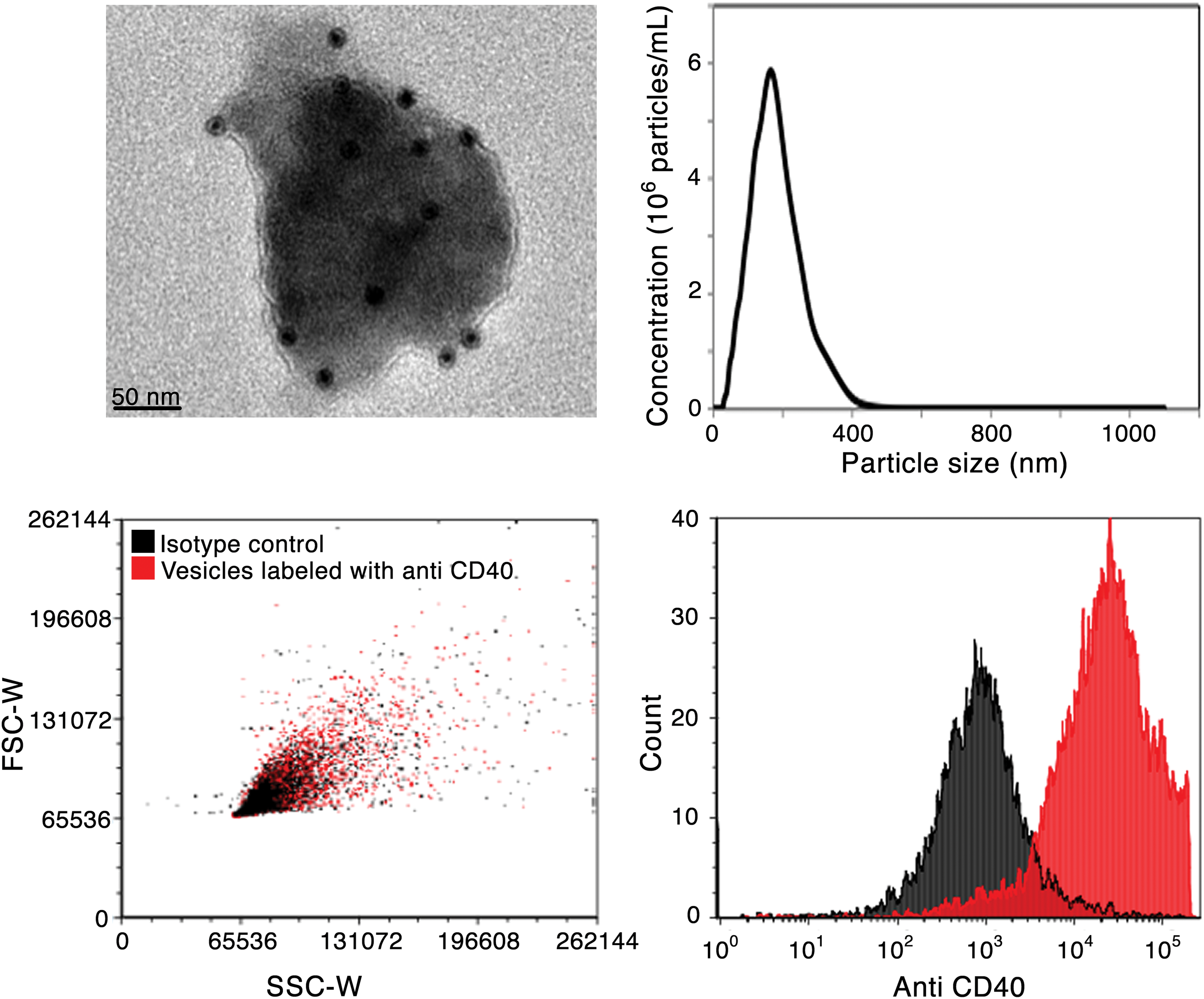
Vesicle characterization. Vesicles isolated from preosteoblast cultures stained positive for CD40 using immunogold transmission electron microscopy (immunoTEM; top left). Nanoparticle tracking analysis indicated that particles were on average 174.2±22 nm (top right). Flow cytometric analysis identified a CD40-positive population (bottom panel), confirming immunoTEM indications of the surface marker. Color images available online at
Particle sizing determined by both TEM and NTA suggests that the particles isolated from the preosteoblasts fall within the size range of microvesicles and not exosomes [21]. Surface markers are often used to further distinguish between these types of particles, with the presence of CD40 again identifying a microvesicle population. As noted earlier, immunogold labeling of the particles revealed the presence of CD40. In addition, FACS analysis also identified a strong CD40-positive population within the isolated particles. Forward versus side scatter (FSC-W vs. SSC-W) as well as count assessments indicated a distinct positive shift in fluorescent signal compared with isotype controls.
Cellular uptake efficiency of vesicles
The interaction and uptake efficiency of preosteoblast-derived particles incubated with undifferentiated ESCs was analyzed qualitatively by high-resolution confocal scanning microscopy at multiple time points (Fig. 2). The vesicle membrane was fluorescently labeled with a cell membrane dye before incubation with adherent stem cells to enable cellular tracking. Within 6 h, a large number of vesicles (red) were detected within all analyzed ESC clusters, indicating high cellular uptake efficiency of the microvesicles. The particles were widely dispersed within the ESC cluster and were primarily co-localized within the nuclei, the site of action of the vesicle cargo, which indicates endosomal escape and cargo delivery of the vesicles within the cell. By 24 h, some evidence of the particles still remained, though fewer were visible through confocal microscopy, possibly due to lysosomal degradation of the fluorescently labeled vesicle membrane within the cells.
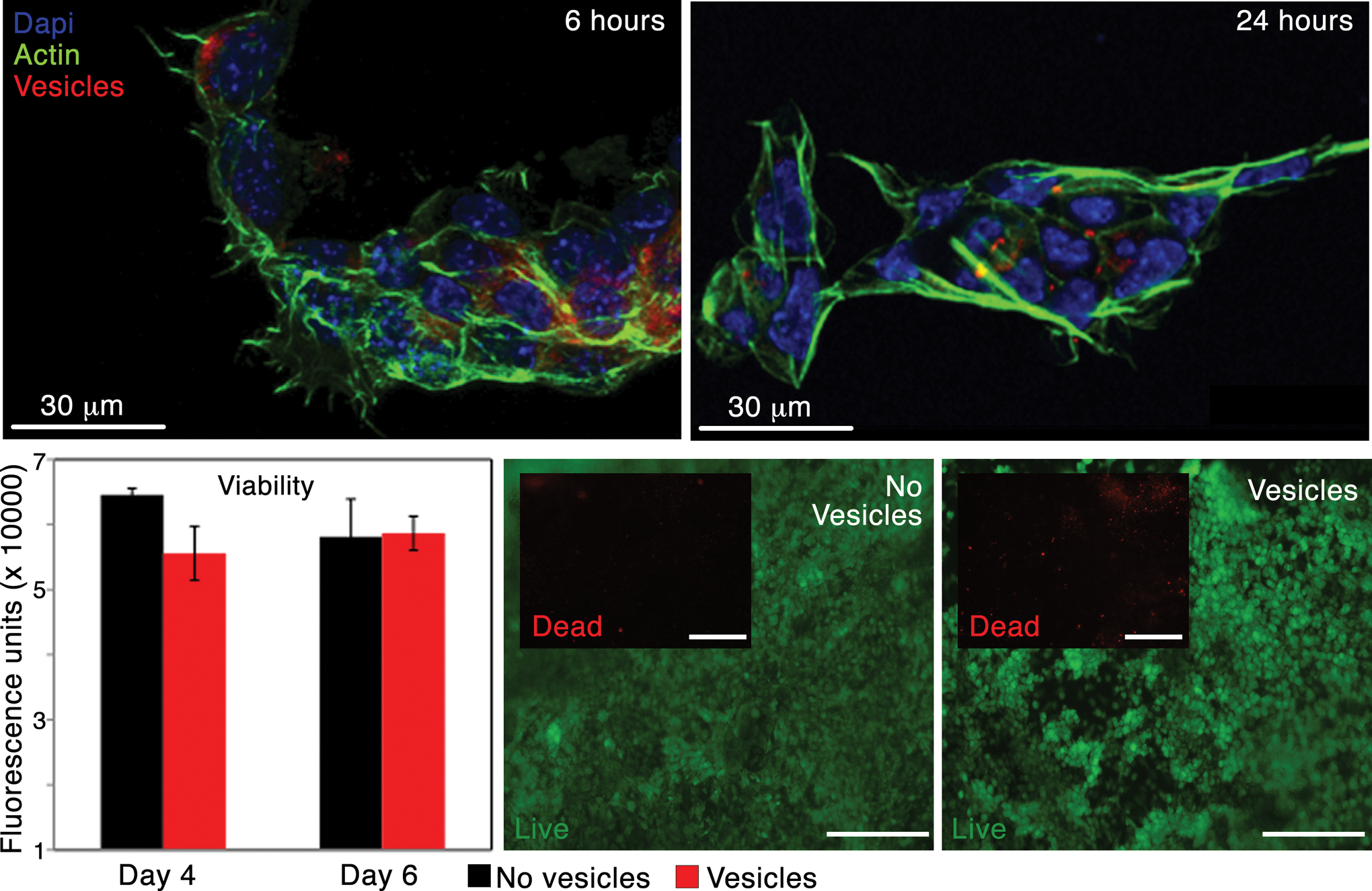
Cell-vesicle interactions. Extracellular vesicles were stained with CellMask™ Orange and visualized on clusters of actin (phalloidin/green)- and nuclei (DAPI/blue)-stained embryonic stem cells (ESCS) after 6 h (top left) and 24 h (top right). The presence of vesicles in ESC culture did not influence viability, as indicated by alamarBlue® (bottom left) and LIVE/DEAD® (bottom right) assays. Scale bars in LIVE/DEAD images are 200 μm and are both from day 6. Live cells are indicated by green fluorescence while dead cells (insets) are indicated by red. Color images available online at
The viability of differentiating ESCs was not affected by the presence of the particles. alamarBlue readings of cells incubated with preosteoblast-derived vesicles did not change significantly compared with untreated cells at either day 4 or 6 after incubation. Further, cells at each time point that were incubated with LIVE/DEAD stain primarily stained green (live) with very few visible red (dead) cells, supporting the alamarBlue results and indicating that the vesicles did not negatively affect ESC viability. These results were consistent across vesicles derived from separate batches of MC3T3s (data not shown).
Alteration of recipient cell behavior
In addition to examining the viability of recipient cells, alterations in the population's gene expression were investigated (Fig. 3). ESCs grown under differentiation conditions (no LIF) were either left untreated or incubated with vesicles, and RNA was collected after 3 or 7 days. Levels of gene expression of pluripotent (oct4, nanog, and sox2) and differentiation (mes/endoderm: sox17, gata6, foxa2; ectoderm: nestin, sox1, and fgf5) markers were analyzed using RT-PCR and expressed as fold change values compared with undifferentiated ESCs (day 0). ESCs incubated with the preosteoblast-derived vesicles expressed higher levels of all pluripotent markers at almost all time points examined. At day 3, oct4 levels were significantly higher in vesicle-treated cells (P<0.01), as were nanog levels (P<0.05) at day 7. In all cases, the fold change was less than one, with the exception of oct4 (1.39±0.3) and sox2 (1.07±0.5) at day 3 in the vesicle-treated population. Taken together, these results suggest that both populations exhibited some extent of differentiation by day 7, but the presence of the vesicles in the ESC culture delayed the onset of differentiation.
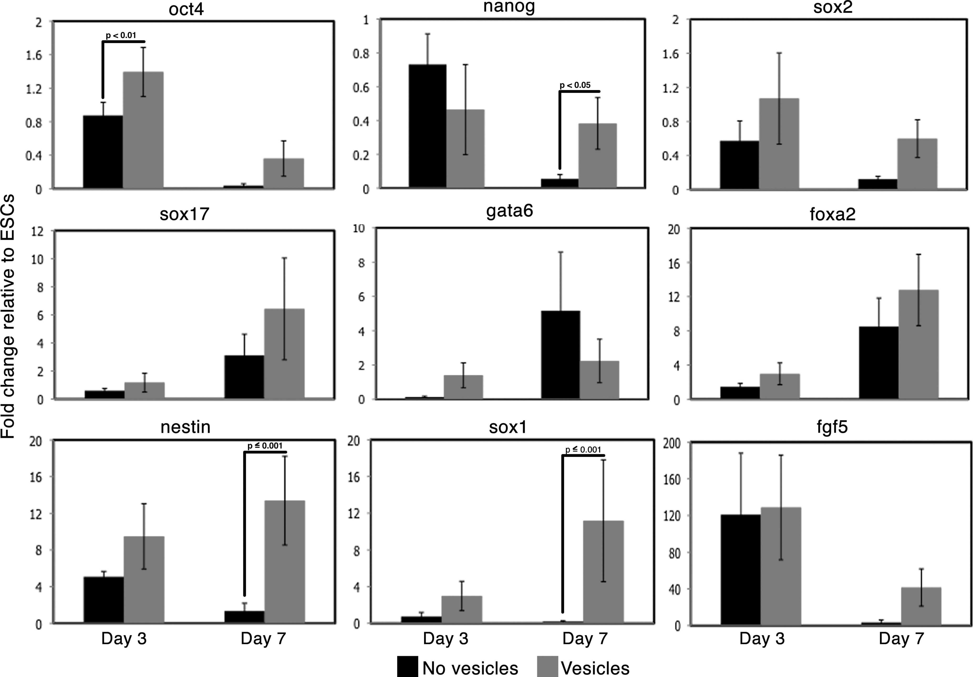
ESC gene expression changes. The influence of extracellular vesicles on differentiation ESCs was monitored using real-time–polymerase chain reaction. Pluripotent genes oct4 and nanog as well as neurectoderm differentiation markers nestin and sox-1 were significantly increased in ESCs treated with vesicles.
Analysis of lineage markers also indicated a modification in the differentiation trajectory of the treated cell population. This panel of markers was specifically chosen to represent each of the three germ lineages, with an up- or down-regulation in any group suggesting that the ESCs were moving toward or away from those lineages, respectively. No significant differences were noted in the mesendoderm markers, although the vesicle-treated cells exhibited higher fold changes at all time points, with the exception of gata6 at day 7 (Fig. 3). However, neurectoderm markers nestin and sox1 displayed significant increases in expression at day 7 in the vesicle-treated cells compared with untreated cells, with nestin fold changes increasing from 1.34±0.8 to 13.39±4.8 and sox1 shifting from 0.18±0.1 to 11.18±6.6 (P≤0.001 in both cases). The gene expression level of fgf5 similarly increased at day 7 from 3.34±2.6 to 41.47±20.3, though the change was not statistically significant. These results were consistent across vesicles derived from separate batches of preosteoblasts (data not shown) and suggest that the presence of the vesicles resulted in a shift toward the neurectoderm lineage in the differentiating ESCs.
Identification of cargo contained within particles
In order to probe why the presence of the vesicles in differentiating ESC cultures resulted in gene expression changes compared with untreated cells, the content of the particles was examined. For initial assessments on the cargo contained within the preosteoblast-derived particles, total DNA, RNA, and miRNA were isolated from multiple batches. On average, the particles isolated from a monolayer of preosteoblasts grown to confluence on a T225 flask contained 7.06±1.6 ng DNA and 1.17±0.3 mg RNA. To further explore the relevance of this content, an miRNA array was utilized to identify which miRNA molecules were most concentrated in the vesicles compared with the preosteoblast population from which they were derived (Fig. 4).
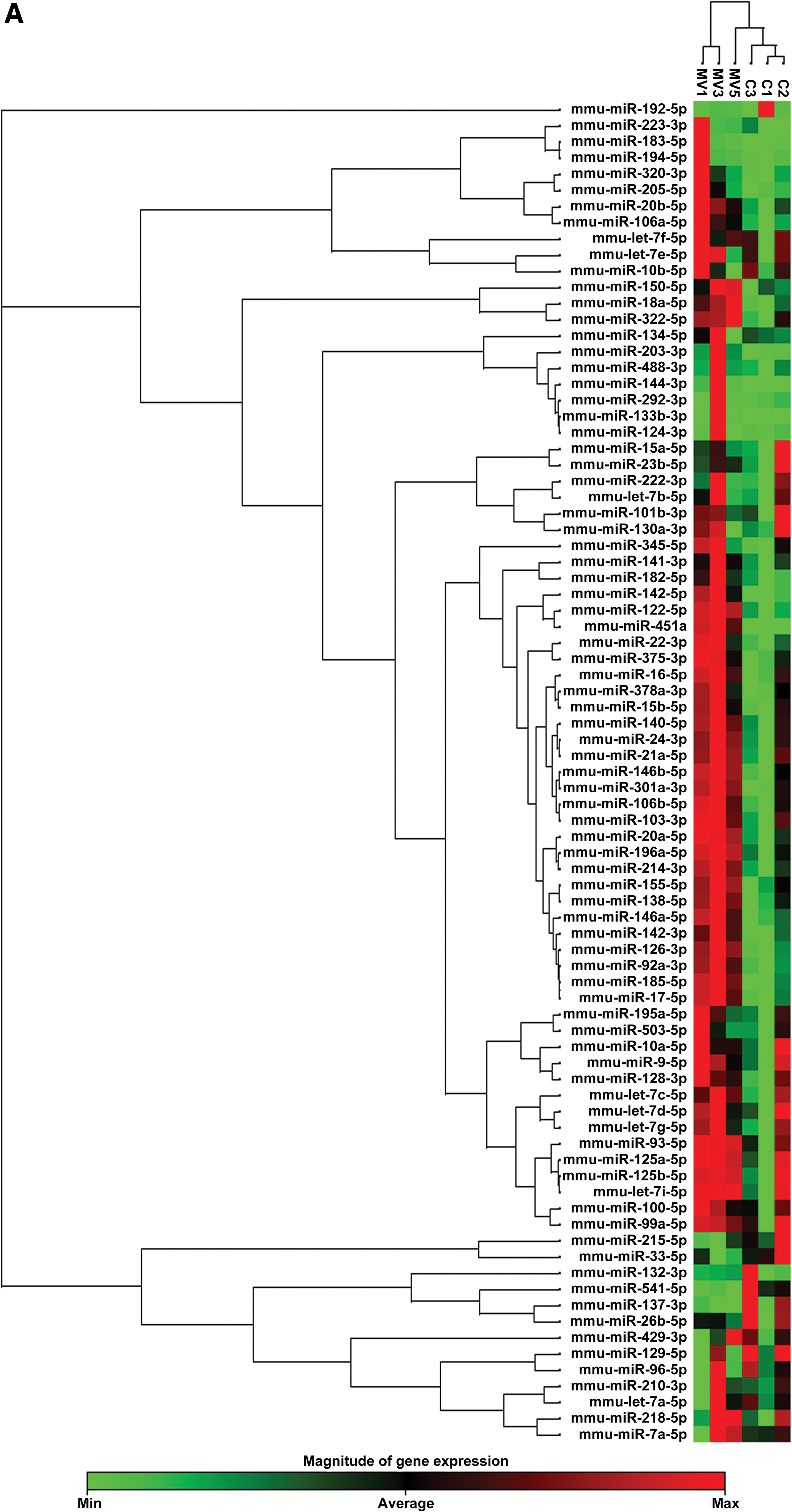
Vesicle cargo characterization. microRNA (miRNA) packaged within the preosteoblast-derived vesicles were analyzed using gene expression arrays for mouse cell differentiation and development.
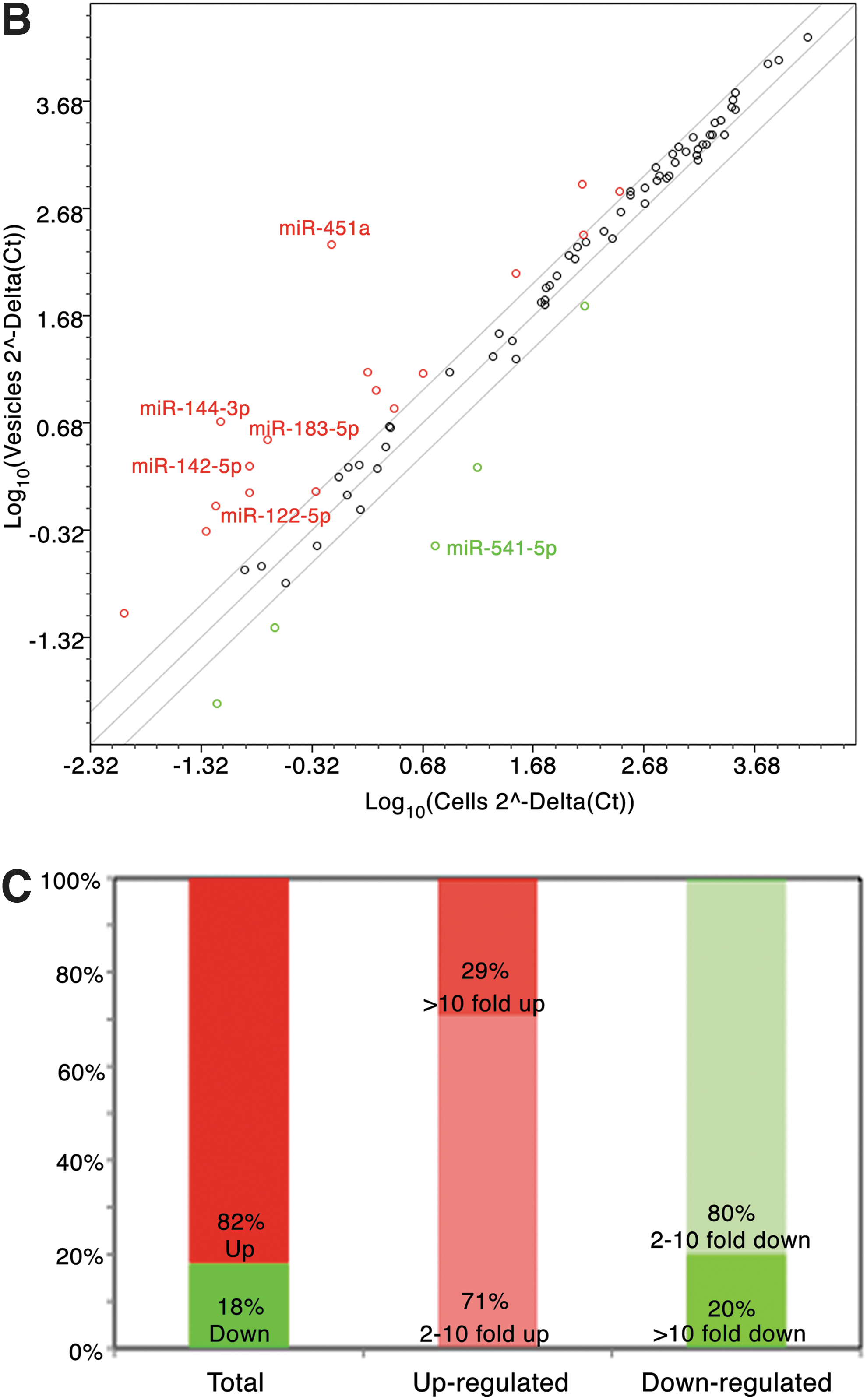
In total, 84 different miRNA genes affiliated with development and differentiation were analyzed in cell and isolated vesicle populations (n=3 in both cases). Heat map analysis clustered individual cell and vesicle groups together, as expected, with the latter demonstrating overall higher expression levels of the miRNAs examined (indicated in red) (Fig. 4A). Cluster analysis suggested that miRNA contained within the cell population was generally expressed at lower levels (green) than its vesicle counterparts, indicating that cells undergo a process that concentrates genetic material within the particles. This trend is corroborated by examining specific Ct values from each group: 44% of the isolated vesicles exhibited miRNA Ct values below 20 (indicating relatively high gene expression), while 37% fell under a Ct of 20 in the cell group. Log scatter plots of the vesicle versus cell populations highlight these differences between the groups (Fig. 4B). Points falling along the diagonal line crossing the center of the graph indicate miRNA expression levels that are equivalent between groups. As corroborated by the heat map cluster analysis, most points (81%; 68/84 genes) fall above this diagonal (fold change >1), suggesting a general increasing expression trend in vesicle miRNAs compared with the cells from which they were derived. The two outer diagonal lines delineate a twofold expression increase or decrease in vesicles as compared with cells. Approximately 20% (17/84 genes) of the analyzed miRNA genes in isolated vesicles exhibited a greater than twofold increase compared with cells (red dots). In contrast, only 6% of genes (5/84) showed greater than twofold decreased expression (green dots). Of the miRNAs exhibiting an increase in fold regulation in vesicles compared with cells, five displayed a 10-fold increase or greater: miR-122-5p (12.1-fold), miR-451a (312.3-fold), miR-183-5p (17.5-fold), miR-144-3p (67.5-fold), and miR-142-5p (14.4-fold). In contrast, only one miRNA (miR-541-5p) was down-regulated more than 10-fold. Figure 4C highlights these differences, showing the differing percentages of total up- and down-regulated genes. In addition, the breakdown of up- or down-regulation is displayed, indicating the percentage of genes within those that are up-regulated (red bar) which exhibit a 2–10-fold increase versus a greater than 10-fold increase. Similarly, the percentages of genes within those that are down-regulated (green bar) are differentiated based on either a 2–10-fold down-regulation or a greater than 10-fold down-regulation (for a list of which genes fall into these categories, see Supplementary Table S2).
Discussion
Particles naturally shed from a variety of cell types have been previously categorized most commonly as apoptotic bodies, microvesicles, exosomes, and others [21]. These “physiological liposomes” have been shown in several cell systems to contain genetic material originating from the host cell and, in many instances, to contain a combination of RNA and miRNA [21]. The extracellular vesicles presented in this study were harbored within the preosteoblast microenvironment and were isolated using ultracentrifugation techniques. They were found to be ∼170 nm in diameter, and immunogold and FACS analyses indicated the presence of CD40 in the population, suggesting that at least a portion of the isolated particles may fall into the “microvesicle” category established by recent literature [21]. Preosteoblast vesicles appeared to impact the maintenance of pluripotency as well as promoted neurectoderm differentiation in treated ESCs. Both mRNA and miRNA were found in the preosteoblast vesicles, and the definitive alteration in gene expression in the recipient ESCs suggests that the vesicles may have responded to this cargo. While the role of mRNA in influencing gene expression is well understood, the impact of miRNA on stem cell pluripotency and differentiation has only recently been investigated and continues to evolve [38 –44].
As previously outlined, ∼20% (17/84) of the miRNAs examined exhibited at least a twofold increase in the vesicles compared with the cells from which they were derived. Previous work in other cell systems has similarly shown an increase in genetic material in vesicles relative to the cells [15,22,45]. Twelve of the seventeen up-regulated miRNAs in the preosteoblast system are affiliated with the mesoderm lineage, which may be explained by its derivation from a mesoderm-derived cell source. It is of importance to note that miRNAs may play roles in multiple lineages, so those associated with the mesoderm may also be important in other developmental processes. Five of the miRNAs that were up-regulated at least twofold [miR-182-5p (2.1-fold), miR-183-5p (17.5), miR-320-3p (2.1), miR-205-5p (2.5), and miR-133b-3p (8.8)] have been implicated in pluripotency, which may explain why ESCs incubated with these vesicles exhibited higher levels of pluripotent genes. In addition, miR-141-3p, which promotes self-renewal in ESCs [46], was up-regulated 8.2-fold; all examined miRNAs in the pro-differentiation let-7 family [41,47] exhibited levels around 1 (0.9–1.5), with no significant increase relative to preosteoblast cells. Interestingly, three of the miRNAs up-regulated at least twofold (miR-182-5p, miR-183-5p, and miR-205-5p) have also been reported to influence development of the ectoderm, specifically in relation to neurological and inner ear development, as well as epidermal differentiation [48]. Given the up-regulation of these miRNAs in the isolated vesicles, it is possible that one or all of them played a role in the neurectoderm differentiation observed in the ESCs treated with vesicles. In addition to miR-183-5p, only two other miRNAs exhibited at least a 15-fold increase in the vesicles: miR-451a (312.3-fold) and miR-144-3p (67.5-fold), both of which have been implicated in erythropoiesis and megakaryopoiesis [49].
Although research is still relatively young regarding the roles of miRNAs and their corresponding effects in pluripotent cells, the work presented here has established the importance of understanding what is contained within naturally shed vesicles, as the genetic material enclosed within them may have significant effects on any recipient cell type. As an example, in other cases where documented protective effects are induced by shed particles, it is possible that miRNA cargo contained within the particles and transferred to the recipient cell play critical roles [30,31]. As demonstrated here, one potential application of this transferrable genetic material is as a means of directing the differentiation of stem cells. It is possible that the stem cell niche in vivo is influenced by particles shed from cells in that environment, which could direct the fate of localized stem cells. The technology of a biomaterials construct in which vesicles are incorporated could be one method of delivering packaged miRNAs to heterogeneous pluripotent populations as a means of controlled modulation at the gene level. In addition to this potential critical application as a means of controlling differentiation, the transfer of genetic material between cells has tremendous potential in the development of therapies. It is currently not well known how cells select the mRNA or miRNA that will be contained within the vesicles, although evidence suggests a non-random packaging [45], but controlling or understanding that process will be critical for manipulating vesicle transfer for therapeutic purposes. As an example, let us consider miR-133b-3p, which was up-regulated almost ninefold in the preosteoblast vesicles. This miRNA belongs to the cluster mmiR-133, which has been described in multiple reports for their potential in the treatment of heart disease [50,51] and promoting angiogenesis [52,53]. In addition, miR-133b has been shown to inhibit the growth of non-small-cell lung cancer [54] and invasion of prostate cancer cells [55]. If properly harnessed, it is possible that this miRNA, along with related molecules, may be able to integrate into critical niches to enhance or provide therapeutic efforts toward the treatment of specific diseases. It is, however, first critical to ensure that the select group of miRNAs are consistently up-regulated and transferred into the vesicles. Future work coupling controlled delivery methods along with vesicle biology may also serve to direct the targeting and uptake of the vesicles to the desired recipient cell population.
The horizontal transfer of genetic material via extracellular vesicle delivery may be considered a potent mediator for directing the differentiation of pluripotent populations. The work presented here identifies particles shed from preosteoblasts that resemble microvesicles and contain both mRNA and miRNA. The presence of these vesicles in differentiating ESC cultures significantly influenced stem cell fate, prolonging expression levels of pluripotent markers while increasing later markers of neurectoderm differentiation. Future work investigating the control of miRNA packaging as well as targeted delivery of the vesicles may significantly enhance the potential of this technology in cellular reprogramming and directed differentiation.
Footnotes
Acknowledgments
The authors would like to acknowledge Kharissa Nitiputri for her contributions to the immunogold labeling of the extracellular vesicles. R.N. was supported by a Whitaker Foundation International Scholarship, and L.S. was supported by a Marie Curie Actions Intra-European Fellowship.
Author Disclosure Statement
No competing financial interests exist.
References
Supplementary Material
Please find the following supplemental material available below.
For Open Access articles published under a Creative Commons License, all supplemental material carries the same license as the article it is associated with.
For non-Open Access articles published, all supplemental material carries a non-exclusive license, and permission requests for re-use of supplemental material or any part of supplemental material shall be sent directly to the copyright owner as specified in the copyright notice associated with the article.