
Abstract
With the recent advances in regenerative medicine, nanotechnology has created a niche for itself as a promising avenue in this field. Innumerable studies have been carried out by researchers using virus-based methodologies for the purpose of epigenetic reprogramming. Although this method is ostensibly safe, nonetheless, they are tagged with the risk of viral genome integration into the host genome or insertional mutagenesis. Transient transfection by the use of nanocarriers is the best way to overcome these problems. This review focuses on some of the significant works carried out by researchers utilizing nanocarrier systems that have shown promising results and thus created a landmark in the epigenetic reprogramming.
Introduction
T
Stem cells are cells that are capable of self-renewal [4]. They are undifferentiated cells that have the potential to differentiate into specialized cell types. Stem cells are basically classified into embryonic stem cells (ESCs) and adult stem cells. However, the recent breakthrough in stem cells in the year 2006 by Kazutoshi Takahashi and Shinya Yamanaka, introduced to the world a novel type of stem cell, namely, the induced pluripotent stem (iPS) cell. In this unprecedented work, mouse embryonic or adult fibroblast cells were induced to pluripotency by introducing four transcription factors (TFs), also known as Yamanaka factors, that are dominantly expressed in ESCs such as Oct3/4, Sox2, Klf4, and c-Myc (OSKM) [5].
ESCs are pluripotent cells that are capable of differentiating into any type of cell. They are exclusively found in the inner cell mass of the blastocyst, which remain undifferentiated and later differentiate into the three germ layers–the ectoderm, mesoderm, and the endoderm layers, which then further differentiate into a wide variety of cell types [6,7]. Generally, an ES cell is identified by its pluripotent nature as well as its endless ability of self-renewal, which is accomplished by manipulations through the addition of growth factors such as the basic fibroblast growth factor or Wnt protein [8 –11] for the maintenance of pluripotency. It can also be accomplished by manipulations through genome editing by zinc finger nucleases and other alternative methods [12 –15] or even with the help of prosurvival cocktails [16,17].
Adult stem cells are, however, multipotent in nature. They have the ability to self-renew and differentiate into a specific type of cell. They possess tissue-specific niches where they reside. These niches are responsible for maintaining the stem cells in their undifferentiated state [18]. It is only during the need of the hour, such as during damage, injury, or disease, that these cells undergo differentiation into a specialized cell type, which possesses a characteristic shape or specialized function, thereby regenerating lost or damaged tissue [19]. Adult stem cells are of several kinds such as hematopoietic stem cells (HSCs), mesenchymal stem cells (MSCs), and neural stem cells (NSCs) to name a few.
So what is epigenetic reprogramming and how is it useful for regenerative medicine? Earlier, it was conceptualized that the mammalian development was unidirectional where it began with a single-celled zygote, which differentiated to form several specialized cell types due to the characteristic epigenetic patterns that they possessed [20,21]. It was only later in 1992 that the term nuclear reprogramming came into picture highlighting the works carried out by several researchers who demonstrated that it was possible to revert the cells from their differentiated state to a less differentiated state such as the conversion of unipotent cells like primordial germ cells to pluripotent embryonic germ cells [22,23]. These works carried out on nuclear transfer, created a significant milestone in the field of nuclear reprogramming [24,25]. It was thus concluded that certain developmental restrictions in differentiated cells were not due to permanent genetic changes, but rather due to reversible epigenetic modifications [26]. This led to a hypothesis that certain factors could have been responsible for the induction of pluripotency if they were forcefully made to express in somatic cells. This is termed as epigenetic reprogramming.
Epigenetic reprogramming has been used to describe the chromatin modifications or remodeling that occurs as a result of erasure and re-establishment of epigenetic marks through histone modification or DNA methylation [2]. These modifications are heritable. The interactions between the TFs and DNA contribute to the identity of a cell, which is manifested in the form of specific patterns of gene expression [27].
Regenerative pharmacology is an amalgamation of works carried out in the field of tissue engineering, gene therapy, and pharmacology wherein experts from these interdisciplinary fields come together with the aim of curing a disease through the restoration of the function of an organ or tissue. Regeneration can be accomplished in three different ways: (1) Reprogramming–where a differentiated cell is induced to pluripotency, (2) Transdifferentiation–where a cell type of one lineage is converted into a cell type of a different lineage (such as brain stem cells converting into blood cells) [28], and (3) Dedifferentiation–wherein a terminally differentiated cell is reverted to a lesser differentiated one [29]. Thus, reprogramming a cell to form an iPS cell by epigenetic modifications with the help of TFs, mRNA, transposons, proteins, or small molecules is a promising approach for the restoration of specific tissues or organs. It is accomplished through simple chromatin modifications without actually altering the DNA sequence. This forms the holy grail of regenerative medicine. This review thus highlights some of the works carried out by researchers in the context of reprogramming and the importance of nanotechnology in regenerative medicine. Figure 1 provides a brief overview of the various methods of reprogramming a cell using viral and nonviral delivery systems. We shall also briefly look into some of the noteworthy applications of nanofibrous scaffolds in the context of reprogramming and conclude the review by outlining the significant epigenetic modifications that take place at the molecular level when reprogramming the cells to induced pluripotency.
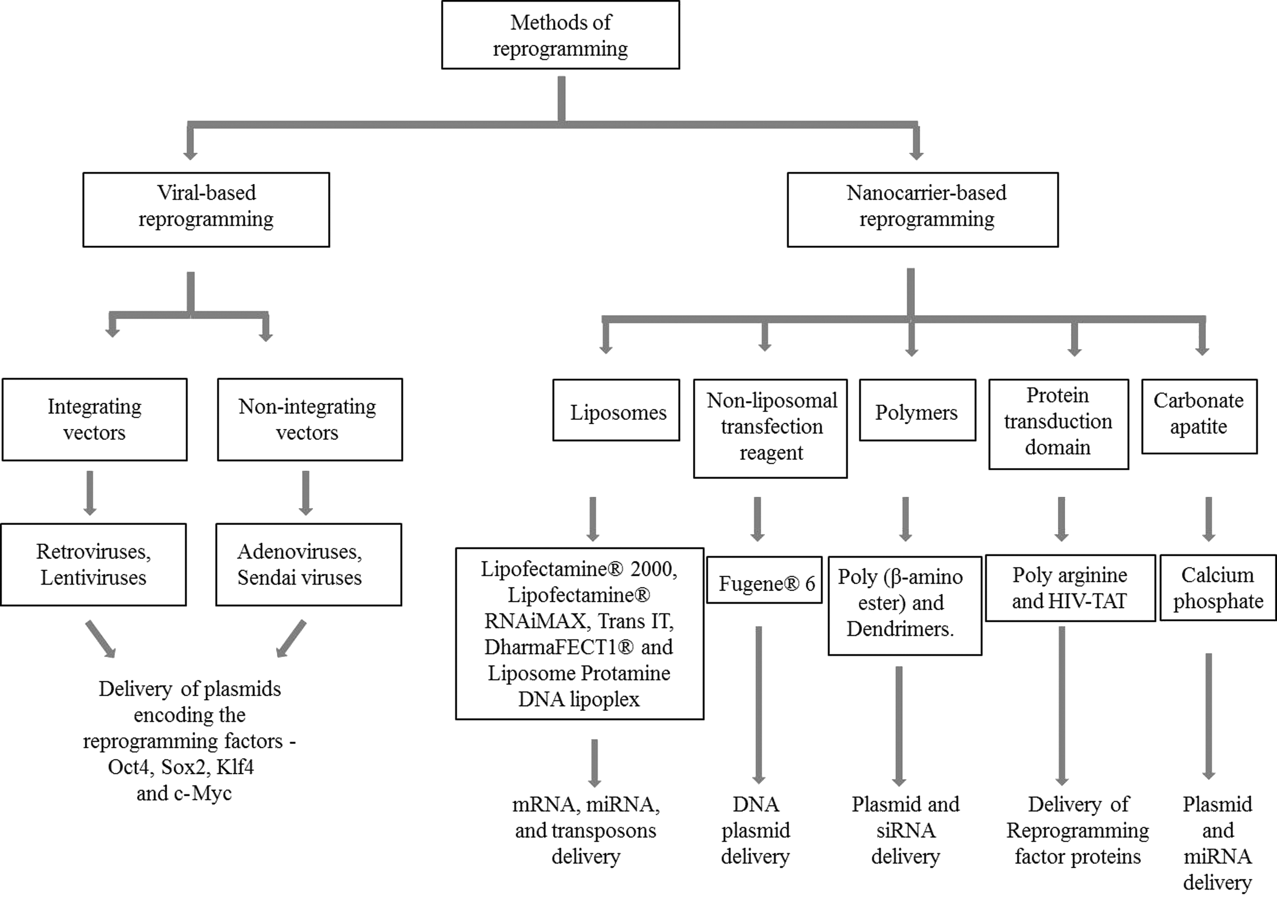
Flow chart outlining the various methods of reprogramming. These methods are broadly classified into viral-based and nonviral or nanocarrier-based reprogramming.
Reprogramming by Viral or Nonviral-Based Methods
A quintessential factor for the successful manipulation of cell behavior lies in the efficient delivery system. It is known that stem cells are quite difficult to transfect [30,31]. Researchers have largely relied on viruses as a vector for carrying genes for efficient reprogramming of cells through transduction. Although the results of viral transduction in providing a stable and long-term expression of a particular gene remain incomparable to any other, the downside of the process remains in the unwanted integration of viral genome into the host cells, which is risky since it could lead to mutations in the gene by increasing the risk of tumor formation, thus nullifying the purpose [32]. Immune response is also another factor of concern.
Nanocarriers gain an upper hand wherein transfection with nanocarriers does not carry any baggage with it regarding these safety issues. They provide transient gene expression without integrating into the host genome. However, there are minor drawbacks such as low transfection efficiency and cytotoxicity. Researchers have been, over the years, improvising the nanocarrier systems for the purpose of delivery. Recent studies focus on not only increasing their efficiency, but also in minimizing their toxicity. Daniel G. Anderson and group at the Massachusetts Institute of Technology employed polymeric nanoparticles and increased their efficiency for the delivery of gene carrying the TF such as Oct4 into human ESCs (hESCs) [33]. Another group reported a study on the inorganic nanocarrier carbonate apatite biofunctionalized with fibronectin and E-cadherin to be successful in delivering DNA to ESCs [34,35].
Viral-Based Reprogramming
Integrating vectors
Retroviruses
Retroviruses are double-stranded RNA viruses. These vectors have been used for gene delivery since it results in a long-term stable expression of the genetic elements. Actively proliferating cells are infected readily and with high efficiency utilizing retroviruses when compared with the cells that have been differentiated such as the myocytes or the neurons [36]. Some of the commercially available retroviral vectors are derived from moloney murine leukemia virus (MoMuLV) and molony murine sarcoma virus (MoMuSV) [37].
One of the most recent works in stem cell research that gained worldwide recognition was, undoubtedly, the work on iPS cells carried out by Shinya Yamanaka and group, wherein the concept that a somatic cell could be reprogrammed into a pluripotent stem cell was shown for the first time by the introduction of TFs through retroviral infection. Reprogramming was carried out in somatic cells by the overexpression of TFs. Of the twenty-four candidate genes of TFs that were responsible for the maintenance of stemness in ESCs, four of them were found to be indispensable for the stem cell maintenance. These TFs, namely OSKM, were introduced to the adult fibroblast cells through retroviral infection [5]. Teratoma (complex masses of cells produced in vivo) or embryoid bodies (aggregates of cells produced in vitro and/or in vivo), a gold standard for the indication of stemness [38], was observed after the introduction of these factors and hence concluded the idea that it was possible to directly generate stem cells from fibroblast cells. This eliminates major concerns regarding ethical issues or the rejection of a tissue because these cells could be isolated from the patient's own cells. Furthermore, they were also successful in generating iPS cells from mouse embryonic fibroblasts (MEFs) as well as adult human dermal fibroblasts using the same TFs [39]. Several works were also carried out by the same group [40] as well as other groups to provide iPS cells that were epigenetically identical to ESCs using this method [41]. It was later reported by further research that it was possible to eliminate the TF c-Myc when reprogramming somatic cells [42,43].
Generating iPS cells from oncogenic TFs has been one of the most efficient methods. However, one of the chief concerns has been an increase in the genetic aberrations, for reasons unknown, of the iPS cells which are absent in parental cells [44,45]. On the addition of these TFs, there is an increase in the growth rate, which in turn increases the metabolic demand for energy and precursors for biosynthesis [46]. There occurs a shift in the metabolism of reprogramming cells from oxidative respiration to oxidative glycolysis [47]. A gradual accumulation of electrons in the electron transport chain takes place which increases their leakage into the cytoplasm as Reactive Oxygen Species (ROS). DNA damage occurs as a consequence of this oxidative stress. Therefore, antioxidants such as N-acetyl-cysteine (NAC) or Vitamin C, also known as ROS scavengers, were used in a study carried out by Ji et al. as they are helpful in preventing this damage. The iPS cells were generated from the fibroblast cells by retroviral transduction. During the reprogramming process, the growth media was supplemented with NAC or Vitamin C which, from the data collected, showed a reduction in the genome instability so as to enhance the quality of iPS cells [48].
However, transduction using retroviruses has some disadvantages. On one hand, the transduced gene was observed to be progressively silenced and on the other hand, the proviral expression in individual clones of cells was found to be intensified during cell differentiation. This problem was predominant in the early embryo or ESCs during their transition from an undifferentiated to a differentiation stage [49]. Recently, it was found that the retroviral vector silencing was a result of the epigenetic mechanisms that were predominantly controlled by a specific insulator such as the D4Z4 element. However, vectors that contain the 3′D4Z4 as well as HS4 insulators were found to block the complete silencing of the transduced gene [50].
Lentiviruses
Lentiviruses belong to a subclass of retroviruses having double-stranded RNA as their genetic material. Lentiviral vectors are also preferred for the reprogramming of cells through transduction since they can infect a broad spectrum of host cells, including the terminally differentiated cells as well as primary cells with very good efficiency and minimal gene silencing [51,52]. Rudolf Jaenisch and group believed that it was possible to directly reprogram human somatic cells to pluripotent stem cells by using a drug-inducible system. Employing doxycycline (dox)-inducible lentiviruses, it was possible to generate secondary somatic cells carrying the four reprogramming factors. These cells were found to be genetically homogeneous which were further reprogrammed to form the pluripotent secondary human iPS [53]. Using this system [54], it was also possible to accomplish the same feat by the expression of Oct4 TF alone along with certain small molecules which were able to replace certain TFs [55].
Recently, it was shown that it is possible to reprogram mouse fibroblasts into cardiac cells, through the process of transdifferentiation, by the expression of only a single transcription factor such as the Oct4 along with a defined cocktail of small molecules consisting of Activin-Linked Kinases 4/5/7 (ALK4/5/7) inhibitor, Glycogen Synthase Kinase 3 (GSK3) inhibitor, parnate (Lysine Specific Demethylase 1A inhibitor), and forskolin (Adenylyl Cyclase activator). The MEFs infected with lentivirus encoding the Oct4 gene and the small molecules were thus found to be converted to spontaneously contracting clusters. Therefore, this method enabled the efficient conversion of mouse fibroblast cells into beating cardiac cells, exhibiting a ventricular phenotype, without crossing over the pluripotent stage [56].
Attempt was made to induce the formation of human neuronal cells designated as iN (induced neural) cells from dox-inducible lentiviral vectors. By forced expression of TFs such as Brn2, Ascl1, and Myt1l, Pang et al. were able to successfully drive mouse fibroblast as well as iPS cells into functional iN cells. Additionally, encouraging results were obtained wherein it was also possible to generate iN cells from fetal and postnatal fibroblasts by combining the basic TF NeuroD1 along with the above-mentioned TFs [57].
Again, the use of lentiviral vectors has been a cause of concern. A recent report by Yamagata et al. in 2012 provided evidence that when HSCs or human CD34+ cells (bone marrow-derived progenitor cells) were transduced with these vectors, unwanted chromatin modifications were introduced through methylation of histone proteins of H3K36me3. Histone methylation was thus found to increase by several folds when the HSCs were transduced with lentiviral vectors [58].
Nonintegrating vectors
Adenoviruses
Unlike retroviruses and lentiviruses, adenoviruses were considered safer since genetic reprogramming was possible without the risk of integration of the viral genome into the host genome [59]. Other than that, these viruses do not cause any severe pathogenicity.
Generation of adeno-iPS cells by the adenoviruses provided evidence that it was possible to reprogram mouse as well as human fibroblast cells from the four Yamanaka factors without the need for any insertional mutagenesis [59,60]. Many researches have been carried out in the recent years using the adenoviruses. However, these will not be discussed in detail.
One of the major concerns regarding the use of adenovirus is the high immunogenicity caused by the virus itself. The viral capsid was found to be the triggering agent [61]. As a result, the transgene expression was silenced or repressed. In the year 1999, after systematic administration of adenoviral-targeted gene therapy to a patient, there was an initiation of a strong cytokine response that rapidly led to multiple organ failure and death of the patient [62,63].
Sendai viruses
Sendai viruses replicate in the cytoplasm of the infected host cells without integrating into the host genome [64]. In 2009, Fusaki et al. successfully attempted to reprogram somatic cells—human fibroblasts, into induced pluripotent cells with the help of Sendai viruses [65]. A similar work was reported to induce iPS cells from cord blood cells by improving the Sendai viral vectors by making them temperature sensitive, which was more effective in eliminating any traces of viral genes in the host cell [66]. A very recent work was carried out to induce iPS cells from HSCs and progenitor cells utilizing this vector [67].
Epstein–Barr viruses
Yu et al. carried out an interesting work on the generation of iPS cells from the human somatic cells using the oriP/EBNA1 (Epstein–Barr Nuclear Antigen1)-based episomal vector, which was derived from the Epstein–Barr virus. These episomal reprogramming vectors are nonintegrating systems that replicate only once per cell cycle. They were introduced into the cells through nucleofection. The human iPS cells so derived were made completely free from the vector and transgene sequences due to the gradual loss of the episomal vectors in the absence of drug selection and, therefore, exhibited typical hESC morphology. However, reprogramming efficiency with this vector was found to be low [68].
Nanocarrier-Based Reprogramming
Nanocarriers have always been the choice for transfecting cells because of the low immunogenicity, the unlimited size of gene that can be used for transfection, as well as the ease with which the transfection could be achieved [69,70]. Many works related to genetic engineering have been successfully achieved utilizing nanoparticles. There is no risk of gene integration or insertional mutagenesis, which is usually the case with viral vectors [71,72]. However, the disadvantage lies in their efficiency which is quite low. Researchers have efficaciously tackled this problem and, thereby, were successful in increasing their efficiency.
Liposomes
Liposomes, most importantly the cationic liposomes, have been the most popular choice for transfecting cells. Liposomes are bilayered vesicles surrounded by phospholipid membrane and are amphiphilic in nature [73]. Cationic liposomes are positively charged and are attracted to the negatively charged cell membrane. They are taken up by the cells through endocytosis and other mechanisms [74]. Among the cationic liposomes, Lipofectamine® has proven to be the gold standard for transfection-related studies [75]. Other lipid-based formulations such as DharmaFECT1® [76] or TransIT [77] have also been utilized for the purpose of reprogramming cells to pluripotency.
Lipid-based formulations
Messenger RNA (mRNA) delivery:
The mRNA carries the genetic sequence required for the synthesis of protein. Synthetic mRNA is considered to be appealing in terms of cell fate manipulation or reprogramming because of its transient expression. Thus, mRNA encoding the required TF can be transfected into cells easily.
In the year 2010, Derrick and colleagues at the Harvard Medical School in Boston, Massachusetts, created a new stride in epigenetic reprogramming by inducing a direct differentiation of the fibroblast cells to iPS cells using synthetic modified RNA [75]. Furthermore, in the same study it was also shown that the RNA-induced iPS cells, referred to as the RiPSCs, were also found to differentiate into terminally differentiated myogenic cells using the same technique. The results were found to be substantially superior to the viral transduction method. mRNA synthesized by in vitro transcription was modified so as to evade innate immune responses due to exogenous RNA and it also possessed an increased half-life in cytoplasm due to the presence of a 5′ guanine cap. For the induction of RiPS cells, mRNA was complexed with the cationic lipid Lipofectamine® RNAiMAX through electrostatic interactions, and transfected daily to the fibroblast cells for 17 consecutive days, after which hESC-like colonies appeared. It was preceded by the formation of compact epitheliod morphology around the first week of mRNA transfection (Fig. 2). Daily transfection did not prove to be toxic to the cells nor did the molecular profile of the transfected cells get altered. For the RiPSC-to-myogenic conversion, the RiPSCs were subjected to a 3-day transfection using Lipofectamine® RNAiMAX complexed with mRNA encoding MyoD. Myogenic cells were induced on the third day (Fig. 2) [75]. Thus, from this work, it was clear that it was also possible to induce iPS cells by simple mRNA transfection.

Schematic illustration of the protocol for the induction of iPS cells namely the RiPSCs (RNA-induced pluripotent cells)
Modified mRNAs can be engineered by various methods, which include the methylation of 5′ end posttranscriptionally or synthesizing highly modified mRNAs through the replacement of all the pyrimidines or the removal of 5′ triphosphate by treatment with a phosphatase enzyme resulting in the generation of a 5′-OH end [78]. A detailed established protocol describing the synthesis of modified mRNA and their successful introduction into the cells for reprogramming has been published recently by the same group [79]. A very recent work was carried out wherein modified mRNA, encoding the enhanced green fluorescent protein (EGFP), was delivered to the primary human as well as mouse NSCs with the help of the cationic lipids, Lipofectamine® 2000 or Trans IT. NSCs serve as attractive candidates for reprogramming since the TFs Sox2 and c-Myc are constitutively expressed and, therefore, require the expression of only Oct4 and Klf4 [80,81]. EGFP was expressed after transfecting the neurosphere cultures with mRNA encoding EGFP, hence opening doors for the possible reprogramming applications using primary cultures as well [77].
Current approaches to reprogramming through mRNA transfection have certain disadvantages. First, mRNA encoding a single transcription/reprogramming factor is a time-consuming process since its expression remains robust for the first 24 h after which a time period of around 2 weeks is typically required for the induction of iPS cells. Second, typically for mRNA reprogramming, feeder layers consisting of mitotically inactivated fibroblast cells are employed which are themselves subject to transfection. This hinders the process of monitoring the cells for reprogramming, thus adding to the complexity of the procedure [82]. Added to that, there is a risk of feeder layer-associated toxicity and contamination [82 –84]. Xenogeneic contaminants from mouse feeder cells restrict their application mainly for transplantation into humans and, therefore, human fibroblast cells were utilized [85]. However, even human feeder cells possessed some form of xenogeneic contamination [82]. Therefore, there arises the need to eliminate these potentially harmful threats that hamper the use of regenerative medicine. In an attempt to eliminate this problem, Warren et al. carried out mRNA reprogramming with a novel approach using a xeno-free protocol that eliminates the need for a feeder layer. Fibroblast cells were transfected with an mRNA transcript comprising of a cocktail of reprogramming factors, such as the 5-factor or 7-factor with the help of the cationic lipid, Lipofectamine® RNAiMAX, to increase the rate of the reprogramming process. This reduced the time to half of what was needed using the traditional approaches [82].
Micro RNA (miRNA) delivery:
miRNAs are noncoding RNAs and consist of 22 nucleotides. They function by regulating the transcription and translation of a gene expression [86]. miRNAs are synthesized in the eukaryotic organisms and are usually encoded by the introns. Once derived from the transcripts, they fold back themselves to form the hairpin loop [87], which is then processed. The processed miRNA is loaded onto the argonaute protein [88] which base pairs with the mRNAs of protein-coding genes thus silencing the function of a particular gene through posttranscriptional repression [89 –92].
The first attempt to reprogram somatic cells through miRNA was carried out by Robert Blelloch and group [76]. Certain miR-290 clusters are known to be of paramount importance in the maintenance of the stemness in mouse ESCs [93]. During the course of differentiation, their expression is downregulated [94]. The present report demonstrated that a subset of the miRNA clusters, which are the ESC cycle regulating miRNAs namely, miR-294, miR-295, and miR-291-3p [95], have the potential to enhance the efficiency of reprogramming along with retroviruses expressing Klf4, Oct4, and Sox2 in mouse embryonic fibroblast cells. The cells were transfected with miRNAs with the help of a lipid-based formulation such as DharmaFECT1 and reprogrammed to pluripotency.
Liposome protamine DNA lipoplex
Transposon delivery:
Transposons are circular plasmid DNA molecules. They are also known as the jumping genes since they are mobile genetic elements that translocate from one DNA site to another by a cut-and-paste mechanism [96]. They are becoming increasingly desirable as nonviral gene delivery systems and have been used to overexpress genes in various types of cells without the risk of toxicity [97]. Hence, transposon-based gene delivery systems are appealing tools for transgene delivery. A transposon system is typically a 2-plasmid system consisting of a helper plasmid and a donor plasmid, which are plasmids coding for the transposase enzyme and the gene of interest, respectively. These plasmids are flanked by terminal repeat sequences [98]. Sleeping Beauty (SB), consisting of the resurgent SB transposase gene [99], and the piggyBac (pB) are the most commonly used transposon systems.
Usually, transposons are delivered into the host cell by means of phage delivery systems or plasmid delivery systems, wherein a suicide/naked plasmid, which does not have the ability to replicate in the recipient cell, is preferred [97,100]. This is transferred by nucleofection/electroporation [101] or transformation. For example, following nucleofection of the SB transposon system, it was possible to transpose OSKM genes so as to reprogram human foreskin fibroblasts as well as MEFs. This protocol was found to be on par with the viral transduction method [101]. However, nucleofection has its own disadvantages given the occurrence of cell death. A novel work was carried out wherein the SB transposon was delivered into the cells with the help of liposome protamine DNA (LPD) nanoparticle. This nanoparticle was engineered by the combination of cationic 1,2-dioleoyl-3-trimethylammonium propane liposome with the cationic polymer, protamine. It forms a tight complex with the DNA through electrostatic interactions [102]. In this study, the LPD nanocarrier was complexed with the anionic SB transposon, anionic cell surface-targeting peptides, such as the homing peptide to target the rat MSCs as well as a nuclear localization signal peptide, for efficient delivery into the nucleus. The LPD was successful in transfecting the rat MSCs with the SB transposon making this delivery system a promising one for future applications in reprogramming cells for the purpose of regenerative medicine [103].
Nonliposomal transfection reagent
FuGENE® 6
Plasmid delivery:
Shinya Yamanaka and group explored the possibilities of generating iPS cells by plasmid transfection. Plasmid constructs carrying the four factors, OSKM, were transfected to the MEFs using the FuGENE® 6 transfection reagent, a nonlipid-based formulation. Two expression plasmids, one complementary DNA (cDNA) containing the three factors–Oct3/4, Sox2, and Klf4 and the second cDNA encoding the fourth factor c-Myc were transfected to MEFs, which resulted in the formation of iPS cells without any plasmid integration into the MEF genome. These cells when transplanted into mice produced teratomas, a hallmark of stem cells, and contributed to the formation of adult chimeras [72].
Polymers
Polymers, synthetic or natural, have gained considerable interest in regenerative medicine due to their compatibility, high efficiency, low immunogenicity, and most importantly, their biodegradability [104]. They have been exclusively used by researchers over the years for the delivery of cargoes, such as nucleic acids like siRNA, for the purpose of gene knockdown [105] or for the delivery of other therapeutic nucleic acids [105,106] or proteins [107] for the development of vaccines [107,108] and other hydrophobic and hydrophilic drugs [109]. Polymers that have been used for the purpose of reprogramming cells are discussed in this study.
Poly (β-amino ester) nanoparticles
Poly (β-amino ester)s (PBAEs) are positively charged polymers, which were synthesized first by Robert Langer and group by the Michael addition reaction consisting of small molecular weight monomers and diacrylate monomers. The polymer backbone consists of ester linkages that can be degraded easily through hydrolysis. PBAEs were found to degrade rapidly at a higher pH such as pH 7.4 [110]. They are biodegradable nanoparticles that can be easily synthesized by the conjugation of amines to diacrylates as well as chemically modified [111 –113]. They are promising biomaterials for efficient transfection, including hard-to-transfect cells such as hMSCs and form stable complexes with nucleic acids that escape endosomal degradation [113]. They have been successfully used by researchers for the purpose of gene delivery [114].
Plasmid delivery:
PBAEs were employed for the reprogramming of human fibroblast cells by serial transfections for the transient expression of a single CAG-driven (a promoter containing a Cytomegalovirus or the CMV enhancer, a chicken beta-actin intron and a rabbit beta-globin gene) polycistronic plasmid expressing the OSKM factors as well as a reporter gene such as GFP. Transfection results concluded the generation of iPS cells in 3–4 weeks, which differentiated successfully into the three germ layers, hence proving a noteworthy application of these polymers in the field of reprogramming [115]. Furthermore, it was also concluded that the transfection efficiency of this polymer was far superior when compared with Lipofectamine® or FuGENE.®
Green and coworkers attempted to transfect IMR-90 human primary fibroblast cells using a feeder-free protocol. This was beneficial in terms of safety as well as efficacy. PBAEs, self-assembled with plasmids encoding the reprogramming factors, were transfected to these cells. Human iPS-like colonies were observed after a period of 36 days. This work clearly highlighted the potential of these polymer nanoparticles in reprogramming cells [116]. In another study, PBAE nanoparticles facilitated the uptake of DNA construct consisting of the TF, Oct4, and the GFP into hESCs. PBAEs due to their positive charges interact with the negatively charged DNA and condense it to form a compact structure that can be easily taken up by the cell [110,117,118]. The crux of the study was to maintain the hESCs in an undifferentiated state, which was made possible by the nonviral gene transfer of Oct4 gene tagged with GFP using the polymer nanoparticles. The level of GFP expression was used as an indicator to track the state of hESCs, that is, if the cells remained in an undifferentiated state or were undergoing differentiation. High expression level/fluorescence of GFP protein in the hESCs indicated that these cells remained in an undifferentiated state [33].
Small interfering RNA delivery:
Gene therapy is an area which has been explored over the years for the treatment of various diseases that stem from the defective genes. RNA interference has been a powerful tool for silencing the expression of a particular gene with high accuracy. Small interfering RNA (siRNA) are duplexes of 21 nucleotides which knockdown a gene in a sequence-specific manner [119]. The mechanism was observed by Fire et al. in the year 1998 in C. elegans [120]. This unique method of silencing genes has been exploited by scientists for the purpose of directing the fate of stem cells for the purpose of regenerative medicine [121]. Although the possibility of using siRNA for reprogramming has not been completely explored, its application can certainly be extended on this front. J.J. Green and group shed some light on the delivery of siRNA to Human Umblilical Vein Epithelial Cells (HUVECs) expressing GFP protein by employing poly (ester amine) nanoparticles. Transfection with the siRNA led to a robust knockdown of the GFP expression in HUVECs [111]. Another study showed that when polyethylene glycol-modified gold nanoparticles were complexed with siRNA and given a final coating of PBAE, they were efficiently taken up by the cells [122]. However, for the purpose of somatic cell reprogramming, siRNA delivery can be exploited to our advantage wherein certain somatic expressing genes could be knocked down utilizing this delivery system. Simultaneously, ES expressing genes could also be delivered through other methods so as to upregulate their expression, thereby increasing their reprogramming efficiency [123].
Dendrimers
Dendrimers are synthetic polymer-based macromolecules formed from monomeric or oligomeric units. The word dendrimer means a tree-like branching structure. They are, therefore, highly branched and globular structures with many arms emanating from a central core [124,125]. Dendrimers have been used for drug as well as gene delivery. By encapsulation of hydrophobic drugs such as the anticancer drugs, delivery could be achieved by dendrimers. DNA was also delivered by poly (amido amine) (PAMAM) dendrimers [126].
Plasmid delivery:
Attempt was made to enhance the efficiency of the generation of iPS cells along with the added advantage of labeling them for imaging as well as for long-term tracing [127]. Pan et al. synthesized magnetic nanoparticles that were modified with various generations of PAMAM dendrimers and were thus designated as dendrimer-modified magnetic nanoparticles (dMNPs). These nanoparticles were 8 nm in diameter and revealed markedly enhanced gene delivery efficiency [128]. Ruan et al. further demonstrated the feasibility of these magnetic PAMAM nanoparticles for enhancing the generation of iPS cells produced by lentiviral transduction. Therefore, utilizing MNPs that were modified with generation 5.0 PAMAM dendrimers, it was possible to enhance the reprogramming efficiency indirectly through enhancement in the preparation efficiency of the reprogramming lentivirus that was produced in 293T cells. Lentiviral titers, based on dMNP transfection, were increased by 10-folds when compared with that of Lipofectamine® 2000-based transfection. The lentiviruses so produced were collected and coincubated with human fibroblast cells for a period of 21 days until the obtainment of ES-like cells that were confirmed to be iPS cells through the RT-PCR and immunostaining analysis. Result was concluded although the formation of teratomas in 6-week old NON-SCID mice upon the injection of iPS cells. Furthermore, the iPS cells generated from human fibroblasts using this lentivirus were further labeled with fluorescent MNPs (FMNPs), thus proving that FMNPs are appropriate for imaging and tracing iPS cells [127].
Protein transduction domain
It has always been a major problem to deliver exogenous proteins intracellularly since these macromolecules are not taken up by the cells due to their large size. Similarly, due to the very same reason, plasmid DNA fails to enter the cells. Therefore, a drug delivery system facilitates their easy uptake into the cells by condensing these macromolecules and protecting them from being degraded by enzymes [129]. It was found that certain naturally occurring proteins had the ability to overcome the cell membrane barrier and enter the cells. These proteins were rich in certain amino acids such as lysine or arginine. Hence, these proteins were termed as the cell penetrating peptides or the cell penetrating peptide (CPPs) and used by many researchers for the purpose of drug delivery [130 –136]. The mechanism of entry of the CPPs has not been clearly understood. However, certain mechanisms have been proposed of which the most popular is through macropinocytosis for arginine-rich CPPs (Fig. 3). The CPP-cargo comes into contact with the proteoglycan receptors which then trigger a Rac signal transduction pathway. Activation of the signal transduction pathway results in the reorganization of actin which then forms the lamellipodia. The lamellipodia then internalizes the CPP-cargo to form a vesicle known as the micropinosome which then releases the contents into the cytosol [137]. However, another protein transduction domain (PTD), the human immunodeficiency virus transactivator of transcription (HIV-TAT) enters the cell through multiplex interactions using the same sequence that is, they can either enter the cell by a direct translocation or alternatively use the receptor-mediated or receptor-independent endocytic pathway [138].

Mechanism of entry of cargoes on employing the arginine-rich cell-penetrating peptide (CPP). The cargo enters the cell through the micropinocytosis pathway.
Poly arginine
Reprogramming factor protein delivery:
Protein transduction utilizing CPP as the PTD, was defined as the novel and effective drug delivery system by Ogawa et al., since this PTD consisted of 11 consecutive arginines (abbreviated as 11R), which was powerful enough to introduce any protein of interest into the cell [139]. It is a nongenetic approach, which eliminates any concerns regarding unwanted gene alteration. Later, Hongyan and coworkers deployed 11R for inducing pluripotent stem cells [140]. Poly arginine, Antennapedia (Ant), or HIV-TAT [140 –143] conjugated with cargoes such as proteins or organic molecules have been used for therapeutic purpose through protein transduction therapy [144 –146]. In this study, 11R was linked to the four TFs, OSKM, through their C-terminal end. These proteins were expressed in Escherichia coli, which were further isolated and purified. The resultant recombinant proteins were added to the MEF cells and tested for cell permeability and stability. These proteins were successfully taken up by the cells and localized into the nucleus. The cells were treated with these proteins in four cycles along with a histone deacetylase (HDAC) inhibitor such as valproic acid (VPA), which is known to affect histone acetylation [147,148], to enhance the efficiency of the reprogramming of MEF to iPS cells [140].
Human immunodeficiency virus transactivator of transcription
Reprogramming factor protein delivery:
Frankel and Pabo in 1988 discovered that by employing TAT protein from the human immunodeficiency virus it was possible to deliver other macromolecules such as proteins, intracellularly. The TAT protein was found to be localized into the nucleus after it was taken up by the cells [149,150]. A region in the TAT protein is found to be positively charged due to the enrichment of basic amino acids, which is recognized as the PTD for the TAT protein [151,152]. Researchers have utilized HIV-TAT PTDs to extend their application toward the generation of iPS cells. Jin and group attempted for the first time to reprogram human foreskin fibroblast cells to iPS cells using five reprogramming factor proteins, OSKM and Nanog, by linking them to the TAT protein (designated as TAT-RFs). The cells were pretreated with VPA before transducing them with TAT-RFs. On the 13th day, iPS cell-like colonies were formed which expressed pluripotent markers on their surface. Furthermore, it was found in the same study that initiation of transduction by TAT was far superior when compared with that of 11R [153].
Interestingly, researchers have recently reported that protein transduction by HIV-TAT was found to increase significantly by a 1,000-fold when it was complexed with cationic liposomes [154]. This novel and powerful combination can be employed to explore and further extend their application in the field of reprogramming.
Carbonate apatite
Phosphate minerals have been coined the term apatite. When the apatite is embedded with high concentrations of OH- ions, it is referred to as the hydroxyapatite crystal. The presence of carbonate in the apatite, limits the growth of the crystal. Carbonate apatite is an inorganic component in the body which is present abundantly in the bone and teeth. Calcium phosphate, the inorganic crystal, exemplifies the hydroxyapatite [155]. The synthesis of calcium phosphate crystals as a nanocarrier as well as their improvement for increasing their efficiency has been studied for many years by researchers [156,157]. Several studies have been carried out using pH-dependent carbonate apatite nanocrystals for efficient and cell-specific targeted delivery of transgenes [158,159].
Calcium phosphate
Plasmid delivery:
Calcium phosphate has long been used for transfection of DNA. The demonstration of this technique was first shown in 1973 by Graham and van der Eb wherein mammalian cell line was transfected with DNA–calcium phosphate precipitate [160]. The DNA gets adsorbed onto the precipitate through strong interactions and enters the cell through the endocytic process [161]. Key challenges when synthesizing the DNA–calcium phosphate precipitate lie in the careful balance of the critical parameters such as the solubility and size of the precipitate as well as the concentration of the DNA. In addition, it is of utmost importance to consider the pH of the medium and the nature of cells, adherent or suspension cells, utilized for transfection, which is discussed elsewhere and beyond the scope of this review [162].
An interesting and noteworthy application of the classic nanocarrier was explored in the field of regenerative medicine thus demonstrating their ability to reprogram human adult adipose-derived stem cells to pluripotency. The cellular uptake of polycistronic plasmid, encoding all the four reprogramming factors as well as GFP, was markedly enhanced after 48 h of the transfection as evidenced by the increase in the GFP marker. iPS colonies, carrying the surface pluripotent markers, were observed and were found to be similar to ESCs. Embryoid bodies were detected in vitro and teratomas were detected in vivo in immunodeficient mice. Moreover, no chromosome abnormalities were observed on karyotype analysis. Neither was any integration of the plasmid with the host genome observed [163]. These promising results are indicative of the fact that transient expression of the genes is the safest way to reprogram cells.
miRNA delivery:
More recently, cancer cells were reprogrammed to pluripotency as evidenced by the work carried out by Hideshi and colleagues. miRNA302, responsible in the reprogramming of fully differentiated cells to pluripotency, was successfully introduced into cancer cells such as the gastrointestinal cancer cell by transfection utilizing the carbonate apatite. miRNA was conjugated with the nanocrystal and delivered in vitro as well as in vivo by intravenous administration. More than 50% of the cells were reprogrammed to iPS cells by this method. In vivo results showed the effect of this miRNA particularly in tumor cells and suppressed the progression of tumor cells. Hence, the application of this method certainly broadens the horizon in the field of reprogramming wherein not only the fibroblast cells, but even cancer cells could be reprogrammed to pluripotency [164].
Nanofiber-Based Scaffolds for Reprogramming
Nanofiber scaffolds are tissue-engineered medical implants and an alternative to allografts and autografts. The fibers that constitute the scaffolds are designed by mimicking the dimensions of the collagen fiber bundles in soft connective tissues with the closest simulation to the native extracellular matrix. Fabrication of the nanofiber matrices can be carried out using different techniques such as molecular self-assembly, electrospinning, freeze-drying or phase separation [165]. These scaffolds are, therefore, apt for 3D cell culture as well as for tissue engineering applications [166]. Encapsulating drugs or growth factors during the fabrication of these matrices produces bioactive versions of these materials.
Certain nanofibrous scaffolds have made their noteworthy applications in the field of adult cell reprogramming. It was Downing et al. who showed for the first time that the biophysical environment plays a significant role in modulating the epigenetic state of cells during the reprogramming of somatic cells toward iPS cells. Parallel microgrooves having a width of 10 μm on the surface of cell-adhesive substrates, were found to remarkably enhance the reprogramming efficiency of mouse fibroblast cells that were transduced with lentiviral vectors encoding the four TFs. It was also demonstrated that the results observed on microgrooves were also found to be pertinent in 3D culture on aligned nanofibrous scaffolds [167].
A recent study carried out by Kim et al. on nanopatterned surface proved that when human dental pulp stem cells were cultured onto the 350-nm nanopatterned surface, it not only enhanced adipogenesis by causing modifications in their morphology and gene expression patterns, but also suppressed the osteogenic differentiation of these cells [168].
Certain nanoscale hydrogels or nanoscale growth substrates were employed for wound healing or for maintaining stem cell niche, respectively. Nanoscale hydrogels such as the carboxymethyl-hexanoyl chitosan (CHC) have been used for biomedical applications. Recently, a thermogelling injectable amphiphatic CHC nanoscale hydrogel was developed. Human iPSCs, generated from human corneal keratocytes, were encapsulated within these hydrogels which helped maintain their stemness as well as increase their viability. These hydrogels could ultimately be delivered to enhance corneal wound healing [169]. Other than the nanoscale hydrogels, nanoscale growth substrates when fabricated by electron beam lithography and processed into poly caprolactone were found to function as a tissue engineering scaffold wherein these nanostructured surfaces demonstrated the possibility of employing themselves as noninvasive tools to help maintain the stem cell niche and act as a reservoir for progenitor cells [170].
Epigenetics of Reprogramming Cells Through the Overexpression of TFs
TFs such as OSKM are required to be overexpressed in cells so as to induce pluripotent cells, namely the iPS cells. Certain changes in molecular mechanisms have been found to be associated with reprogramming using TFs. At the molecular level, the binding of TFs causes an alteration in the packaging of DNA into the nucleosomes through histone modification, DNA methylation. It was shown that completely reprogrammed iPS cells possessed transcriptional patterns that were strikingly similar to those in ESCs, functionally as well as molecularly. The promoter regions of Nanog as well as Oct4, the essential pluripotent genes that were expressed in ESCs, were found to be demethylated. Global epigenetic remodeling through histone methylation, including histones H3 lysine 4 (H3K4) and H3K27, was found to occur in iPS cells as well [40,41,171,172]. A probabilistic mechanism for the role of c-Myc in reprogramming somatic cells to pluripotency could be a result of the global histone acetylation [173], which in turn alters the chromatin to a more relaxed structure in a way that would allow other reprogramming factors to gain more access to their target site of action on the chromatin. As discussed earlier, some researchers introduced the HDAC inhibitor, VPA, to the cells so to increase the efficiency of reprogramming. At a molecular level, addition of VPA, therefore, has the advantage of reprogramming somatic cells without the need for reprogramming factors such as c-Myc or Klf4 [147].
In almost all the studies mentioned above, fibroblast cells were reprogrammed to iPS cells with the help of reprogramming factors, OSKM, either indirectly by gene expression or directly by the delivery of the reprogramming factor proteins into the cells. It is, however, known that not all the cells are reprogrammed into iPS cells. Some cells revert back to their original state whereas some undergo apoptosis. Enhanced expression of the reprogramming factors causes the fibroblast-associated marker, Thy-1, to be repressed [174,175]. Next, a mesenchymal-to-epithelial transition takes place, which is explained in terms of cell-to-cell contact wherein the mesenchymal transition represents contact inhibition between the cells and the epithelial transition represents cells that are in close contact with each other as well as highly proliferative in nature. Downregulation of the mesenchymal gene, Snail, was followed by an upregulation of epithelial TF gene, E-cadherin. The subsequent event occurs with a simultaneous upregulation of the ESC marker, Stage-Specific Embryonic Antigen 1 (SSEA-1), including the alkaline phosphatase marker as well [176]. Ultimately, several pluripotent-associated genes undergo an activation cascade, starting with Nanog and Sall4 followed by Utf1 and later Sox2 [177] to give rise to iPS cells.
Concluding Remarks and Future Perspectives
Epigenetic reprogramming of cells can be carried out in several different methods by viral transduction methods or the nonviral transfection methods by utilizing nanocarriers. However, the nonintegrating methods are safer and are found to be on par with the viral transduction. Exploiting nanotechnology for the purpose of generation of iPS or differentiated cells through epigenetic reprogramming, makes this field an exciting platform for translating the work from bench to bedside for the purpose of regenerative medicine.
Footnotes
Acknowledgment
The authors would like to thank the University Grants Commission (UGC), New Delhi, for the fellowship.
Author Disclosure Statement
The authors declare no potential conflicts of interest.