
Abstract
Stem-cell based gene therapy is a promising novel therapeutic approach for inoperable invasive tumors, including brainstem glioma. Previously, we demonstrated the therapeutic potential of human adipose tissue-derived mesenchymal stem cells (hAT-MSC) genetically engineered to express a secreted form of tumor necrosis factor-related apoptosis-inducing ligand (sTRAIL) against brainstem glioma. However, safety concerns should be comprehensively investigated before clinical applications of hAT-MSC.sTRAIL. At first, we injected stereotactically low (1.2 × 10⁵ cells/18 μL), medium (2.4 × 10⁵/18 μL), or high dose (3.6 × 10⁵/18 μL) of hAT-MSC.sTRAIL into the brainstems of immunodeficient mice reflecting the plan of the future clinical trial. Local toxicity, systemic toxicity, secondary tumor formation, and biodistribution of hAT-MSC.sTRAIL were investigated. Next, presence of hAT-MSC.sTRAIL was confirmed in the brain and major organs at 4, 9, and 14 weeks in brainstem glioma-bearing mice. In the 15-week subchronic toxicity test, no serious adverse events in terms of body weight, food consumption, clinical symptom, urinalysis, hematology, clinical chemistry, organ weight, and histopathology were observed. In the 26-week tumorigenicity test, hAT-MSC.sTRAIL made no detectable tumors, whereas positive control U-87 MG cells made huge tumors in the brainstem. No remaining hAT-MSC.sTRAIL was observed in any organs examined, including the brainstem at 15 or 26 weeks. In brainstem glioma-bearing mice, injected hAT-MSC.sTRAIL was observed, but gradually decreased over time in the brain. The mRNA of human specific GAPDH and TRAIL was not detected in all major organs. These results indicate that the hAT-MSC.sTRAIL could be applicable to the future clinical trials in terms of biosafety.
Introduction
S
Based on the merits of MSC, numerous previous studies have been concentrated on the possible therapeutic benefits of MSC-based gene therapies against various brain malignancies. In particular, MSC genetically programmed to express a modified tumor necrosis factor (TNF)-related apoptosis-inducing ligand (TRAIL) showed impressive preclinical treatment results. The modified TRAIL was engineered to be eliminated, rather than remain in the cell membrane without loss of activities. TRAIL is a potent cell apoptosis-inducing agent [4 –8], which is currently being tested in early phase clinical trials [9] along with its derivative agents [10]. Glioma cells are especially susceptible to TRAIL therapy compared with other cancer types [11,12].
The previous positive preclinical experimental results of MSC-based TRAIL gene therapies against brain malignancies ensures promising future for clinical applications of the treatments. Nevertheless, for the clinical purposes, proper and extensive preclinical validation of biosafety of the cell-based gene therapies is important than effectiveness. Undesirable and possible fatal adverse effects of genetically modified stem cells include secondary malignancies by transplanted stem cells and systemic toxic effects of therapeutic genes such as secreted TRAIL (sTRAIL), which might overweigh expected therapeutic results and limit clinical availabilities significantly. In spite of the clinical implications, few studies have proposed optimal protocols to confirm the biosafety of genetically modified stem cells and addressed the corresponding preclinical results.
In a previous study, we demonstrated that the injection of the human adipose tissue-derived MSC secreting TRAIL (hAT-MSC.sTRAIL) into the brainstem harboring glioma cells led to a significant tumor volume reduction and survival benefit [13]. In this study, the in vivo safety of the stem cells, including toxicity, tumorigenicity, and biodistribution has been fully investigated to fill in the gap between the preclinical efficacy study and clinical application of the hAT-MSC.sTRAIL.
Materials and Methods
Cell cultures
Anterior chest subcutaneous fat tissues were obtained in the vagus nerve stimulator-insertion surgery for intractable epilepsy with written informed consent under a protocol approved by the Institutional Review Board (IRB) of the Seoul National University Hospital (IRB number: 0606-049-176) (n = 3, one 13 and two 8 year old boys). hAT-MSC were cultured at the semiclosed bench in the “Center for Biological Therapy” of the Seoul National University Hospital, and characterized as described previously [13]. The characterized hAT-MSC were used at early in vitro passages (<5 passages). The human glioma cell line, U-87 MG, and human normal astrocyte cells (Lonza) were cultured in Dulbecco's modified Eagle's medium with 10% fetal bovine serum and 1% penicillin/streptomycin.
Establishment of hAT-MSC expressing sTRAIL
An sTRAIL DNA construct [13] was synthesized (Bioneer) and inserted into the pLenti7.3/V5-DEST vector (Invitrogen-Life Technologies). For the electroporation, the Neon Transfection System (Life Technologies) was used. pLenti7.3.CMV.sTRAIL.GFP was transfected into hAT-MSC according to the developer's protocol at the semiclosed bench in the “Center for Biological Therapy.” Transfection efficiency was observed by a fluorescence microscope and then confirmed by real-time–polymerase chain reaction (RT-PCR) against sTRAIL. sTRAIL levels were also quantified in the medium using the TRAIL/TNFSF10 Quantikine ELISA Kit (R&D Systems) according to the manufacturer's protocol.
Animals
Six-week-old male and female Bagg Albino/c nude (BALB/c-nu) mice (Orient Bio) were studied for injection volume test (n = 27), brainstem glioma model (n = 10), and 26-week tumorigenicity study (n = 120). Severe combined immunodeficiency (SCID, Animal Resources Center, WA, Australia) mice (n = 96) were studied for 15-week subchronic toxicity study. The mice were acclimated for 1 week before the initiation of the study. The mice had free access to rodent diet (Purina) and tap water, and were housed in an animal room maintained at 22°C ± 2°C, 40%–60% humidity, and 12-h light cycle. All studies were conducted under appropriate veterinary supervision at the Biomedical Research Institute of Seoul National University Hospital with approval of the Institutional Animal Care and Use Committee (IACUC). All studies were performed in compliance with the Good Laboratory Practices (GLP) for toxicity test guidance issued by the Ministry of Food and Drug Safety (MFDS, 2005).
Determination of injection volume into the brainstem
To determine the maximal safe volume for injection into the brainstem, pilot studies were performed. Various volumes of phosphate-buffered saline (PBS; 3–30 μL) were injected into the brainstem of mice (n = 27). Mice were anesthetized by intraperitoneal (i.p.) injection of 20 mg/kg Zoletil and 10 mg/kg xylazine. The skull was exposed and a small burr hole was made using a 26-gauge needle. The stereotactic coordinates were 1.5 mm to the right of midline on the lambdoid suture and 5.0 mm depth from the skull surface [13 –15]. PBS was injected using a 30-gauge Hamilton syringe at injection rate of 1 μL/min. After injection, the needle was removed at a speed of 1 mm/min and sterile bone wax was applied to the hole. Mice were observed for 14 days.
15-week subchronic toxicity and 26-week tumorigenicity study and short-term TRAIL secretion level in normal mice
Toxicity and tumorigenicity studies were conducted under GLP conditions with approval of the IACUC (SNUH-IACUC No. 13-0379) according to the Organization for Economic Cooperation and Development (OECD) test guideline 408 (OECD, 1998) [16].
For 15-week toxicity study, SCID mice were randomly divided (12/sex/group, total n = 96) into saline control and low- (1.2 × 10⁵ hAT-MSC.sTRAIL/18μL/mouse), medium- (2.4 × 10⁵/18 μL/mouse), and high (3.6 × 10⁵/18 μL/mouse)-dose groups. After the cell injection, general condition and body weight were monitored daily and weekly, respectively. Daily feed intake was calculated once a week. Hematology, clinical chemistry, and urinalysis were performed at the last week of the experiment. At 15 weeks after the injection, brain, heart, liver, kidney, lung, adrenal gland, urinary bladder, thyroid gland, parathyroid gland, trachea, esophagus, pancreas salivary gland, duodenum, jejunum, ileum, colon, rectum, preputial gland, clitoral gland, skin, pituitary gland, bone marrow, prostate, seminal vesicle, ovary, thymus, uterus, vagina, tongue, larynx, skeletal muscle, spleen, and sciatic nerve were harvested. Organ weight, histopathological change, and biodistribution of human cells were measured for each organ.
For 26-week tumorigenicity study, BALB/c-nu mice were divided (11–13/sex/group, total n = 120) into low- (1.2 × 10⁵ hAT-MSC.sTRAIL/18μL/mouse), medium- (2.4 × 10⁵/18 μL/mouse) and high (3.6 × 10⁵/18 μL/mouse)-dose groups. Negative (3.6 × 10⁵ normal human astrocyte cells/18 μL/mouse) and positive (1 × 10⁵ U-87 MG/18 μL/mouse) control groups were also prepared. After the cell injection, general condition, body weight, and daily feed intake were monitored with the same observation interval with the 15-week toxicity study. Mice that died within 1 week after the injection due to surgical stress were excluded from the tumorigenicity study. At 26 weeks after the injection, same kinds of organs with the 15-week toxicity study were harvested. Organ weight, histopathological change, and biodistribution of human cells were measured for each organ.
To measure the short-term TRAIL secretion in the site of injection during the early phase of study, BALB/c-nu mice were divided (5/group, total n = 30) into saline control (saline/18 μL/mouse) and high (3.6 × 10⁵ hAT-MSC.sTRAIL/18μL/mouse)-dose groups. At 1, 3, 7 days after hAT-MSC.sTRAIL injection, mice were sacrificed. The entire brain was fixed, dehydrated, embedded, and sectioned [8]. Immunohistochemistry was performed using anti-human TRAIL (1:500; Abcam) as previously described [17].
Biodistribution of hAT-MSC.sTRAIL in brainstem glioma-bearing mice
For biodistribution study of hAT-MSC.sTRAIL in brainstem glioma-bearing mice, xenograft transplantation was adopted as described previously [13]. The stereotactic coordinates were 1.5 mm to the right of midline on the lambdoid suture and 5.0 mm deep from the skull surface. U87MG glioma cells (4 × 10⁴ cells in 3 μL PBS) were injected. Three and 13 days after the tumor cell injection, hAT-MSC.sTRAIL was labeled with chloromethylbenzamido-DiI (Molecular Probes) for 30 min and then injected into the brainstem. The mice were perfused under deep anesthesia at 4 (n = 7), 9 (n = 2) and 14 (n = 1) weeks after hAT-MSC.sTRAIL transplantation. The entire brain was prepared as described previously. All of the results were determined by a pathologist (Dr. Woo Ho Kim) without information of the experimental group.
Urinalysis, hematology, and serum biochemistry
At the last week of the 15-week toxicity study, urinalysis was performed for six males and six females per group using fresh urine. pH, specific gravity, leukocyte, nitrite, protein, ketone body, urobilinogen, bilirubin, glucose, and occult blood were analyzed by a urine analyzer (CLINITEK Advantus; Siemens Diagnostics).
At the end of the 15-week toxicity study, all mice were anesthetized with isoflurane, and then blood was collected from the posterior vena cava. An automatic hematology analyzer ADVIA 2120i (Siemens Diagnostics) was used to measure total white blood cell (WBC), red blood cell, hemoglobin, hematocrit, platelet, mean corpuscular volume, mean corpuscular hemoglobin, mean corpuscular hemoglobin concentration, and differential WBC. Biochemical parameters, including blood urea nitrogen, creatinine, total cholesterol, total protein, albumin, albumin/globulin ratio, total bilirubin, alkaline phosphatase, aspartate transaminase, alanine transaminase, gamma-glutamyltransferase, triglyceride, glucose, K, Cl, Na, Ca, and P, were measured by Automatic Chemistry Analyzer 7070 (Hitachi).
Histopathological assessments
During necropsy, all mice were observed gross pathological changes of organs and tissues. The organs were removed and fixed in 10% neutral formalin, except for testis and epididymis, which were fixed in Bouin's solution, and the eyes with the Harderian glands, which were fixed in Davidson solution (30 mL 95% ethyl alcohol +20 mL formalin +10 mL glacial acetic acid +30 mL distilled water). The nasal cavity, sternum, and femora were treated with a decalcification solution for up to 3 weeks. All tissues were routinely processed, embedded in paraffin, cut, and stained with Hematoxylin and Eosin (H&E) for histopathological examination through light microscopy.
Immunofluorescence and fluorescence microscopy analysis for human cells
In normal mice, sectioned tissues (brain, heart, liver, kidney, lung, pancreas, testis and ovary) were deparaffinized, rehydrated, incubated in antigen retrieval solution (Invitrogen), permeabilized, blocked, and stained with an anti-human nuclei antibody (1:250; Millipore). Alexa 488-conjugated goat anti-mouse immunoglobulin-G (1:200; Invitrogen) was used for secondary antibodies. The tissue slides were mounted with an antifading solution containing 4′-6-diamidino-2-phenylindole (DAPI; Vector Laboratories). Sham-operated mice were used as negative controls to eliminate possible artifact from tissue section autofluorescence and U-87 MG cell-injected mice were used as positive controls. The biodistribution of human cells was observed using fluorescence microscopy.
In brainstem glioma-bearing mice, the biodistribution of DiI-labeled hAT.MSC.sTRAIL was evaluated using fluorescence microscopy.
Real-time quantitative PCR
In brainstem glioma-bearing mice, the major organs (heart, liver, kidney, lung, genitalia, and lymph node) were harvested by liquid nitrogen processing for RNA extraction. For real-time quantitative PCR (RT-qPCR), total RNAs from hAT-MSC.sTRAIL and major organs were extracted using the Pure Link RNA Kit (Invitrogen). RT-qPCR was performed by TaqMan probes (Applied Biosystems) in an ABI 7500 system. TaqMan probes for human specific glyceraldehyde 3-phosphate dehydrogenase (GAPDH, Hs99999905), TRAIL (Hs00921974), and mouse GAPDH (Mm99999915) were used and the reactions by ABI TaqMan Gene Quantitation assay protocol were performed. The comparative threshold cycle (ΔCt) method calculated the gene expression of hGAPDH and human sTRAIL, normalized to mouse GAPDH. All experiments were conducted in triplicate.
Statistical analysis
All of the values are expressed as mean ± SD. Statistical analysis was performed by a one-way ANOVA, followed by a multiple comparison procedure with a Tukey/Duncan test using a SPSS software version 19 (SPSS, Inc.). P values <0.05 were considered to be statistically significant.
Results
hAT-MSC expressing human sTRAIL
The hAT-MSC primarily cultured from the anterior chest subcutaneous fat tissues (n = 3, one 13 and two 8-year-old boys) satisfied all definition criteria for hAT-MSC: expression of typical MSC surface markers (CD73, CD90, and CD105 ≥ 90%; CD14, CD34, and CD45 ≤ 1%, Supplementary Fig. S1A; Supplementary Data are available online at
At P2, an sTRAIL gene [13] in the pLenti7.3/V5-DEST vector was delivered to hAT-MSC by electroporation. The expression vector also harbors green fluorescent protein (GFP) for the validation of successful transfection and future chase of transplanted hAT-MSC in vivo. Successful transfection was defined as GFP-positive cell >60% under a fluorescence microscope. When the quality control criteria were satisfied (Supplementary Fig. S1C), the expression of sTRAIL was validated further by RT-PCR for sTRAIL mRNA (Supplementary Fig. S1D) and TRAIL ELISA against culture supernatants. Especially, 7.9 ± 2.2, 63.3 ± 3.0, and 116.6 ± 18.1 pg/mL TRAIL was detected in the culture supernatant at 1, 2, and 3 days after transfection, respectively (Supplementary Fig. S1E). TRAIL was not detected in the culture medium of the negative control and hAT-MSC transfected with an empty vector (data not shown).
Determination of injection volume into the brainstem
Next, we addressed the maximal safe injection volume into the brainstem of mouse for the in vivo preclinical studies against hAT-MSC.sTRAIL. Various volumes of PBS (3–30 μL) were injected into the brainstem of mice (Supplementary Table S1). Clinical signs of the mice were monitored daily for 14 days. All mice receiving doses of 18 μL or less survived to the experimental endpoint with overall weight gain and without any clinical symptoms. However, some of the mice receiving doses higher than 21 μL started to die between 3 and 6 days after the injection, regardless of their weight. Injecting 21 μL led to the death of one of three mice; 24 μL led to the death of two of three mice; 30 μL led to the death of two of two mice (Supplementary Table 1). These results suggest that 18 μL serves as a maximum volume for survival when considering a safe injection volume into the brainstem.
Significantly different from saline control group (P < 0.01).
Significantly different from saline control group (P < 0.05).
A/G, albumin/globulin ratio; ALT, alanine transaminase; ALP, alkaline phosphatase; AST, aspartate transaminase; BUN, blood urea nitrogen; hAT-MSC.sTRAIL, human adipose tissue-derived mesenchymal stem cells with a secretory form of tumor necrosis factor-related apoptosis-inducing ligand; HCT, hematocrit; HGB, hemoglobin; MCH, mean corpuscular hemoglobin; MCHC, mean corpuscular hemoglobin concentration; MCV, mean corpuscular volume; PLT, platelet; RBC, red blood cell; SCID, severe combined immunodeficiency; TB, total bilirubin; TC, total cholesterol; TP, total protein; γGT, gamma-glutamyltransferase; TG, triglyceride; WBC, white blood cell.
15-week toxicity study
Local and systemic toxicity of hAT-MSC.sTRAIL was assessed by a 15-week toxicity study, according to the OECD test guideline 408 (OECD, 1998) [16]. Low- (1.2 × 10⁵/18 μL), medium- (2.4 × 10⁵/18 μL), and high (3.6 × 10⁵/18 μL)-dose of hAT-MSC.sTRAIL were analyzed independently.
15-week toxicity study: general observation, body weight, and daily feed intake
After the cell injection, mice were monitored for 15 weeks under the GLP conditions. During the observation period, no mortalities, behavioral symptoms, or clinical signs were observed in the saline control mice and the hAT-MSC.sTRAIL-injected groups. There was no statistically significant difference in body weight between the negative control and the hAT-MSC.sTRAIL-treated groups in each week (n = 12 for each group, Fig. 1A). No significant difference in mean daily food or water consumption was observed between the negative control and the hAT-MSC.sTRAIL-injected groups throughout the experimental period (Supplementary Fig. S2).

The body weight changes of the male and female SCID mice injected with hAT-MSC.sTRAIL for
15-week toxicity study: hematology, clinical chemistry, and urinalysis
There were some sporadic, statistically significant changes in some hematological parameters at 15 weeks after the injection (Table 1). The WBC level in the males of the low-dose group was significantly higher than that of the negative control group. WBC differential showed significant decreases in the percentage of lymphocytes in the females of the low- and high-dose group. Although the changes were statistically significant, magnitudes of alteration were not high (within ±100%) and no dose dependency was observed, indicating that these differences were not specifically associated with the hAT-MSC.sTRAIL injection. Moreover, no statistically significant changes were found in the hAT-MSC.sTRAIL groups for the other hematology parameters. In serum biochemistry (Table 2) and urinalysis (Supplementary Table S2), notable alterations were not detected.
Significantly different from saline control group (P < 0.05).
15-week toxicity study: organ weights and histopathological changes
There were no statistically significant differences in the absolute organ weights and relative organ weights (organ-to-body weight ratios) between the negative control and the hAT-MSC.sTRAIL-treated groups, except the adrenal glands in female mice of the low-dose group (Supplementary Table S3). The absolute weights and relative weights of both right and left adrenal glands were significantly increased in the female mice of the low-dose group compared with those of the negative control group. However, no macroscopic and histopathological findings were observed in the adrenal glands. Histopathological examination revealed pericardial calcification in the heart in the negative control group and the hAT-MSC.sTRAIL groups with similar incidences and severities, indicating that these spontaneous lesions were not related to the hAT-MSC.sTRAIL injection (Data not shown).
26-week tumorigenicity study
Secondary tumor formation by injected hAT-MSC.sTRAIL was monitored for 26 weeks (6 months).
26-week tumorigenicity study: general condition and body weight
After cell injection [low (1.2 × 10⁵/18 μL), medium (2.4 × 10⁵/18 μL), and high (3.6 × 10⁵/18 μL) dose, n = 11–13 per each sex per each group], BALB/c-nu mice were monitored for 26 weeks under the GLP conditions. During the entire period of the experiment, two female mice in the normal human astrocyte negative control group died (Fig. 2). One, three, and one male mice in the low-, medium-, and high-dose group, respectively, also died. However, the death for these mice were not considered as the transplanted cell-related because histological lesions were transient and showed no dose dependency. In particular, we did not find any human cells in all organs of these mice. In contrast, in the U-87 MG-positive control group, all male and female mice were dead before 7 weeks after the cell injection due to excessive tumor growth. The survival rate was significantly different between the positive control (U-87 MG) and the hAT-MSC.sTRAIL groups, whereas there were no significant differences among the negative control and hAT-MSC.sTRAIL groups.
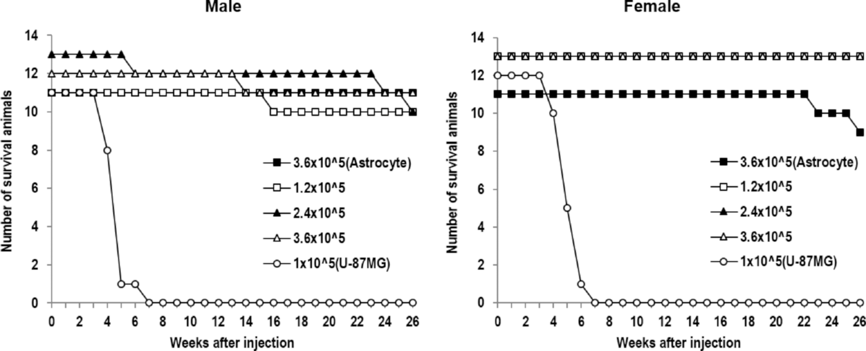
The survival of male and female SCID mice injected with hAT-MSC.sTRAIL at 26 weeks.
No adverse clinical signs were noted in any of the hAT-MSC.sTRAIL groups. There was no statistically significant change in the body weight of male mice in any of the hAT-MSC.sTRAIL groups compared with those of the negative control group. However, the body weight of the female mice of the low-dose hAT-MSC.sTRAIL group significantly increased compared with that of the negative control group at 16 and 24 weeks (Fig. 1B). The body weight of the female mice of the medium-dose hAT-MSC.sTRAIL group was also significantly higher than that of the negative control group at 24 weeks after injection. The increase in body weight was transient indicating intermittent body weight changes with little relationship to the hAT-MSC.sTRAIL injection.
26-week tumorigenicity study: organ weights and histopathological changes
The absolute and relative organ weights at 26 weeks after the injection are summarized in Supplementary Table S4. Significant decreases in absolute and relative liver weights were observed in the female mice of the high-dose hAT-MSC.sTRAIL group. The absolute and/or relative adrenal gland weights also significantly increased in the female mice of the medium-dose group. These changes were sporadic and were not correlated to the number of transplanted cells. Moreover, there was no evidence of tumor in the organs that showed increased weight. On the other hand, brain tumor formation was observed in the positive control U-87 MG group, which excluded the mice of the positive control group from the organ weight analysis.
Histological analysis showed that all male and female mice injected with 1.0 × 10⁵ U-87 MG glioma cells developed tumors in the brainstem (Supplementary Table S5 and Fig. 3). In contrast, no mice in any of the hAT-MSC.sTRAIL groups developed a tumor, indicating that hAT-MSC.sTRAIL did not induce tumor up to 3.6 × 10⁵ cells/mouse.
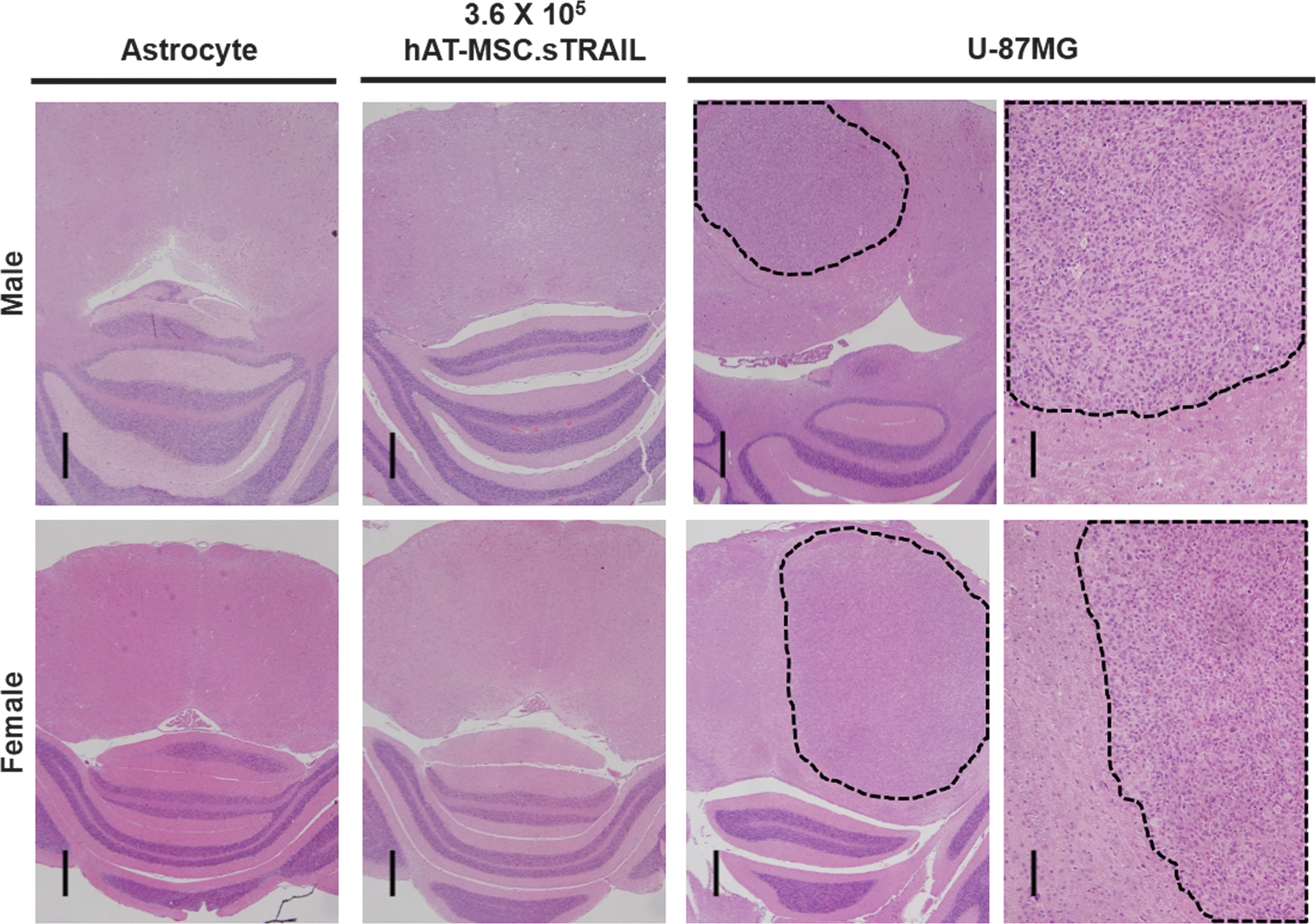
Representative histological images of the mouse brain. Dotted lines indicate the transplanted cell regions. Scale bars indicate 1.0 mm. Color images available online at
Biodistribution of hAT-MSC.sTRAIL in normal mice
To confirm whether the hAT-MSC.sTRAIL remains in the brain tissues or migrates to the other organs, we examined histological sections of various organs of the male and female mice of the high-dose hAT-MSC.sTRAIL group at 15 and 26 weeks after the transplantation.
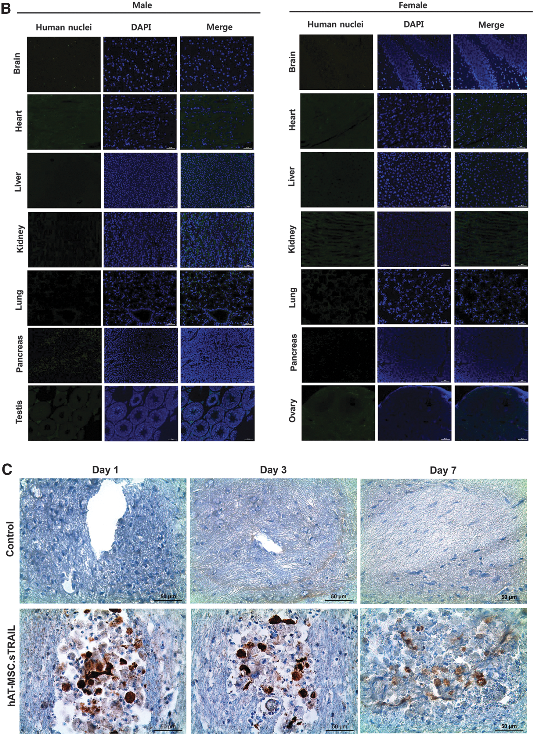
The tissue slides were stained with human nuclei-specific antibody to detect human cells. In all organs, we could not detect any human cells and abnormalities such as tissue damage and tumor formation at both 15 (Fig. 4A) and 26 weeks (Fig. 4B) after the injection. Our data suggest that intracranially injected hAT-MSC.sTRAIL does not migrate into the major organs out of the brain in normal mice.

The biodistribution of hAT-MSC.sTRAIL in normal mice. Various organs and tissues from male and female mice in the high-dose group (3.6 × 10⁵ hAT-MSC.sTRAIL/18μL) were harvested and stained for human nuclei. No human nuclei were detected in any of the tissues evaluated, including the brain, heart, liver, lungs, kidneys, testes, and ovaries at
The hAT-MSC.sTRAIL in brainstem for a short period of time
We evaluated TRAIL secretion levels by the hAT-MSC.sTRAIL at the site of injection during the early phase of study. Immunohistochemistry results displayed strong TRAIL expression at the injection site of brain at 1 and 3 days, but weak expression at 7 days (Fig. 4C). No TRAIL expression was observed in the control group. This result indicated that hAT-MSC.sTRAIL could actively produce sTRAIL until at least 1 week after injection.
Biodistribution of transplanted hAT-MSC.sTRAIL in brainstem glioma-bearing mice
In brainstem glioma-bearing mice, DiI-labeled hAT-MSC.sTRAIL mainly localized to the tumor foci at 4 weeks (Fig. 5A). However, the numbers of DiI-positive cells gradually decreased at 9 and 14 weeks. RT-qPCR was performed to quantify the biodistribution of cells (hAT-MSC.sTRAIL) and gene (sTRAIL) in the major organs. The expression level of human-specific GAPDH (Fig. 5B) and sTRAIL (Fig. 5C) were below the limit of detection in all major organs. No abnormalities, including tissue damage or tumor formation were found in major organs after hAT-MSC.sTRAIL injection in brainstem glioma-bearing mice.
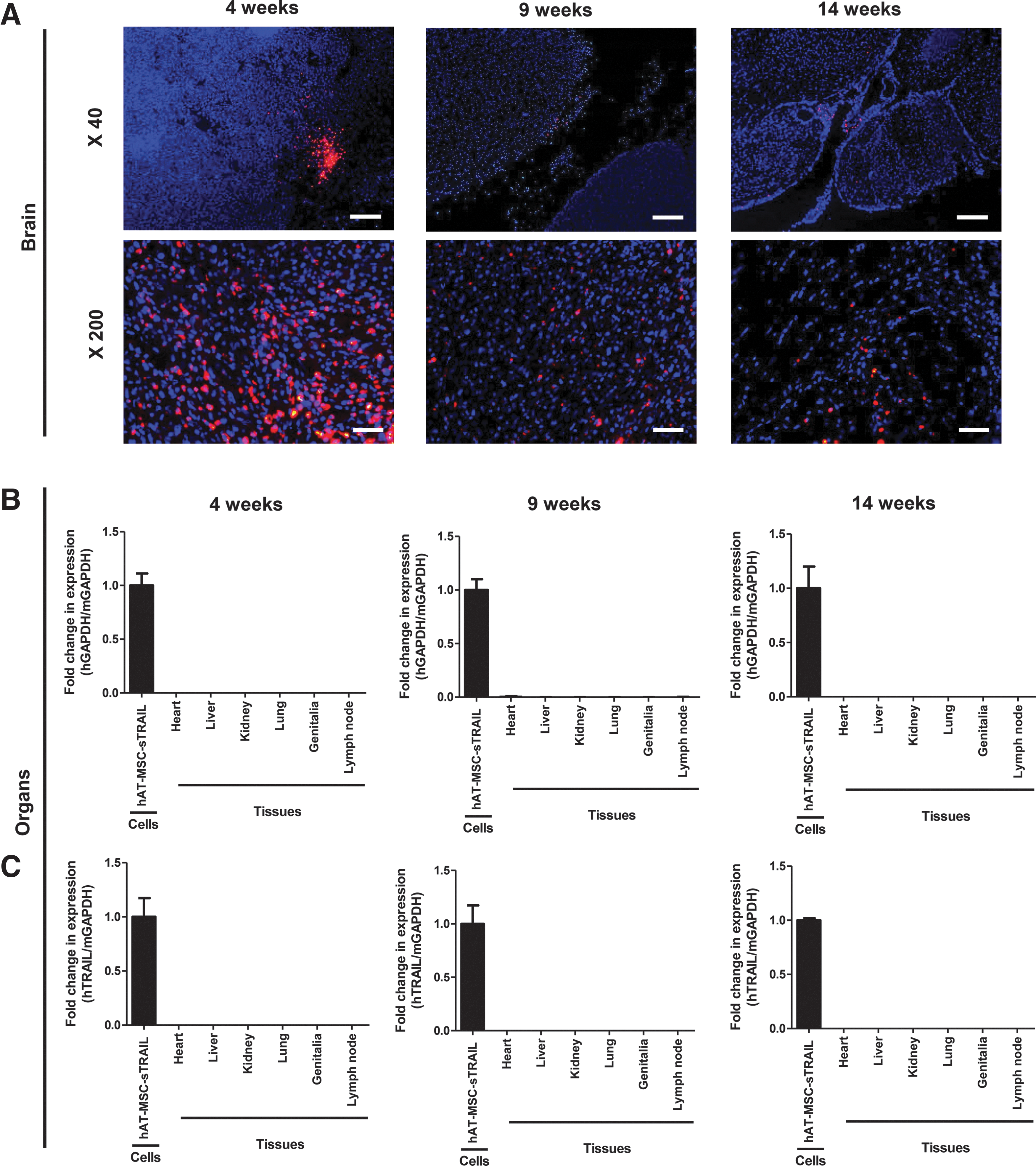
The biodistribution of hAT-MSC.sTRAIL in brainstem gliomas-bearing mice.
Discussion
Human clinical trial for brainstem glioma treatment using hAT-MSC.sTRAIL has been designed as follows. The enrolled patients will first undergo harvest of their fat tissue at the time of biopsy for diagnosis, from which MSC will be cultured. An Ommaya reservoir will be inserted into the tumor mass simultaneously with the biopsy procedure, as the route of direct access to the tumor. The patients will undergo standard treatment, including chemotherapy and radiation. The treatment using hAT-MSC.sTRAIL will be applied to those in whom the standard treatment has failed. Previously cultured autologous hAT-MSC will be genetically engineered to express sTRAIL at this stage of trial. Then, injection of those cells will be done through the Ommaya reservoir. The injection number of hAT-MSC.sTRAIL will increase according to the approved clinical trial protocol.
The purpose of the preclinical study was to identify (1) doses for phase I clinical trial, (2) local toxicity, (3) systemic toxicity, (4) secondary tumor formation, and (5) biodistribution. Based on the clinical trial synopsis, hAT-MSC.sTRAIL was locally transplanted to the brainstem of mouse. Since mouse version of the Ommaya reservoir is not available, local injection of hAT-MSC.sTRAIL was performed using a stereotactic device, as previously reported [13]. Moreover, we identified the maximal safe injection volume into the brainstem of mouse to avoid local toxicity by injection itself. Previous clinical trials for stem cell therapeutics against various spinal cord diseases used 1 × 10⁶–1 × 10⁸ stem cells for single transplantation, which indicated that 1 × 10⁷–5 × 10⁷ would be optimal for the future clinical trial of hAT-MSC.sTRAIL [18 –20]. The low- (1.2 × 10⁵ hAT-MSC.sTRAIL/mouse), medium- (2.4 × 10⁵), and high (3.6 × 10⁵)-dose groups are equivalent to 1.42 × 10⁷, 2.84 × 10⁷, and 4.26 × 10⁷ for human children (20 kg) [21], respectively.
Local and systemic toxicity of hAT-MSC.sTRAIL into the brainstem was monitored for 15 weeks in this study. There were minor differences between the negative control group and the hAT-MSC.sTRAIL groups in the body weight, daily food/water consumption, hematologic parameters, serum biochemistry, and organ weights. However, those rare differences were transient without any trends and dose dependencies. The sporadic statistical significances could result from expected false positive results in the multiple statistical comparisons; with the P < 0.05 setting, five false positive statistical significances could be observed in 100 analyses theoretically [22,23]. Since no obvious local and systemic toxicity was detected in this preclinical examination, injection dose of hAT-MSC.sTRAIL could be elevated to 5.0 × 10⁷ in the future phase I clinical trial.
Secondary tumor formation of hAT-MSC.sTRAIL that was locally injected into the brainstem was monitored for 26 weeks in normal mice. Since the lifespan of mouse is about 2 years, 6 months would be equivalent with 20 years in human given that the lifespan of human is 80 years. Moreover, the standard observation duration of tumorigenic potential of cells has been 6 months in various animal models [24,25]. In the 26-week tumorigenicity study, we could not observe any tumors derived from hAT-MSC.sTRAIL, whereas the positive control U-87 MG human glioma cells made huge brain tumors in all mice. The results demonstrated that hAT-MSC.sTRAIL has little in vivo tumorigenic potential. Although several articles have provided relevant data for protumorigenic potential of MSCs [26 –30], the clinical setting of hAT-MSC-sTRAIL transplantation would be different from the experimental conditions. Patients harbor established brainstem glioma and have a life expectancy of less than 1 year in spite of modern multimodality treatment. Moreover, we have not observed any increased tumor growth when hAT-MSC-sTRAIL was injected into brainstem glioma in the previous research [13]. Therefore, although MSCs might have protumorigenic potential at the initial stage of tumor formation, treatment effects of AT-MSC-sTRAIL against established brainstem glioma would surpass the potential protumorigenic activities in the progression of brainstem glioma.
In the biodistribution assay, no residual hAT-MSC.sTRAIL was observed in the normal mice at 15 and 26 weeks after the local transplantation, which indicates that bioavailability of hAT-MSC.sTRAIL might be shorter than 15 weeks and that possible tumorigenesis after 26 weeks would be minimal. The survival of hAT-MSC might not be sustainable without tumor or stimuli [8]. In brainstem glioma-bearing mice, we evaluated whether the hAT-MSC.sTRAIL remained in brain and major organs at 4, 9, and 14 weeks. DiI-labeled hAT-MSC.sTRAIL was observed in the tumor foci at 4 weeks, but decreased at 9 and 14 weeks. No mRNA of human GAPDH and sTRAIL was detected in major organs other than brain by RT-qPCR.
Given that bioavailability of hAT-MSC.sTRAIL is shorter than 15 weeks, it could be expected that treatment effects of hAT-MSC.sTRAIL against brainstem gliomas could be short term. In this case, various repeated injections of hAT-MSC.sTRAIL through the Ommaya reservoir are possible alternative therapeutic options for brainstem glioma patients. However, additional preclinical tests using multiple transplantations of hAT-MSC.sTRAIL should be performed before clinical trials for alternative protocols.
In this study, we preclinically examined various expected side effects of stem cell-based gene therapy using hAT-MSC.sTRAIL as a model system. Those carefully designed tests have demonstrated that hAT-MSC.sTRAIL would have little short- and long-term side effects in the clinical trials. Based on the preclinical results, clinical trials would be developed more reasonably with less risk. In addition, present results could be used as a reference of preclinical tests for newly developed stem cell-based gene therapeutics.
Footnotes
Acknowledgments
This study was supported by a grant from the Korea Health Technology R&D Project, Ministry of Health & Welfare, Republic of Korea (A120446), and by the Seoul National University Hospital Research Fund (30-2014-0270).
Author Disclosure Statement
No competing financial interests exist.
References
Supplementary Material
Please find the following supplemental material available below.
For Open Access articles published under a Creative Commons License, all supplemental material carries the same license as the article it is associated with.
For non-Open Access articles published, all supplemental material carries a non-exclusive license, and permission requests for re-use of supplemental material or any part of supplemental material shall be sent directly to the copyright owner as specified in the copyright notice associated with the article.